DOI: 10.5935/2359-4802.20180092
ORIGINAL ARTICLE
Mailing Address: André Luiz Lisboa Cordeiro
Rua Japão, 94. Postal Code: 44052-022, Caseb, Feira de Santana, BA - Brazil. E-mail: [email protected]
Behavior of Pulmonary Function after Hospital Discharge in Patients Submitted to
Myocardial Revascularization
André Luiz Lisboa Cordeiro,1,2 Letícia Gardênia Romualdo da Silva,2 Milena Oliveira Pinto,2 Jaclene da Silva Araújo,2
André Raimundo Guimarães,3 Jefferson Petto1,4,5,6
Escola Bahiana de Medicina e Saúde Pública,1 BA - Brazil Faculdade Nobre,2 BA - Brasil
Instituto Nobre de Cardiologia,3 BA - Brasil Universidade Salvador,4 BA - Brasil Faculdade Adventista da Bahia,5 BA - Brasil Faculdade Social da Bahia,6 BA - Brasil
Manuscript received on June 03, 2018; reviewed on August 07,2018; accepted on August 20,2018.
Abstract
Background: Coronary artery bypass grafting (CABG) is a type of surgery where there is a compromise in one or more coronary arteries, with the objective of restoring function to the areas that have been compromised in the heart, possibly leading to respiratory compromise and postoperative complications. The return time of the pulmonary function to the preoperative condition is still indeterminate in the literature.
Objective: To describe the behavior of pulmonary function after hospital discharge in patients submitted to CABG.
Methods: This is a prospective cohort study. Only patients undergoing MRI, whose lung function was evaluated preoperatively, at hospital discharge and 30 days after surgery, were evaluated. This evaluation consisted of maximum inspiratory pressure (MIP) and expiratory (MEP), vital capacity (VC) and expiratory flow peak (EFP).
Results: A total of 30 patients were evaluated, of which 18 (60%) were males, mean age 62 ± 9 years. A reduction in lung function from preoperative time to hospital discharge was observed in all variables. There was improvement in MIP (88 ± 9 vs 109 ± 5, p < 0.001), MEP (67 ± 10 for 90 ± 8, p < 0.001) and EFP (310 ± 59 for 390 ± 32, p < 0.001), high for review. At the time of the review, no variables returned to their preoperative value: MIP (116 ± 5 for 109 ± 5, p = 0.43), MEP (111 ± 8 for 90 ± 8, p < 0.001), VC (45 ± 12 for 39 ± 7, p = 0.33) and EFP (430 ± 40 for 390 ± 32, p < 0.001).
Conclusion: It is concluded that MRI surgery reduces lung function and is not reestablished after 30 days of the procedure. Being the expiratory muscular force and the peak of expiratory flow the most affected. (Int J Cardiovasc Sci. 2018; [online].ahead print, PP.0-0)
Keywords: Cardiovascular Diseases/physiopathology; Coronary Artery Bypass, Myocardial Infarction; Myocardial Revascularization; Patient Discharge.
Introduction
Coronary artery bypass grafting (CABG) is a type of cardiac surgery where one or more impaired coronary arteries receive saphenous or mammary grafts and whose surgical goal is to reestablish a connection to areas that have been affected in the heart. Studies show that, in Brazil, in 2010, 21,000 coronary artery bypass grafting surgeries were performed and they have been some of
the most performed cardiac procedures in recent years.1
Coronary artery bypass grafting (CABG) improves the patients’ quality of life, but there are associated factors that may compromise respiratory function. There is a great chance that these patients will develop postoperative complications, including the following most common ones: pulmonary edema, pneumothorax,
pleural effusion, atelectasis and pneumonia.2
Pulmonary function is significantly reduced in the immediate postoperative period and the reason
Operative conditions such as cardiopulmonary bypass (CPB), surgical incision, anesthesia, the patient’s hemodynamics, type and duration of the procedure, length of drainage or pain can cause lung disorders. All of these components seem to lead to decreased pulmonary volumes and capacities, thoracic expansion
and respiratory muscle dysfunction.2,4
The efficacy of respiratory physiotherapy is as an important marker for cardiac rehabilitation both inside
and outside the intensive care unit.5 The physiotherapist
contributes directly to the best prognosis, prevention and treatment in patients who undergo coronary artery bypass grafting, where rehabilitation treatments are applied, such as: manual, respiratory and pulmonary reexpansion
maneuvers, postural advice and bed positioning.6
There is still little information on the behavior of lung function after cardiac surgery related to the time required to reestablish the lungs to preoperative values. Therefore, the objective of this study was to describe the behavior of pulmonary function after hospital discharge in patients undergoing CABG.
Methodology
This is a prospective study involving patients from a reference hospital in Feira de Santana - BA, Brazil, from August 2017 to April 2018. This study was approved by the research ethics committee of Faculdade Nobre (FAN) of Feira de Santana-Bahia under opinion no. 2.088.639. All patients signed an Informed Consent Form.
The inclusion criteria were individuals of both sexes, aged equal to or above 18 undergoing coronary artery bypass grafting procedure via median sternotomy and extracorporeal circulation. The exclusion criterion were patients who were readmitted to the hospital, patients who did not return for the review, who found it hard to understand or to collaborate, who present post-surgical complications, hemodynamic instability, previous cardiac surgery, previous neurological symptoms, cardiac arrhythmia, and who remained at least 6 days in hospital.
After meeting all inclusion criteria, the patients had their pulmonary function assessed preoperatively. This evaluation consisted of maximal inspiratory pressure (MIP) and maximum expiratory pressure (MEP), and vital capacity (VC).
Respiratory muscle strength was tested with the patient sitting on a chair. Initially, the patient was asked to breathe close to the current volume and, after three breaths, to perform a maximal forced expiration (residual volume) and
then a maximal static inspiration sustained for 3 seconds with nasal occlusion in order to get the MIP measure.
Subsequently, to measure the PEM, the patient had to breathe close to the current volume for three cycles and perform a maximal inspiration (total pulmonary capacity) followed by a maximum sustained expiration for 3 seconds. Both maneuvers were repeated at least three times with an interval of one minute and the highest value, which cannot be the one from the last measurement, was recorded. These measurements were performed using an analogue Instrumentation Industries manovacuometer model MV - 120, with a range of 0 to
120 cmH2O.
Vital capacity was measured using the Wright Mark 8 analogue ventilator (Ferraris) with a 35 mm display, two 0-1 l/min and 0100 l/min dials. The patients are required to perform a maximal inspiration up to the total lung capacity and then a slow maximal expiration until the residual volume is reached. All patients should be sitting and perform three measurements, with a one-minute interval between them, adopting the highest value obtained as a reference.
After the initial evaluations, the patients were referred to the surgical center where they were always handled by the same medical team and were later sent to the intensive care unit (ICU) where they were managed according to the unit’s routine, which consists of non-invasive ventilation, breathing exercises and positive end-expiratory pressure. Finally, they were admitted to the ward after discharge from the ICU.
Upon hospital discharge, the patients had their lung function evaluated again. Note that no researcher has influence on the procedures adopted during the hospitalization or decision on hospital discharge.
One month after the surgery, the patients were evaluated for respiratory muscle strength and vital capacity upon their return to medical reevaluation. The evaluations at the two moments are always carried out by the same examiner.
Statistical analysis
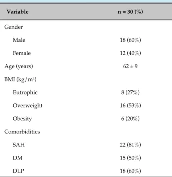
Table 1 - Clinical characteristics of the patients who had coronary artery bypass grafting
Variable n = 30 (%)
Gender
Male 18 (60%)
Female 12 (40%)
Age (years) 62 ± 9
BMI (kg/m2)
Eutrophic 8 (27%) Overweight 16 (53%)
Obesity 6 (20%)
Comorbidities
SAH 22 (81%)
DM 15 (50%)
DLP 18 (60%)
BMI: body mass index; SAH: systemic hypertension; DM: diabetes mellitus; DLP: dyslipidemia.
Table 2 - Surgical characteristics of patients that had coronary artery bypass grafting
Variable Mean and standard
deviation
CPB time (minutes) 88 ± 15
MV time (hours) 7 ± 3 Number of grafts 2 ± 0.8 Number of surgical drains 2 ± 0.4
CPB: cardiopulmonary bypass; MV: mechanical ventilation.
Results
Between August 2017 and April 2018, 30 patients who were admitted for coronary artery bypass grafting at Instituto Nobre de Cardiologia were evaluated. Of these, 18 (60%) were men and the average sample age was 62 ± 9. Other data related to the patient’s clinical characteristics are presented in table 1.
The patients had CPB time of 88 ± 15 minutes and average MV time of 7 ± 3 hours. Other data related to the patients’ surgical characteristics are shown in table 2.
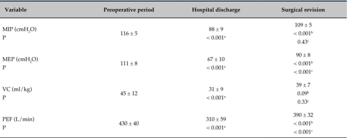
Table 3 shows the three evaluation phases in the variables. There was a significant reduction in pulmonary function behavior at hospital discharge compared to the preoperative period and surgical revision. At hospital
discharge, MIP (cmH2O) dropped to 88 ± 9 and the
preoperative value was 116 ± 5 (p < 0.001), as well as in the surgical revision with 109 ± 5 (p < 0.001). In MEP
(cmH2O), the variables also dropped compared to the
preoperative period. At hospital discharge, the value was 67 ± 10 (p < 0.001) and, at surgical revision, it was 90 ± 8 (p < 0.001).
As for the variables of vital capacity (ml/kg), the preoperative period obtained 45 ± 12, and there was as a decrease in the hospital discharge figures (31 ± 9,
p < 0.001) and surgical revision figures (39 ± 7, p = 0.33). At the peak expiratory flow (L/min), the preoperative period was 430 ± 40. At hospital discharge, the value dropped to 310 ± 59 (p < 0.001) and, at surgical revision, it was 390 ± 32 (p < 0.001).
Discussion
Based on the results reported, it can be seen that coronary artery bypass grafting surgery may lead to reduced pulmonary function and it is not reestablished even after one month of the surgical procedure.
According to Barros et al.,7 inspiratory and peripheral
muscle strength is often described as reduced after cardiac surgery. As analyzed in this study. Patients undergoing cardiac surgery repeatedly develop postoperative pulmonary dysfunction, always presenting severe reduction in pulmonary volumes, impaired respiratory
mechanics and increased respiratory work.8
According to Cavenaghi et al.,9 pulmonary
dysfunction after cardiac surgery is normal, in which the patient has severely reduced pulmonary volumes and capacities, decreased lung compliance, poorer respiratory mechanics and increased respiratory work.
For Soares et al.,10 some intraoperative factors that may
interfere with and justify the impairment of pulmonary function, such as the use of CPB, the degree of sedation, the intensity of surgical manipulation and the number of pleural drains, are indicated as some responsible for altering respiratory mechanics.
Table 3 - Behavior of pulmonary function at different operative moments
Variable Preoperative period Hospital discharge Surgical revision
MIP (cmH2O)
P 116 ± 5
88 ± 9 < 0.001a
109 ± 5 < 0.001b
0.43c
MEP (cmH2O)
P 111 ± 8
67 ± 10 < 0.001a
90 ± 8 < 0.001b
< 0.001c
VC (ml/kg)
P 45 ± 12
31 ± 9 < 0.001a
39 ± 7 0.09b
0.33c
PEF (L/min)
P 430 ± 40
310 ± 59 < 0.001a
390 ± 32 < 0.001b
< 0.001c
a: Comparison of preoperative period with hospital discharge; b: Comparison of hospital discharge with surgical revision; c: Comparison of preoperative period with surgical revision. MIP: maximum inspiratory pressure; MEP: maximum expiratory pressure; VC: vital capacity; PEF: peak expiratory flow. All analyses were conducted using the Student’s paired t-test.
discharge and surgical revision. These findings were evidenced in other studies on pulmonary function after cardiac surgeries, which showed that pulmonary function remains 25% to 30% lower even after 3.5
months of surgery.11,12
Urell et al.,13 state that respiratory muscle strength is
not compromised two months after cardiac surgery. In
the study by Jonsson et al.,14 it was evidenced that after
two months of cardiac surgery there is an increase in pulmonary function (vital capacity, functional residual capacity and total lung capacity) associated with increased physical activity level and, consequently, functionality. These results were not replicated in this study and may be related to the smaller sample size than the study
by Jonsson et al.,14 Another cause for this difference in
results may lie in the possibility of pain impacting lung function for up to 45 days due to the scarring process. As the patients in this study were evaluated after thirty days, there may be some interference, as opposed to the
study by Jonsson et al.14
Pain has a significant negative correlation with the decreased values in the variables of the study of
Baumgarten et al.15 In this study, pain was not evaluated,
but this may be a possible explanation for a reduction in the variables of this study.
Mueller and Lima16 state in their study that most
patients that underwent cardiac surgery complain of intense postoperative pain, being one of the main causes
of pulmonary complications due to the lower thoracic expansion, thus generating a shallow breathing.
Static and dynamic complacencies that reflect pulmonary function may be significantly reduced even
after one year of surgery.17 In the study by Annoni et
al.,1 patients undergoing coronary artery bypass grafting
presented increased expiratory muscle strength and, consequently, peak expiratory flow, in addition to showing improvement in quality of life. As opposed to the results of this study, which shows inconsistent responses, since the PEF, MIP, MEP and VC values were smaller, thus showing loss of muscle strength.
These differentiated results can be associated with several factors. In the study by Annoni et al.,1 all patients
were followed up by physiotherapy that prescribed individualized exercises at least twice a day. All patients were encouraged to perform a 6-minute walk test (6MWT) over 50 meters with medium intensity
in the preoperative period. According to Laizo,18 the
6-minute walk test (6MWT) is a method that has been used to evaluate functional capacity and is also used as a predictor of morbidity and mortality in patients with cardiovascular and respiratory diseases.
1. Annoni R, Silva WR, Mariano MS. Análise de parâmetros funcionais pulmonares e da qualidade de vida na revascularização do miocárdio. Fisioter Mov. 2013;26(3):525-36.
2. Oh HC, Han JW, Choj JW, Kim YH, Hwang HY, Kim KB. Concomitant off-pump coronary artery bypass and non-cardiovascular surgery. J Thorac Dis. 2016;20(8):2115-20.
3. Roncada G, Dendale P, Lisen L, Hendrikx M, Hansen D. Reducion in pulmonary function after CABG surgery is related to postoperative inflammation and hypercortisolemia. Int J Clin Exp Med. 2015;8(109):38-46.
4. Jensen L, Yang L. Risk factors for postoperative pulmonary complications in coronary artery bypass graft surgery patients. Eur J Cardiovasc Nurs. 2007;6(3):241-6.
5. Roque V, Machado Vaz I, Maia M, Rocha A, Araújo V, Maciel MJ, Parada F. Preditores da capacidade funcional em doentes coronários. Revista da Sociedade Portuguesa de Medicina Física e de Reabilitação. 2013; 23(1).:29-37.
6. Mello DLV. Intervenções fisioterapêuticas em pacientes submetidos a revascularização do miocárdio: Uma revisão de literatura. [Internet]. [Citado em 2017 dez 10]. Disponível em: https://www.repositorio. bahiana.edu.br:8443/jspui/handle/bahiana/446
7. Barros GF, Santos CS, Granado FB, Costa PT, Límaco RP, Gardenghi G. Treinamento muscular respiratório na revascularização do miocárdio. Rev Bras Cir Cardiovasc. 2010; 25(4):483-90.
8. Renault JA, Costa R, Rossetti MB. Fisioterapia respiratória na disfunção pulmonar pós-cirurgia cardíaca. Rev Bras Cir Cardiovasc. 2008;23(4):562-9.
9. Cavenaghi S, Ferreira LL, Marino LHC, Lamari NM. Fisioterapia respiratória no pré e pós-operatório de cirurgia de revascularização do miocárdio. Rev Bras Cir Cardiovasc. 2011;26(3):455-61.
10. Soares GM, Ferreira DC, Gonçalves MP, Alves TG, David FL, Henriques KM, et al. Prevalência das principais complicações pós-operatórias em cirurgias cardíacas. Rev Bras Cardiol. 2011;24(3):139-46.
11. Ferreira GM, Haeffner MP, Barreto SS, Dall’Ago P. Espirometria de incentivo com pressão positiva expiratória é benéfica após revascularização miocardio. Arq Bras Cardiol. 2010; 94(2):230-5.
12. Wynner R, Botti M. Postoperative pulmonary dysfunction in adults after cardiac surgery with cardiopulmonary bypass: clinical significance and implications for practice. Am J Crit Care. 2004;13(5):384-93.
13. Urell C, Emtner M, Hedenstrom H, Westerdahl E. Respiratory muscle strength is not decreased in patients undergoing cardiac surgery. J Cardiothoracic Surg. 2016;11:41.
14. Jonsson M, Urell C, Emtner M, Westerdahl E. Self-reported physical activity and lung function two months after cardiac surgery – a prospective cohort study. J Cardiothoracic Surg. 2014;9:59.
15. Baungarten MCS, Garcia GK, Frantzeski MH, Giacomazzi CM, Lagni VB, Dias AS, et al. Comportamento da dor e da função pulmonar em pacientes submetidos a cirurgia cardíaca via estornotomia. Rev.Bras. Cir Cardiovasc. 2009;24(4):497-505.
16. Mueller XM, Tinguely F, Tevaearai HT, Revelly JP, Chioléro R, von Segesser LK. Pain location, distribution, and intensity after cardiac surgery. Chest. 2000;118(2):391-6.
References
The limitations of this study include the lack of a sample calculation, failure to evaluate confounding variables such as pain and lack of correlation between pulmonary function behavior and clinical and functional outcomes.
Conclusion
It can be concluded that patients undergoing coronary artery bypass grafting surgery present significant worsening of pulmonary function in the postoperative period, causing significant reduction in respiratory, expiratory and peak expiratory flow, not returning to normal even after one month of the procedure.
Author contributions
Conception and design of the research: Cordeiro ALL, Silva LGR, Pinto MO. Acquisition of data: Silva LGR, Pinto MO. Analysis and interpretation of the data: Cordeiro ALL. Statistical analysis: Cordeiro ALL. Writing of the manuscript: Silva LGR, Pinto MO. Critical revision of the manuscript for intellectual content: Cordeiro ALL, Guimarães AR, Petto J. Writing of the manuscript: Araújo
JS. Critical revision of the manuscript for intellectual content: Araújo JS.
Potential Conflict of Interest
No potential conflict of interest relevant to this article was reported.
Sources of Funding
There were no external funding sources for this study.
Study Association
This article is part of the thesis of Doctoral submitted by André Luiz Lisboa Cordeiro, from Escola Bahiana de Medicina e Saúde Pública.
Ethics approval and consent to participate
17. Westerdahl E, Jonsson M, Emtner M. Pulmonary function and health-related quality of life 1-year follow up after cardiac surgery. J Cardiothoracic Surg. 2016;11(1):99.
18. Laizo A, Delgado FE, Rocha GM. Complicações que aumentam o tempo de permanência na unidade de terapia intensiva na cirurgia cardíaca. Rev Bras Cir Cardiovasc. 2010;25(2):166-71.