1. MsC; Physiotherapist. 2. Specialist; Physiotherapist.
This study was carried out at Ministro Costa Cavalcanti Hospital, Foz do Iguaçu, PR, Brazil.
Christiane RIEDI1,Cintia Teixeira Rossato MORA2,Taissa DRIESSEN2,Mônica de Castro Guimenes COUTINHO2, Diogo Mattos MAYER2,Fabio Luiz MORO2,Carla Regina Camargo MOREIRA2
RBCCV 44205-1220
Relação do comportamento da força muscular com as complicações respiratórias na cirurgia cardíaca
Relation between respiratory muscle strength with
respiratory complication on the heart surgery
Correspondence address: Christiane Riedi
Rua Barra Mansa, 116 - Jardim Ipê - Foz do Iguaçu, PR Brazil - CEP 85869-686.
E-mail: [email protected]
Article received on June 4th, 2010.
Article accepted on September 23rd, 2010. Abstract
Objective: Verifying the respiratory muscle strength in
cardiac surgery and the relationship with the postoperative pulmonary complications.
Methods: Sixty-three adult patients undergoing elective
cardiac surgery were enrolled in this study. In the day of the surgery all patients underwent respiratory muscle strength test. In the postoperative a multidisciplinary team took care all patients and they received physiotherapy treatment twice a day following the usual care of the hospital. In the fifth day after surgery, the patients repeat the respiratory muscle strength test and were examined for pulmonary complications on the postoperative period.
Results: A significant lower inspiratory muscle strength
(P=0.001) and expiratory muscle strength (P=0.0001) was found in the postoperative period, however it was not statistic significantly the relationship between the inspiratory muscle strength (P=0.58) and expiratory muscle strength (P=0.4), preoperatively and the presence of postoperative pulmonary complication.
Conclusions: The preoperative respiratory strength could
not be considered a predictor of postoperative pulmonary complication.
Descriptors: Cardiac surgical procedures. Postoperative
Complications. Muscle strength.
Resumo
Objetivo: Verificar o comportamento da força muscular
respiratória na cirurgia cardíaca e sua relação com as complicações respiratórias pós-operatórias.
Métodos: Participaram do estudo 63 indivíduos
submetidos à cirurgia cardíaca. No dia da cirurgia, foi realizada a avaliação da força muscular respiratória e, no pós-operatório, os pacientes foram acompanhados pela equipe multidisciplinar e submetidos à intervenção fisioterapêutica duas vezes ao dia, seguindo o protocolo padrão do hospital. No quinto dia de pós-operatório, foi realizada a reavaliação e análise das complicações respiratórias.
Resultados: Apesar da diminuição significativa da força
muscular inspiratória (P= 0,001) e expiratória (P=0,0001) no pós-operatório, não foi observada relação estatisticamente significativa entre a força muscular inspiratória (P=0,58) e expiratória (P=0,4) pré-operatória e a presença de complicações pós-operatórias.
Conclusão: A força muscular respiratória pré-operatória
não pode ser utilizada como preditor de complicações respiratórias pós-operatória.
Descritores: Procedimentos cirúrgicos cardíacos.
INTRODUCTION
The postoperative complications in cardiac surgery are still frequent, despite the development of minimally invasive techniques and improvement of care given to patients [1,2]. Complications included pulmonary, present in up to 70% of cases are defined as an abnormality that occurs in the postoperative period, which produces significant dysfunction, affecting the clinical outcome [3,4].
The most common respiratory complications are atelectasis (24.7%), pneumonia, hypoxemia and pleural effusion (47.5%). Physiotherapy has its established role in prevention and treatment of complications and intervening early aiming at recovering lung function, encourage physical activity and consequently improve the patient’s quality of life [3,4].
The physiotherapeutic follow-up is important since the preoperative period, as it enables the profissional with a thorough assessment of physical and respiratory conditions of patients and intervene therapeutically in the preoperative period, when necessary [3-6].
Among the assessment tools, measurement of respiratory muscle strength is often used, because of its easy implementation and low cost. Classification (grading) of respiratory strength has been widely studied in order to identify the presence of respiratory muscle weakness, as this interferes with the respiratory mechanics, which may hinder the rehabilitation of these patients [3-6].
Based on this, this study aims to investigate whether the presence of respiratory muscle weakness in the preoperative period may be indicative of respiratory complications in the postoperative perido and to assess its behavior after the surgical procedure.
METHODS
Sample
During the period October 2008 to October 2009 were evaluated 76 individuals. Of these, two patients were excluded due to unstable angina, three patients had their surgeries suspended and eight died postoperatively, participating 63 subjects. Among patients who died, two suffered pulmonary complications, two, neurological, two deaths occurred in the operating room and two for postoperative cardiac dysfunction.
The study was approved by a Human Research Ethics Committee, under protocol 221/2007.
Inclusion and Exclusion Criteria
Patients referred to the service of Cardiac Surgery of the Ministro Costa Cavalcanti Hospital who underwent CABG, valve replacement, aneurysm repair and atrial septal
defect were included. Of these, were excluded emergency surgery, patients with unstable angina and who were not in physical or cognitive conditions for the tests.
Preoperative evaluation
The preoperative evaluation was performed on the day of surgery by a hospital physiotherapist, who was also responsible for implementing the guidelines on the surgical procedure and postoperative period. This included an interview, which aimed to identify key risk factors, then the evaluation of measures of respiratory muscle strength and peak flow.
To assess respiratory muscle strength, an analog manometer was used (Wika MV300 model) with range of +/ -300 cmH2O.
Tests of respiratory muscle strength were performed with the patient positioned sitting with the inferior limbs pending and feet resting. The nostrils were occluded with a nose clip and the nozzle of the equipment attached to the mouth.
Maximum inspiratory pressure (MIP) was measured starting at 2-3 breaths at tidal volume with the hole open, and requested a breath as complete as possible until residual volume (RV), and the patient instructed to notify this time to then request a maximal inspiration with the hole occluded until the total lung capacity (TLC), where the individual should remain firmly supported by 2 seconds [7].
To evaluate the muscle expiratory pressure (MEP), the patient was instructed to breath at tidal volume and was requested a deep breath until TLC, warning the appraiser such moment, with the hole occluded to perform a maximum expiration at the level of RV, which should be sustained for 2 seconds [7].
Both maneuvers were repeated three times, with interval freely performed by the patient and recorded the best measure. For the calculation of the projected pressure was used the equations proposed by Neder et al. [8].
After surgery, patients were followed postoperatively by the multidisciplinary team. The physiotherapy intervention was performed twice daily for a period of approximately 20 minutes, following the standard protocol of postoperative cardiac surgery of the hospital.
During the time of hospital stay, patients were evaluated daily by the multidisciplinary team in order to identify the postoperative respiratory complications, which were recorded by the physiotherapist in a specific form.
Determination of postoperative complications
The preoperative values were compared with those predicted in the literature by means of t test for unpaired samples and with values obtained postoperatively by means of t test for paired samples.
RESULTS
Patients included in the study had a mean age of 54.05 ± 13.6 years, these 32% (20) were female and 68% (43) male, seven (11%) were smokers, 25 (40% ) were ex-smokers (abstinence for more than 6 months), 11 (17%) had pulmonary disease, 43 (68%) were hypertensive, 13 (21%) diabetics and 29 (43%) had dyslipidemia, according to Table 1.
Chart 1. Classification of postoperative respiratory complications.
Type 1
Type 2:
Type 3
Type 4
• Dry cough
• Microatelectasis and temperature greater than 37.5 degrees without documented cause
• Dyspnea without documented cause
• Productive cough not assigned to specific cause
• Bronchospasm (wheezing) and need to
change the treatment
• Hypoxemia with signs and symptoms of
wheezing and dyspnea
• Atelectasis with radiological confirmation associated to temperature greater than 37.4°C or abnormal pulmonary findings • Hypercapnia with need of treatment • Pleural effusion requiring thoracentesis
• Suspected pneumonia (radiological
evidence without confirmation of bacterial meningitis)
• Pneumonia with radiological evidence and confirmation in the bacterioscopy • Pneumothorax
• Reintubation with mechanical ventilation period not exceeding 48h
• Ventilatory failure: ventilator dependency in the postoperative period exceeding 48h • Intubation with subsequent dependence on mechanical ventilation for more than 48h
Table 1. Characteristics of patients undergoing cardiac surgery.
Risk factors
Smoking Ex-Smoking Respiratory disease Arterial Hypertension Diabetes
Dyslipidemia BMI Normal Overweight Obesity I Obesity II Obesity III
Sim
7 (11%) 25 (40%) 11 (17%) 43 (68%) 13 (21%) 29 (43%)
31.2% 41.6% 20.8% 2% 4%
Não
56 (89%) 38 (60%) 52 (83%) 20 (32%) 50 (79%) 34 (54%)
Complicações Respiratórias 2 (28.5%) 13 (52%) 7 (63%) 20 (46.5%)
3 (23%) 14 (48.2%)
5 (25%) 11 (42.3%)
5 (38.4%) __ 3 (66.3%)
BMI: body mass index
Postoperative evaluation
The revaluation was performed at 5 days postoperatively, with the patient already admitted to the ward. If patients were still in the Coronary Care Unit, this was performed later, repeating the same maneuvers performed preoperatively.
Statistical analysis
For statistical analysis, in order to compare the preoperative respiratory muscle strength with the complications, it was determined in the limit of MIP in 80
cmH20. This value was determined, because the ATS/ERS
[10], in the guidelines for pulmonary function testing, affirms that values less than this may be indicative of weakness. With MIP subdivided into higher and lower than - 80 cmH2O, it was performed using the McNemar test, the investigation of the influence of values recorded preoperatively with the complications encountered postoperatively.
For comparison of MEP, the analysis was based on the study of Hulzebos et al. [9], who found that values greater than 75% predicted are positive indicators of decreased complications.
Among the risk factors, former smokers and patients with respiratory disease had higher respiratory complications at 52% and 63% respectively (Table 1).
When analyzed the body mass index (BMI) of these individuals, it was found that 41.6% were overweight, 20.8% presented with grade 1 obesity, obesity grade 2 2% and 4% for obesity grade 3, the remaining individuals (31.2%) had normal BMI. By correlating the pulmonary complications with BMI, we found that 41% presented overweight, while 25% had normal BMI.
Among the surgeries performed, 55% had CABG, 24%, valve replacement, 10%, other surgeries and 11% associated surgeries. Of these, 68% were performed with cardiopulmonary bypass (CPB) with a mean of 81.7 ± 44.34 minutes and 32% without CPB. The average time of anesthesia was 281 ± 60 minutes.
When assessing inspiratory muscle strength, the
and 91.5 ± 52.2 cmH2O in the postoperative, where it is observed a decrease of 11% of preoperative in relation the postoperative, a value statistically significant (P=0.001). In relation to the predicted value using the equation of Neder et al. [8], there were no significant differences between preoperative values and predicted for age, which was on average 101.5 ± 14.5 cm H2O (p = 0.84) (Table 2).
observed that irrespective of the type of surgery, the most common complications were type II.
DISCUSSION
By analyzing the profile of patients undergoing cardiac surgery in this study, we observe the presence of different comorbidities and risk factors. Smoking was present in 56% of the cases and diabetes in 16%, data a little smaller than those found in the study of Feier et al. [11], who evaluated the profile of patients undergoing cardiac surgery in 1991-1992 and 2001-2002 and found 63% of smokers and 36.4% of diabetics in the latest period. In relation to respiratory diseases, Feier et al. [11] found 6.5%, while in this study, we found 14%.
Despite these differences, data from this study are more similar to those found by Feier et al. [11] in 2001-2002 when compared with those collected in 1991-1992, which shows a changing pattern in these patients.
In relation to BMI, there was a greater number of patients with overweight (41.6%), this finding becomes important when analyzing the study by Kajimoto et al. [12], who assessed the 10-year survival of diabetic patients with metabolic syndrome who underwent cardiac surgery and found that the metabolic syndrome regardless of diabetes is a factor of decreased survival of patients.
In another study assessing the ventilatory profile of patients undergoing coronary artery bypass grafting, Morsch et al. [13] also reported similar data with an average BMI of 27, emphasizing that a sedentary lifestyle and obesity are risk factors for these cardiovascular patients.
By linking respiratory muscle strength preoperatively with complications in the postoperative, it seems that this was not statistically significant, which differs from the data found by Hulzebos et al. [9], who analyzed the pulmonary complications based on risk factors preoperatively, considering four main risk factors: age over 70 years, productive cough, smoking in the last eight weeks prior to surgery and diabetes mellitus. Two factors were also considered as protective for pulmonary complications, inspiratory capacity and maximal expiratory pressure, when they were greater than 75% predicted [6].
In this study, when evaluating the risk factors, we found that patients with respiratory diseases and ex-smokers presented the highest frequency of complications, since diabetic patients showed only 23%. This may be due to the possible difference in the profile of the patients, because in the study by Hulzebos et al. [9] were included only CABG, and it was reported the use or not of cardiopulmonary bypass.
In another study, Hulzebos et al. [14] used inspiratory muscle training in order to prevent pulmonary complications in high risk patients undergoing coronary artery bypass Table 2. Behavior of respiratory muscle strength.
Measurement MIP (cm of H2O) MEP (cm of H2O)
Pre 106.2± 49.42 89.18±30.18
Post 91.5±52.2* 66.8±22.11
Predicted 101.5±14.5† 105.23±19.5
MIP: maximal inspiratory pressure, MEP: maximal expiratory pressure; * statistically significant between pre and post, † statistically significant between pre and predicted
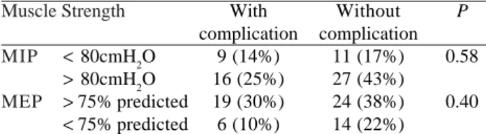
Table 3. Relationship between MIP, MEP and respiratory complications.
Muscle Strength
MIP
MEP
< 80cmH2O > 80cmH2O > 75% predicted < 75% predicted
With complication
9 (14%) 16 (25%) 19 (30%) 6 (10%)
Without complication
11 (17%) 27 (43%) 24 (38%) 14 (22%)
P
0.58
0.40
MIP: maximal inspiratory pressure, MEP: maximal expiratory pressure
The mean maximum expiratory pressure preoperatively
was 89.18 ± 30.18 cm H2O in the postoperative period of
66.8 ± 22.11 cmH2O and the expected age of 105.23 ± 19.5
cmH2O, resulting in 23% reduction (P=0.0001) compared to
pre- and postoperative period and 14.8% (P=0.0001) between preoperative MEP with the predicted.
Among patients undergoing cardiac surgery, 37 (59%) did not present any respiratory complication, five (8%) had complications of type 1, 12 (19%) type 2, 6 (9%) type 3 and 3 (5%) type 4.
In the comparison of patients with respiratory complications and those who did not, with preoperative MIP and MEP, there was no statistically significant difference (Table 3).
grafting. The trained group (140 patients) showed 18% of postoperative complications and the untrained group (139 patients) showed 35% of pulmonary complications, suggesting that the greater inspiratory muscle strength the lower the risk of complications. This result was not found in this study and may be related to the smaller patient number and profile, because in the study were included only patients who underwent CABG.
Fuster et al. [15] evaluated spirometry in 1412 patients undergoing cardiac surgery, of whom 39% had abnormal spirometry, obstructive 368, 127 restrictive and 55 mixed. In patients with obstructive FEV1 <60%, there were more complications, and mortality rate 24.6% vs 1.4%, among other complications with pneumonia, sepsis, prolonged mechanical ventilation and reintubation with a higher incidence in that group [8 ]. In this study, the population with lung disease was relatively small, being only seven (14%) of the sample, but without access to pulmonary function test.
There was a reduction in the values of MIP and MEP in the postoperative period of these patients which was confirmed by Stein et al. [6] who when assessing respiratory muscle strength in pre- and postoperative period of cardiac surgery, compared to the conventional protocol with another added EPAP, observed that in both groups there was a decrease in respiratory muscle strength, which was higher in control. Importantly, in this same study, Stein et al. [6] found a significant positive correlation between respiratory muscle strength and distance walked during the 6-minute walk test, indicating that the respiratory muscle strength is related to the functional capacity of the patients.
Morsch et al. [13] also found a decrease in respiratory muscle strength postoperatively of 33.7% for MEP and 35.5% for MIP, values less than the values of this study, on which was obtained decrease of 24.7 and 11% respectively. It should be noted that in the study of Morsch et al. [13], only patients undergoing CABG with CPB were assessed, which may have interfered negatively in respiratory muscle strength. The CPB and the lack of ventilation during this period are decisive factors for pulmonary complications, this is probably due to changes in mechanical properties of the respiratory system, resulting from changes in compliance and lung resistance [16,17].
This decrease in muscle strength found in this study is consistent with a study published in 2005 by Ambrozin and Cataneo and [5], which evaluated the parameters of mechanical ventilation of patients in the immediate postoperative period of cardiac surgery and found compliance decreased in most of these, resistance increased in 1/3 of them and gas exchange (PaO2/FiO2) decreased by 50% of individuals assessed. There was no statistical difference between patients with respiratory symptoms, pulmonary disease or smoking, demonstrating that the
worsening of mechanical ventilation is related to the surgical procedure [5].
When compared to respiratory muscle strength with the predicted by the equation of Neder et al. [8], there was statistical difference only for MEP data which differ from those of van der Palen et al. [18], who found decreased muscle strength in patients with acute myocardial infarction, considering it as an independent risk factor. One possible reason to explain this finding is that in our studied sample we included patients with valve and aortic disease, which may have a positive effect on the values of respiratory muscle strength.
In another study, Lavietes et al. [19] investigated the respiratory muscle strength in patients with diastolic heart dysfunction and found that these patients had a decrease of this strenght as compared to healthy individuals. In this study, the patient’s ventricular function was not assessed. In the study by Stein et al. [6], patients undergoing cardiac surgery have achieved a MIP predicted in the preoperative period of 60% and 68% for the control and treated group, and a MEP of 82% and 89% respectively. Data that differ from this study, which showed no change in MIP found when compared with the predicted and MEP was found 76.3% of predicted. This may have occurred because of the age of individuals in this study was lower than that of Stein et al. [6], considering that the older the lesser strength found. It is worth noting that in both studies, we used the same reference equation.
CONCLUSION
No relationship was found between respiratory muscle strength in the preoperative period and postoperative complications, and it is not possible in this population to use measures of muscle strength as predictors of complications.
Patients who had higher numbers of respiratory complications were those who underwent coronary artery bypass grafting, ex-smokers, with presence of respiratory diseases and increase in body mass index.
REFERENCES
1. Sarmento GJV. Fisioterapia respiratória no paciente crítico: rotinas clínicas. 2ª ed. São Paulo:Manole;2009. p.339.
submetidos a operação de revascularização do miocárdio. Rev Bras Cir Cardiovasc. 2005;20(3):317-22.
12. Kajimoto K, Kasai T, Miyauchi K, Hirose H, Yanagisawa N, Yamamoto T, et al. Metabolic syndrome predicts 10-year mortality in non-diabetic patients following coronary artery bypass surgery. Circ J. 2008;72(9):1481-6.
13. Morsch KT, Leguisamo CP, Camargo MD, Coronel CC, Mattos W, Ortiz LDN, et al. Perfil ventilatório dos pacientes submetidos a cirurgia de revascularização do miocárdio. Rev Bras Cir Cardiovasc. 2009;24(2):180-7.
14. Hulzebos EH, Helders PJ, Favié NJ, De Bie RA, Brutel de la Riviere A, Van Meeteren NL. Preoperative intensive inspiratory muscle training to prevent postoperative pulmonary complications in high-risk patients undergoind CABG surgery: a randomized clinical trial. JAMA. 2006;296(15):1851-7.
15. Fuster RG, Argudo JA, Albarova OG, Sos FH, López SC, Codoñer MB, et al. Prognostic value of chronic obstructive pulmonary disease in coronary artery bypass grafting. Eur J Cardiothorac Surg. 2006;29(2):202-9.
16. Szeles TF, Yoshinaga EM, Alencar W, Brudniewski M, Ferreira FS, Auler Jr JOC, et al. Hipoxemia após revascularização miocárdica: análise dos fatores de risco. Rev Bras Anestesiol. 2008;58(2):124-36.
17. Malbouisson LMS, Brito M, Carmona MJC, Auler Jr JOC. Impacto hemodinâmico de manobra de recrutamento alveolar em pacientes evoluindo com choque cardiogênico no pós-operatório imediato de revascularização do miocárdio. Rev Bras Anestesiol. 2008; 58(2):112-23.
18. Van der Palen J, Rea TD, Manolio TA, Lumley T, Newman AB, Tracy RP, et al. Respiratory muscle strength and the risk of incident cardiovascular events. Thorax. 2004;59(12):1063-7.
19. Lavietes MH, Gerula CM, Fless KG, Cherniack NS, Arora RR. Inspiratory muscle weakness in diastolic dysfunction. Chest. 2004;126(3):838-44.
3. Yánez-Brage I, Pita-Fernández S, Juffé-Stein A, Martínez-G o n z á l e z U , P é r t e g a - D í a z S , M a u l e ó n - Martínez-G a r c í a A . Respiratory physiotherapy and incidence of pulmonary complications in off-pump coronary artery bypass graft surgery: an observational follow-up study. BCM Pulm Med. 2009;9:36.
4. Herdy AH, Marcchi PL, Vila A, Tavares C, Collaço J, Neibauer J, et al. Pre- and postoperative cardiopulmonary rehabilitation in hospitalized patients undergoing coronary artery bypass surgery: a randomized controlled trial. Am J Phys Med Rehabil. 2008; 87(9):714-9.
5. Ambrozin ARP, Cataneo AJM. Aspectos da função pulmonar após revascularização do miocárdio relacionados com risco pré-operatório. Rev Bras Cir Cardiovasc. 2005;20(4):408-15.
6. Stein R, Maia CP, Silveira AD, Chiappa GR, Myers J, Ribeiro JP. Inspiratory muscle strength as a determinant of functional capacity early after coronary artery bypass graft surgery. Arch Phys Med Rehabil. 2009;90(10):1685-91.
7. Souza RB. Pressões respiratórias estáticas máximas. J Pneumol. 2002;28(supl 3):S155-65.
8. Neder JA, Andreoni S, Lerario MC, Nery LE. Reference values for lung function tests. II. Maximal respiratory pressures and voluntary ventilation. Braz J Med Biol Res. 1999;32(6):719-27.
9. Hulzebos EH, Van Meeteren NL, De Bie RA, Dagnelie PC, Helders PJ. Prediction of postoperative pulmonary complications on the basis of preoperative risk factors in patients who had undergone coronary artery bypass graft surgery. Phys Ther. 2003;83(1):8-16.
10. American Thoracic Society/European Respiratory Society. ATS/ERS Statement on respiratory muscle testing. Am J Respir Crit Care Med. 2002;166(4):518-624.