Intra-operative Complications During Third Molar Removal
Universidade Fernando Pessoa
Faculdade de Ciencias de Saúde
Intra-operative Complications During Third Molar Removal
Universidade Fernando Pessoa
Faculdade de Ciencias de Saúde
Intra-operative Complications During Third Molar Removal
“Trabalho apresentado à Universidade Fernando Pessoa
como parte dos requisitos para obtenção do grau
de Mestre em Medicina Dentária”
________________________________
Abstract
The third molar, or wisdom tooth, is the most commonly impacted tooth and its removal is one of the most frequently carried out procedures in oral surgery. Its removal can be advocate for a lot of reasons such as associated pathology and prosthetics considerations among others but the management of asymptomatic disease-free impacted third molar remain questionable.
Most third molars surgeries are performed without difficulties, however sometimes this common procedure can result in several complications.
The aim of this work is to provide an overview of the impacted third molar surgery, summarize its intra-operatives complications and their management and discuss about some alternatives proposed in the current literature.
Keywords: "Impacted third molar", "Third Molar Surgery", "Wisdom Tooth Removal”, "Complication Third Molar Surgery"; "Complication Wisdom Tooth Removal".
Resumo
O terceiro molar, ou dente de siso, é o dente mais comumente impactado e sua remoção é um dos procedimentos mais frequentemente realizados em cirurgia bucal. Sua remoção pode ser indicada para muitas razões, como a patologia associada e considerações proteticas, entre outras, mas a abordagem do terceiro molar impactado assintomático e sem doença permanece questionável. A maioria das cirurgias do terceiro molar são realizadas sem dificuldades, no entanto, por vezes, este procedimento comum pode resultar em várias complicações.
O objetivo deste trabalho é fornecer uma visão geral da cirurgia de terceiro molar impactado, resumir suas complicações intra-operativas e seu gerenciamento e discutir sobre algumas alternativas propostas na literatura atual.
Palavras-chave: “Terceiro molar impactado/retido“, “Terceiro molar cirurgia", “Ablação dente de siso", “Complicação terceiro molar cirurgia", “Complicação dente de siso ablação".
GENERAL INDEX
I. INTRODUCTION 1
Materials and Methods II. DEVELOPMENT 2
1. The Impacted Third Molar 2
1.1 Definitions………2
1.2 Etiology………2
1.3 Classifications………..3
2. Surgical Procedure 3
2.1 Indications and Contraindications .………..3
i. Indications ii. Relatives contraindications iii. Prophylactic indication 2.2 Preoperative Evaluation………4
2.3 Surgical Difficulty………5
2.4 Surgical Protocol………..6
i. Flap design ii. Bone removal iii. Tooth sectioning iv. Debriding, irrigation and sutures 3. Intraoperative Complications 8
3.1 General Complications..………8 i. Damage to adjacent tooth
ii. Soft tissue injuries
iv. Hemorrhage
v. Subcutaneous or submucosal emphysema vi. Fracture of alveolar process
vii. Temporomandibular joint disorder
3.2 Specific Maxillary Complications..………10
i. Maxillary tuberosity fracture ii. Oroantral communication iii. Tooth displacement into maxillary sinus or infra temporal fossa 3.3 Specific Mandibular Complications..……….12
i. Mandibular fracture ii. Tooth displacement into adjacent structures iii. Nerve damage III. DISCUSSION 13
IV. CONCLUSION 15
V. REFERENCES/BIBLIOGRAPHY 16
VI. APPENDICES 19
1. Appendix 1: The radiographic classifications of the third molar……….19
2. Appendix 2: American Society of Anesthesiologists’ (ASA) physical status classification system………20
3. Appendix 3: Predictive variables associated with surgical difficulty during impacted third molar removal ……….……21
4. Appendix 4: Mandibular tooth sectioning technique depending of impacted tooth angulation ..………23
I - INTRODUCTION
An impacted tooth is a mature or immature tooth whose eruption process has stopped. The third molar, or wisdom tooth, is the most commonly impacted tooth. (Haroun, 2008; Juodzbalys et Daugela, 2013)
Its removal can be advocate for a lot of reasons such as associated pathology and prosthetics considerations among others. (NICE, 2000; Hupp et al, 2014)
There is many presentations of the surgical situation of impacted third molars than in any other dental surgical procedure. Generally, the surgical approach leads to adequate access to the underlying bone and tooth through the soft tissue flap. The bone should be removed using atraumatic and aseptic technique. Then, the tooth may be sectioned and removed from the socket. Finally, the wound have to be cleaned, to provide a good healing environment, before the suture of the flap. (Miloro et al, 2004)
Even if removal of impacted third molar is one of the most frequently carried out procedures in oral surgery, it's inevitably associated with complications, such as any surgery. (Contar et al, 2010; Marciani, 2012)
Knowledge of the possible complications allows the surgeon to correctly inform the patient, and also, a better management when complication arise. (Brauer, 2009)
•
Materials and MethodsFor the elaboration of this work, a first search was conducted between October 2017 and January 2018 on the NCBI database using the following keywords: "Impacted third molar", "Third Molar
Surgery", "Wisdom Tooth Removal”, "Complication Third Molar Surgery"; "Complication Wisdom Tooth Removal". Were included only articles published since 2008, in humans, in
English, French and Portuguese; for a result of 2461 articles. A selection by title and abstract reading allowed the selection of 27 articles. Further research, using the same criteria, was conducted between January and February 2018, on other databases such as: B-on, Researchgate. Some bibliographical references of the articles selected as well as 3 books were also used.
II - DEVELOPMENT
1. The Impacted Third Molar 1.1 - Definitions
An unerupted tooth is a mature tooth which has failed to erupt on chronological age of eruption and whose pericoronary capsule do not have communication with oral cavity. An unerupted tooth can be covered or not by bone, but is always under oral mucosa. (Haroun, 2008)
An impacted tooth is a mature or immature tooth whose eruption process has stopped. It can be due to an obstacle. If the tooth still has an eruptive potential, the removal of the obstacle allows the tooth to continue its eruption. If the tooth is mature, retention may progress to inclusion. (Haroun, 2008)
1.2 - Etiology
The third molar, or wisdom tooth, is the most commonly impacted tooth. Its impaction can have several not completely understand etiology, like hereditary factors and anatomicals characteristics among others. (Juodzbalys et Daugela, 2013)
The relation of bony arch length to the mesiodistal widths of the teeth in the arch can explain the impaction of the third molar. Indeed a larger sized teeth are encountered in patients with impacted third molar. (Miloro et al, 2012) A dentoalveolar discrepancy anterior increase the probability of posterior arch size discrepancy, which can explain the impaction too. (Lakhani et
al, 2011) A reduced retromolar space, distance between the second molar to the ramus, is one
of the most commonly etiology of the impacted lower third molar. (Alhaija et al, 2010)
Juodzbalys et Daugela suggest also some characteristics from the third molar development to explain its impaction. The mal position of the germ, the root angulation, an unfavorable path of eruption and lack of eruption force can cause the impaction of the tooth. (Juodzbalys et Daugela, 2013) More specifically, an inadequate germ rotation during the root formation or a delayed maturation can avoid a correct eruption of the tooth. (Miloro et al, 2012)
Also, Sebbar et Bourzgui, (2011) in their study observed that an initial angulation between the germ of the lower third molar and the mandibular plane inferior than 40° induce impaction of the tooth.
1.3 -Classifications
The classifications of the third molar are based on radiographic analysis. The most commonly used, are Winter’s system (1926) and Pell and Gregory’s system (1946). (Hupp et al, 2014) They are illustrated in the Appendix 1. (Winter, 1926, Pell et Gregory, 1946, Fragiskos, 2007)
• Winter’s Classification : Angulation of the tooth (Winter, 1926) : Determined by the angle formed between the long axis of the third molar and the long axis of the second molar (Appendix 1, Figure 1).
• Pell and Gregory’s Classes : Horizontal classification (Pell et Gregory, 1946) : Based on the mesiodistal diameter of the lower third molar and the distance between the distal face of the second molar and the ramus which correspond to the retromolar space (Appendix 1, Figure 2). • Pell and Gregory’s Positions : Vertical classification (Pell et Gregory, 1946) : Determined by
the position of the third molar regarding the occlusal plane and the second molar (Appendix 1, Figure 3).
2. Surgical Procedure
2.1 - Indications and Contraindications i - Indications
The National Institue for Health and Care Excellence (NICE), recommends to remove the impacted third molar in case of pathological symptoms such as : unrestorable caries, pulpal or peri-apical pathology, cellulites, abscess and osteomyelitis, tooth/root resorption, fracture of the tooth, follicular disease (cyst, tumor) and recurrent pericoronitis. The extraction is also advocate if the tooth is under the way or impeding a surgery (tumor resection, jaw reconstruction). (NICE, 2000) It looks acceptable to consider prosthetics considerations (tooth under the prosthesis), management of a pain of unexplained origin in the retromolar region, prevention or treatment of jaw fracture and orthodontics necessities as indications for impacted third molar removal. (Hupp et al, 2014) The french High Health Authority (HAS) also recommends the extraction in case of future cervico-facial radiotherapy, and in a young patient with infectious risk (bacterial endocarditis). (Haroun, 2008)
ii - Relatives contraindications (Miloro et al, 2012; Hupp et al, 2014)
The removal of impacted third molar is contraindicated because the surgical sequelae outweigh the potential benefits. They involve the patient's physical status :
-
Extremes of age : It has been suggested that removal of the tooth bud around 9 years can be completed without complication and for this reason should be done at this moment. However, the general consensus is that this is not a prudent approach because there is unable to predict the future impaction of the tooth at this age. The surgical removal should be deferred until definitive diagnosis can be made. On the other hand, the most common contraindication for impacted third molar removal is advanced age. The bone characteristics (density, flexibility) increase the bone removal necessity. Furthermore, older patients respond with more sequelae.-
Compromised medical status : If the patient’s cardio-vascular or respiratory function or immune system are seriously compromised or in case of serious acquired or congenital coagulopathy, the surgeon should consider leaving the tooth in the alveolar process.-
Potential excessive damage to adjacent structures : Occasionally an impacted tooth is positioned such that its removal may seriously compromise adjacent nerves, teeth, sinus, and other vital structures. This risk have to be weighed by the surgeon before the surgery.iii - Prophylactic removal indication
Prophylactic removal of impacted third molar is defined as surgical removal of impacted third molar in the absence of signs and symptoms of disease affecting the tooth or nearby structures. A lot of practitioner believe that the removal is justified to avoid future complications associated with this teeth and their late surgery.(Ghaeminia et al, 2016)
2.2 - Preoperative Evaluation
The primary success of the surgical procedure and a correct management of complications when they arise depend on correct preoperative evaluation and planning. The practitioner must discover the presence or history of medical problems that may affect the safe delivery of the procedure, as well as any conditions affecting the health of the oral and maxillofacial regions. Commonly the preoperative evaluation include the anamnese of the patient, a physical
examination and a radiographic evaluation. (Fragiskos, 2007; Hupp et al, 2014) The anamnese of the patient include the biographic data, patient’s medical history, patient’s systems review, laboratory evaluation and social and family medical histories. If obtaining the history is done well, the physical examination and laboratory evaluation of a patient usually play minor roles in the presurgical evaluation. The physical examination of the patient focuses on the oral cavity and, to a lesser degree, on the entire maxillofacial region. (Hupp et al, 2014) The radiographic examination provides the information to correctly plan the surgery. The main radiographic techniques used in oral surgery are : panoramic radiograph, periapical and occlusal projections. Also, other projections of the face and neck may be used. (Fragiskos, 2007) The result of the medical evaluation allow to assign a physical status classification. The most commonly used is the American Society of Anesthesiologists’ (ASA) physical status classification system. The classification is shown in the Appendix 2. (ASA, 2014)
Once an ASA physical status class has been determined, the dentist can decide whether required treatment can be safely and routinely performed in the dental office. If a patient is not ASA class I or a relatively healthy class II patient, the practitioner generally has the following four options: (1) modifying routine treatment plans by reduction measures, pharmacologic anxiety-control techniques, more careful monitoring of the patient during treatment, or a combination of these methods; (2) obtaining medical consultation for guidance in preparing patients to undergo ambulatory oral surgery; (3) refusing to treat the patient in the ambulatory setting; or (4) referring the patient to an oral and maxillofacial surgeon. (Hupp et al, 2014)
2.3 - Surgical Difficulty
The surgical difficulty is, in the current literature, most correctly reflected by the surgical technique employed and the length of surgery. The surgical technique refers to the actions employed for extraction such as : the use of elevator alone, the necessity of bone removal and the need of tooth sectioning. Which are considered as low, moderate and high surgical difficulty, respectively. (Carvalho et Vasconcelos, 2011) There is no international model about the length of surgery but everyone agrees to say there is a correlation between surgical difficulty and surgical time; both increasing simultaneously. (Renton et al, 2001; Akadiri et al, 2008; Carvalho et
Vasconcelos, 2011) Different models have been created in purpose to determine the difficulty of impacted lower third molar removal. MacGregor (1979) made the first attempt to create a model for assessing surgical difficulty: The WHARFE system.It’s an analysis of 6 different parameters: Winter’s classifications, Height of the mandible, Angulation of the second molar, Root shape and morphology, Follicle development, Exit path. Each parameter has a score and the difficulty evaluation is based on the total radiographic scoring of an impacted tooth. (MacGregor, 1979) In the same way, Pederson (1988) has proposed a difficulty index based on the Winter and the Pell and Gregory classifications. However, this method has recently been found to be inadequate for the determination of surgical difficulty. (Bali et al, 2013)
Both systems are exclusively based on radiographic variables whereas current literature suggest three categories of predictive variables associated with surgical difficulty of impacted third molar removal: Radiographic, operative and demographic variables. (Akadiri et Obiechina, 2009) The predictive variables and their influence on the impacted third molar surgery are exposed in the Appendix 3. (Akadiri et al, 2008; Akadiri et Obiechina, 2009; Carvalho et al, 2013; Carvalho et Vasconcelos, 2011; Carvalho et Vasconcelos 2014; Hupp et al, 2014; Miloro et
al, 2012; Renton et al, 2001)
2.4 - Surgical Protocol
Generally, the surgical approach leads to adequate access to the underlying bone and tooth through the soft tissue flap. The bone should be removed using atraumatic and aseptic technique, limiting as much as possible the amount of bone removed and damaged. Then, the tooth may be sectioned and removed from the socket. Finally, the wound have to be cleaned mechanically and by irrigation, to provide a good healing environment, before the suture of the flap. (Miloro et al, 2012) The removal of impacted third molar can be done under local anesthesia, sedation, or general anesthesia. Usually, infiltration anesthesia placed in the area overlying third molar impactions is used in addition to nerve block anesthesia. (Farish et Bouloux, 2007)
i - Flap design
lower third molar surgery is the envelope flap, which involve a sulcular incision from the first to the second molar and a distal relieving incision along the external oblique ridge to the ramus. The flap can extends to the ramus with lateral divergence of the posterior extension to avoid lingual nerve injury. (Farish et Bouloux, 2007; Coulthard et al 2014) If the surgeon requires more access, a release incision can be made at the second molar's mesial aspect, creating a three-cornered flap. The flap is pushed laterally to the external oblique ridge and held in this position with an Austin or Minnesota retractor. (Monaco et al, 2009; Miloro et al, 2012)
The envelope flap is also commonly used for upper third molar surgery. It extends from the angle of the distobuccal line angle of the second molar to the first molar. When the occlusal surface of the impacted tooth is at or superior to the middle portion of the second molar root, a release incision at the distal aspect of the first molar, although rarely used, can be helpful. (Farish et Bouloux, 2007; Miloro et al, 2012)
ii - Bone removal (Rafetto et Synan, 2012)
Most frequently, bone removal is made using high-speed low-torque air-driven handpiece, but a chisel can also be used. The bone on the occlusal, buccal, and cautiously on the distal aspects of the impacted tooth is removed down to the cervical line. It is advisable not to remove any bone on the lingual aspect because of the likelihood of damage to the lingual nerve.
For maxillary teeth, bone removal is done primarily on the lateral aspect of the tooth down to the cervical line to expose the entire clinical crown. Usually, it can be done with a Potts elevator, periosteal elevator, or chisel using hand pressure.
iii - Tooth sectioning (Farish et Bouloux, 2007; Hupp et al, 2014)
The direction of the mandibular impacted tooth dividing depend on the angulation of the impacted tooth. The sectioning technique in accordance with the angulation of the tooth is shown in the Appendix 4. It can be performed also with a chisel, a piezo instrument or a bur on handpiece which provides a more predictable sectioning plane. Generally, the tooth is divided on three-quarter of the bucco-lingual way with a bur and then split on the last quarter with a straight elevator to prevent injury to the lingual cortical plate and lingual nerve.
The maxillary third molar are rarely sectioned because of the characteristics of the underlying bone. He is thin and relatively elastic. Indeed, in cases of patient with thicker bone, the surgery is usually completed by additional bone removal. However, the tooth should never be sectioned by a chisel. His struck can dislocate the tooth into the infra-temporal fossa or maxillary sinus.
iv - Debriding, irrigation and sutures (Farish et Bouloux, 2007; Miloro et al, 2012)
After removal of the tooth from the alveolar process, the surgical site must be cleaned of all debris. To do this, the method consist to mechanically débride the socket using periapical curette. The rough sharp edges of the bone and some rest of the dental follicle have to be eliminated. The irrigation of the socket and wound is made using 30-50 ml of saline or sterile water. The flap can normally be closed by primary intention posteriorly at the second molar. Additional sutures can be necessary. Frequently, upper third molar sites do not require suturing.
3. Intra-operative Complications
Most third molars surgeries are performed without intra- or post- operative adverse event, however sometimes this common procedure can result in several complications. (Contar et al, 2010) A surgical complication is any unexpected event in a particular surgical situation that requires additional management beyond that originally planned. (Carvalho et Vasconcelos, 2011) Knowledge of the possible complications allows the surgeon to correctly inform the patient, and also, a better management when complication arise. (Brauer, 2009) The management of intra-operative complications is summarize in Appendix 5.
3.1 - General Complications
i - Damage to adjacent tooth. (Bouloux et al, 2007; Fragiskos, 2007; Marciani 2012) When great amount of force is used during the luxation attempt and elevation of the impacted molar, particularly when adjacent tooth is used as a fulcrum, fractures, luxation, dislocation of the tooth and restoration fracture can happen. In case of luxation or partial avulsion, the tooth should be stabilized for 40-60 days. After that, if the tooth is still symptomatic, an endodontic treatment is required. In case of dislocation, it must be repositioned and stabilized for 3-4 weeks.
ii - Soft tissues injuries (Fragiskos, 2007; Marciani, 2012)
The most common soft tissues injuries are abrasion, laceration and burn. They usually are located on the cheeks, the floor of the mouth, the palate and the retro molar area. Also, the corner of the mouth, the lips and the gingiva can be touched. Generally due to an inadequate mastery of surgical technique, inept manipulation of an instrument and poor condition of the instrument. When injuries are small and localized, any treatment is required. Healing can be facilitate with ointment. In case of extensive injury, associated with hemorrhage, the surgery is stoped, and the surgeon should control the bleeding and suture the wound.
iii - Broken instrument in tissues (Fragiskos, 2007)
Breakage of an instrument (anesthesia needle, bur, tip of the elevator) in the tissues are the result of repeated use of the instrument and/or excessive/inadequate surgical force. After precise radiographic location, the broken piece is removed surgically at the same time as the extraction.
iv - Hemorrhage (Bouloux et al, 2007; Fragiskos, 2007; Kamoh et Swantek 2012)
The cause of hemorrhage can be either local and systemic in nature. Systemic condition such as coagulopathy and local condition such as trauma of vessel and artery especially in case of arteriovenous malformation which can lead to massive intra-operative bleeding.
The main means to stop bleeding are compression with gauze, ligation, suturing, electrocoagulation and the use of various hemostatic agents depending on the extent of the hemorrhage. A meticulous surgical technique with avoidance of the inferior alveolar neurovascular bundle and palatal artery and particular care at the distolingual aspect of the mandible can spare massive bleeding.
v - Subcutaneous or submucosal emphysema (Fragiskos 2007; Romeo et al, 2011)
This complication is a result of air entrance into connective tissue, resulting in distention of the overlying skin or mucosa. This happen when the air from the high-speed hand-piece is forced into sort tissue and invades the adjacent tissues. Elevation of large flap can raise the risk of inducing emphysema. There is no specific treatment, it usually resolved in 7-10 days.
vi - Fracture of alveolar process ( Fragiskos, 2007)
This complication occur if extraction movements are abrupt and awkward, and in case of ankylosis of the tooth in the alveolar process. Fracture of the lingual plate in lower impacted third molar removal is significant, because the lingual nerve may also be damaged.
When small and reflected from the periosteum, the fragment is removed. Irrigation with saline solution and sutures are needed. If the broken part is still attached to the periosteum, then fragment stabilization and suture of the mucoperiosteum are advised.
vii - Temporomandibular joint disorder (Bouloux et al, 2007; Marciani 2012)
In surgical procedure of impacted third molar, the temporomandibular joint can be overload or dislocated. Some pre-existing internal disturbance, a lengthy procedure with excessive opening mouth or a loss of patient's protective mechanism due to general anesthesia, associated to injudicious and excessive surgical forces can lead to injury one or both temporomandibular joints. During the surgery, the mandible have to be stabilized when forces are applied and mouth opening should not exceed the patient's comfortable interincisal distance.
The classic technique for reduction of bilateral dislocation involve placement of the thumbs on the occlusal surfaces of the teeth, while the rest of the fingers surround the body of the mandible. Pressure is then exerted downward and simultaneously upwards and posteriorly until the condyles are replaced in its original position. The ipsilateral approach, which focuse on only one side of the jaw at a time, consist of placing the thumb of the dominant hand also on the occlusal surface to apply pressure. The other hand is used to direct the condyle and to stabilize the patient’s head. If the dislocation is bilateral, the approach is repeated on the other side. (McGoldrick et Stassen, 2010) A pre-operative examination of the temporomandibular region, movement of the mandible, and muscle tone can be useful for the surgeon. (Contar et al, 2010)
3.2 - Specific Maxillary Complications
i - Maxillary tuberosity fracture (Bouloux et al, 2007; Fragiskos, 2007; Marciani 2012) A maxillary tuberosity fracture may occur due to sinus pneumatization which leads to weakening of the bone, a decreased bone resistance and the ankylosis of the tooth that presents greater
resistance to movement during extraction attempt. In these cases, a forceful instrumentation and distal luxation forces leads to tuberosity fracture. The use of a periosteal elevator to ensure separation of the periodontal ligament from the tooth is advised to reduce the risk of fracture. When the bone segment is totally reflected from the mucoperiosteum and a oroantral communication occur, the tooth is first removed, the bone is smoothed and the wound sutured. If the bone fragment is not reflected, it is repositioned and the mucoperiosteum sutured. The extraction of the tooth have to be delayed for 1.5-2 month, the time of bone healing. Antibiotic therapy and nasal decongestants are prescribed.
ii - Oroantral communication (Bouloux et al, 2007; Fragiskos, 2007)
An oroantral communication is any opening between the maxillary sinus and the oral cavity. It may be the result of closely relationship between the impacted tooth, its possible periapical lesions and the maxillary sinus. It also can be due to extensive fracture of the maxillary tuberosity and to the displacement of the tooth/root into the sinus.
Oroantral communication smaller than 2mm in diameter closes without any treatment. The removal of a small part of the alveolar bone can be necessary to suture and close the communication. Larger oroantral communication require specific surgical technique.
iii - Tooth displacement into maxillary sinus or infra temporal fossa.
Maxillary third molar displacement is related to patient age, surgical access, maxillary sinus anatomy, tooth position and anatomy, and the presence of odontogenic cysts or tumors (Marciani, 2012). An oroantral communication may be the sign of the tooth being displaced into the maxillary sinus whereas a limited mouth opening may probably indicate a displacement into the infratemporal fossa which lead to restricting the mandibular movement. (Selvi et al, 2011)
An excessive force and incorrect surgical technique are commonly the cause of displacement into the maxillary sinus while a distal elevation without retractor behind the tuberosity within the flap results in posterosuperior displacement into the infratemporal fossa. (Bouloux et al, 2007)
If the tooth cannot be removed immediately after the complication arise by suction, any attempt to find the tooth must be avoided. Antibiotic treatment and nasal decongestants are required.
Should this complication arise for a general practitioner dentist, the operator should not persist and rather should refer the patient urgently to an oral and maxillo-facial surgeon. (Bouloux et al, 2007; Fragiskos, 2007; Gómez-Oliveira et al, 2010; Selvi et al, 2011)
3.3 - Specific Mandibular Complications
i - Mandibular fracture (Bouloux et al, 2007; Brauer, 2009; Marciani 2012)
The mandibular fracture occur when the bone is not strong enough to withstand the forces acting on it. The reduction of bone strength may be caused by metabolic disease (osteoporosis) and association with cyst and tumor. Mandibular fracture can also be associated with mandibular atrophy and increased age. Generally the fracture is a consequence of improper instrumentation and excessive force to the bone. A deeply ankylosed impacted tooth in close relation with the inferior alveolar canal can leads to the fracture even without excessive surgical forces.
The danger of immediate jaw fracture can be avoided by sectioning the tooth in such a way as to minimize bone removal and force cause by instrumentation. (Chrcanovic et Custodio, 2010) Usually, a mandibular fracture is nondisplaced or minimally displaced. The aim of the treatment after removing the impacted tooth, is to restore anatomical and functional characteristics. Anatomic reduction of the fracture and immobilization with intermaxillar fixation or rigid internal fixation during 4-6 weeks, allows correct bone consolidation. (Grau-Manclus et al, 2011)
ii - Tooth displacement into adjacent structures (Bouloux et al, 2007; Selvi et al, 2011) Anatomic considerations, such as a distolingual angulation of the tooth, thin or dehisced lingual cortical plate, and excessive or uncontrolled force, are factors that can lead to displacement of mandibular third molar into adjacent spaces. Mandibular third molars can be iatrogenically displaced into the sublingual, submandibular, pterygomandibular, and lateral pharyngeal spaces. The first attempt to deliver the tooth on the same surgery consist on palpation, localization of the tooth, and extension of the flap to get access on the tooth. A delayed intervention carries the risk of infection, thrombosis, and interference with nerves. In this situation, the general practitioner must refer his patient as soon as possible to an oral and maxillo-facial surgeon.
iii - Nerve injury (Fragiskos, 2007; Contar et al, 2010; Marciani, 2012)
Nerve trauma may cause sensory disturbances in the innervated area : Anesthesia, hypesthesia, paresthesia or dysesthesia. It consist on a compression, strangulation and in worst cases partial or total section of the nerve. The infra alveolar nerve, lingual nerve and mental nerve are concerned. It can occur during the administration of anesthetic solution; when incisions are not judiciously created; in case of bad mastery of flap retraction; when the tooth are in direct contact with the infra alveolar canal; if the lingual plate is perforated or fractured; in case of displacement of a root tip inside the infra alveolar canal; during debridement of periapical lesion that are in direct relation with the mandibular canal; in the case of inadvertent suturing of the nerve.
Careful sectioning of the tooth, avoiding perforation of the lingual plate and being aware of the variable positioning of the lingual nerve are essentials to avoid nerve injury during impacted lower third molar surgery. There is no particular therapy in case of nerve damage. The treatment is usually palliative. Fortunately, most of these injuries undergo spontaneously recovery.
III - DISCUSSION
General agreement exists that removal of wisdom teeth is appropriate if signs or symptoms of disease related to the wisdom teeth are present. The management of asymptomatic disease-free impacted wisdom teeth is more questionable. A lot of practitioner believe that the removal is justified to avoid future complications associated with this teeth and their late surgery. Indeed an impacted tooth almost never has a functional role. Most third molar are removed over a lifetime, and the majority of surviving third molar have associated pathology such as caries or periodontitis in patients over the age of 74 years. Also, the risk of complications increase with age. (Ghaeminia et al, 2016) However, in its Guidance on the Extraction of Wisdom Teeth, the NICE advocate retention of asymptomatic impacted third molar for two reasons : to avoid adverse effects and costs of the removal and because there is no scientific evidence of the surgery's benefits compared to the retention of these teeth. (NICE, 2000) With their review, Ghaeminia et al, (2016) conclude that there is no sufficient evidence in the current literature to support the surgical removal or retention of asymptomatic disease-free impacted third molar.
Most third molar surgeries are performed without any complications. But as any surgery, the risk of complication still exist. (Bouloux et al, 2007) In the literature, the frequency of complication associated with third molar removal is between 2.6% and 30.9%. In the majority of cases, these are minor complications which occurred after the surgery. (Brauer, 2009) The mandibular impacted third molar is associated with an increased frequency of complications relative to maxillary third molar. (Contar et al, 2010) Major complications such as nerve disturbance or fracture aren’t common. The incidence of nerve disturbance relative to lower impacted third molar surgery is around 3%. (Miloro et al, 2012) But the incidence of infra alveolar nerve (IAN) permanent injury is 0,3%. For the lingual nerve transitory damage the incidence is around 0,4% in impacted lower third molar removal, and the incidence of permanent damage is even lower. (Brauer, 2009) Intra-operative unexpected hemorrhage is encountered in 0,7% of the cases according to the literature. And oro-antral communication has an incidence inferior than 0,25% during upper impacted third molar surgery. (Bouloux et al, 2007)
In order to reduce or avoid certain complications, some authors have proposed alternatives to conventional surgical technique. Coronectomy or removal of the crown only, has been presented as a way to reduce neurological complications in high-risk patient. This technique is based on the fact that a retained health root generally heal without complications. However, there is no established protocol and an intra-operative root mobilization, who can conduce to nerve injury, is not totally avoided. Also, the risk that retained roots migrate or became symptomatic and need second surgery as well as possible complications exist. (Martin et al, 2015; Monaco et al, 2015) Mavrodi et al, (2015) proposed modification of the common surgical technique. After initial luxation of the tooth, the surgeon gradually expand the socket with a straight elevator in the mesiobuccal aspect of the impacted tooth, which lead the application of the elevator on the buccal surface. This allows a lingual elevation and an extraction with a lingual inclination of the tooth. In their study, the rate of complications seems to be similar with the control group except for the lingual nerve injury which is lower with the new approach. They also found that the technique decrease the length of surgery, excessive bone removal and even tooth sectioning. The pericoronal ostectomy technique involve removal of pericoronal bone and subluxation of the impacted lower third molar to improve its eruptive potential and avoid nerve injury. The purpose
is to lead the tooth to move away from the IAN. After 6 to 8 weeks, the more coronally positioned tooth can be elevated with little risk of nerve injury. This technique seems to be efficient in shallow mesioangular and vertical impactions. (Tolstunov et al, 2011)
The orthodontic extrusion is proposed as a method to avoid mandibular nerve injury in extraction of a deeply impacted third molar and to prevent or limit such periodontal problems. This technique is a combined orthodontic–surgical approach for extracting third molars that are in close contact with the mandibular canal, using, after surgical exposure of the tooth, an orthodontic appliance to forcefully move the tooth in an occlusal direction; or to gradually remove horizontally impacted third molars with sequential sectioning of the parts of the crown to promote movement of the tooth away from the nerve. (Bonetti et al, 2007; Bonetti et al, 2008)
IV - CONCLUSION
An impacted tooth is a mature or immature tooth whose eruption process has stopped. The third molar, or wisdom tooth, is the most commonly impacted tooth.
The general consensus advocate the removal of impacted third molar in case of associated pathology and other health necessities. On the other hand, there is no sufficient evidence in the current literature to support the surgical removal or retention of asymptomatic disease-free impacted third molar.
Most impacted third molars surgeries are performed without difficulties, however sometimes this common procedure can result in several complications. The mandibular impacted third molar is associated with an increased frequency of complications relative to maxillary third molar. In the majority of cases, these are minor complications which occurred after the surgery.
Major complications such as nerve disturbance, oro-antral communication, or fracture aren’t common.
In order to reduce or avoid certain complications, some alternatives to conventional surgical technique were proposed. However, further studies are needed, to truly evaluate their effectiveness.
V - REFERENCES/BIBLIOGRAPHY
• Akadiri OA, et al. (2008) Relative Impact of Patient Characteristics and Radiographic Variables on the Difficulty of Removing Impacted Mandibular Third Molars. The Journal of Contemporary Dental Practice, 9(4), pp. 51-58. • Akadiri OA, Obiechina AE. (2009) Assessment of Difficulty in Third MolarSurgery - A Systematic Review.
Journal of Oral and Maxillofacial Surgery, 67(4), pp. 771-774.
• Alhaija ES, et al. (2010) Mandibular third molar space in different antero-posterior skeletal patterns. European Journal of Orthodontics, 33(5), pp. 570-576.
• American Society of Anesthesiologists’ (ASA). (2014) ASA Physical Status Classification System. [Em linha]. Disponível em <https://www.asahq.org/resources/clinical-information/asa-physical-status-classification-system>. [Consultado em 12/02/2018].
• Bali A, et al. (2013) Is Pederson Index a True Predictive Difficulty Index for Impacted Mandibular Third Molar Surgery? A Meta-analysis. Journal of Oral and Maxillofacial Surgery, 12(3), pp.359-364.
• Bonetti GA, et al, (2007) Orthodontic Extraction: Riskless Extraction of Impacted Lower Third Molars Close to the Mandibular Canal. Journal of Oral and Maxillofacial Surgery, 65(12), pp. 2580-2586.
• Bonetti GA, et al, (2008) Orthodontic extraction of mandibular third molar to avoid nerve injury and promote periodontal healing. Journal of Clinical Periodontology, 35(8), pp. 719-723.
• Bouloux GF, et al. (2007) Complications of Third Molar Surgery. Oral & Maxillofacial Surgery Clinics of North America, 19(1), pp. 117-128.
• Brauer HU. (2009) Unusual complications associated with third molar surgery: a systematic review. Quintessence International, 40(7), pp. 565-572.
• Carvalho RW, et al (2013) Assessment of Factors Associated With Surgical Difficulty During Removal of Impacted Maxillary Third Molars. Journal of Oral and Maxillofacial Surgery, 71(5), pp.839-845.
• Carvalho RW, Vasconcelos BC. (2011) Assessment of Factors Associated With Surgical Difficulty During Removal of Impacted Lower Third Molars. Journal of Oral and Maxillofacial Surgery, 69(11), pp. 2714-2721. • Carvalho RW, Vasconcelos BC. (2014) Is Overweight a Risk Factor for Adverse Events during Removal of
Impacted Lower Third Molars? The Scientific World Journal, 2014. [Em linha]. Disponível em <https:// www.hindawi.com/journals/tswj/2014/589856/>. [Consultado em 25/01/2018].
• Chrcanovic BR, Custódio AL. (2010) Considerations of mandibular angle fractures during and after surgery for removal of third molars: a review of the literature. Journal of Oral and Maxillofacial Surgery, 14(2), pp. 71-80. • Contar CMM, et al. (2010) Complications in third molar removal: A retrospective study of 588 patients. Medicina
Oral, Patologia Oral Y Cirugia Bucal, 15(1), pp. 74-78.
• Coulthard P, et al. (2014) Surgical techniques for the removal of mandibular wisdom teeth.Cochrane Database of Systematic Reviews, 2014(7).
• Farish SE, Bouloux GF. (2007) General technique of third molar removal. Oral & Maxillofacial Surgery Clinics of North America, 19(1), pp. 23-43.
• Frangiskos FD. (2007) Oral Surgery. Berlin, Heidelberg, New York, Springer-Verlag.
• Ghaeminia H, et al. (2016) Surgical removal versus retention for the management of asymptomatic disease-free impacted wisdom teeth. Cochrane Database of Systematic Reviews, 2016(8).
• Gómez-Oliveira G, et al. (2010) Delayed removal of a maxillary third molar from the infratemporal fossa. Medicina Oral, Patologia Oral Y Cirugia Bucal, 15(3), pp. 509-511.
• Grau-Manclús V, et al. (2011) Mandibular Fractures Related to the Surgical Extraction of Impacted Lower Third Molars: A Report of 11 Cases. Journal of Oral and Maxillofacial Surgery, 69(5), pp. 1286-1290.
• Haroun A. (2008) Recommandations de l'HAS sur l'avulsion des troisièmes molaires. Bulletin de l’Union Nationale pour l’Intérêt de l’Orthopédie Dento-Faciale, 37, pp. 22-37.
• Hupp JR, et al. (2014) Contemporary Oral and Maxillofacial Surgery. Sixth Edition. Saint-Louis, Elsevier Mosby. • Juodzbalys G, Daugela P. (2013) Mandibular third molar impaction: review of literature and a proposal of a
classification. Journal of Oral & Maxillofacial Research, 4(2), e1.
• Kamoh A, Swantek J. (2012) Hemostasis in Oral Surgery. Dental Clinics of North America, 56, pp. 17-23.
• Lakhani MJ, et al. (2011) Anterior arch crowding--a possible predictor for mandibular third molar impaction. Journal of Ayub Medical College Abbottabad, 23(1), pp. 63-65.
• MacGregor AJ. (1979) The radiological assessment of ectopic lower third molars. Annals of the Royal College of Surgeons of England, 61, pp. 107-113.
• Marciani RD. (2012) Complications of Third Molar Surgery and Their Management. Atlas of Oral & Maxillofacial Surgery Clinics of North America, 20(2), pp. 233-251.
• Martin A, et al. (2015) Coronectomy as a surgical approach to impacted mandibular third molars: a systematic review. Head & Face Medicine, 2015, 11:9 [Em linha]. Disponível em <https://www.ncbi.nlm.nih.gov/pmc/ articles/PMC4397866/>. [Consultado em 01/02/2018].
• Mavrodi A, et al. (2015) Influence of two different surgical techniques on the difficulty of impacted lower third molar extraction and their post-operative complications. Medicina Oral, Patologia Oral Y Cirugia Bucal, 20(5), pp. 640-644.
• McGoldrick DM, et Stassen LFA. (2010) Management of acute dislocation of the temporomandibular joint in dental practice. Journal of the Irish Dental Association, 56(6), pp. 268-270.
• Miloro M, et al. (2012) Peterson’s Principles of Oral and Maxillofacial Surgery. Third Edition. Shelton (CT), People's Medical Publishing House-USA (PMPH-USA).
• Monaco G, et al. (2009) Mandibular Third Molar Removal in Young Patients: An Evaluation of 2 Different Flap Designs. Journal of Oral and Maxillofacial Surgery, 67(1) pp. 15-21.
• Monaco G, et al. (2015) Coronectomy of mandibular third molars: a clinical protocol to avoid inferior alveolar nerve injury. Journal of Cranio-Maxillo-Facial Surgery, 43(8), pp. 1694-1699.
• National Institute for Health and Clinical Excellence. (2000) NICE Technology Appraisal Guidance, TA1. Guidance on the Extraction of Wisdom Teeth. London, National Institute for Health and Clinical Excellence. • Pell GJ, Gregory GT. (1933) Impacted mandibular third molars: classification and modified technique for
removal. Dental Digest, 39, pp. 330–338.
• Rafetto LK, Synan W. (2012) Surgical Management of Third Molars. Atlas of Oral & Maxillofacial Surgery Clinics of North America, 20(2), pp. 197-223.
• Renton T, et al. (2001) Factors predictive of difficulty of mandibular third molar surgery. British Dental Journal, 190(11), pp. 607–610.
• Romeo U, et al. (2011) Subcutaneous Emphysema During Third Molar Surgery: A Case Report. Brazilian Dental Journal, 22(1), pp. 83-86.
• Sebbar M, Bourzgui F. (2011) Facteurs prédictifs de l’éruption des dents de sagesse. Revue De Stomatologie Et De Chirurgie Maxillo-Faciale, 112(5), pp. 263-268.
• Selvi F, et al. (2011) Delayed Removal of a Maxillary Third Molar Accidentally Displaced Into the Infratemporal Fossa. Journal of Craniofacial Surgery, 22(4), pp. 1391-1393.
• Tolstunov L, et al. (2011) Pericoronal Ostectomy: An Alternative Surgical Technique for Management of Mandibular Third Molars in Close Proximity to the Inferior Alveolar Nerve. Journal of Oral and Maxillofacial Surgery, 69(7), pp. 1858-1866.
• Winter GB. (1926) Principles of exodontia as applied to the impacted third molar. St Louis: American Medical books.
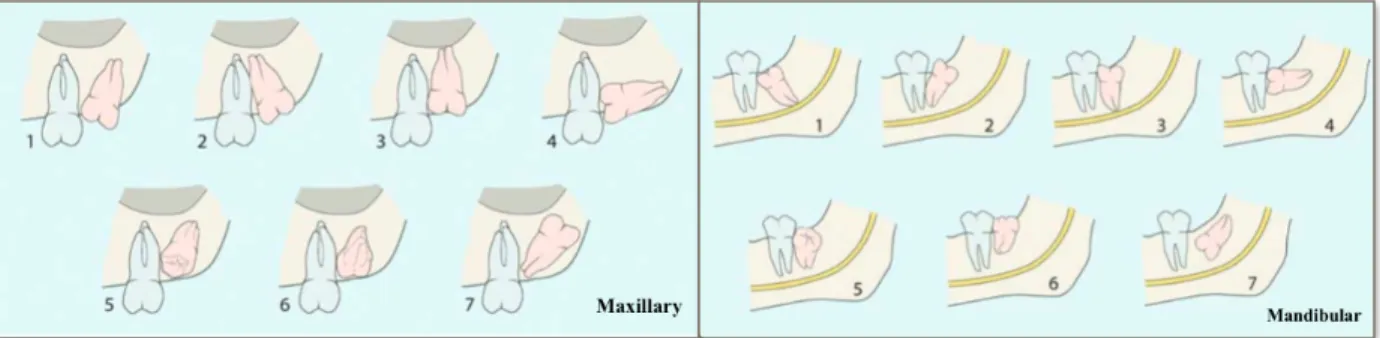
Appendix 1 : The radiographic classifications of the third molar. ( Winter, 1926; Pell et Gregory, 1946; Fragiskos, 2007)
Winter’s Classification
Figure 1 : Classification of impaction of third molar according to Archer (1975) and Kruger (1984). (Fragiskos, 2007)
1) Mesioangular. 2) Distoangular. 3) vertical. 4) Horizontal. 5) Buccoangular. 6) Linguoangular. 7) Inverted.
Pell & Gregory’s Classification Figure 2 : Pell and Gregory’s Classes (1946). (Fragiskos, 2007)
1) Class I: The space is greater or equal to
the mesiodistal diameter of the third molar. 2) Class II: The space is less than the mesiodistal diameter of the third molar. 3) Class III: The third molar is totally embedded in the ramus.
Figure 3 : Depth of impaction according to Pell and Gregory (1946). (Fragiskos, 2007)
1) Position A : The tooth is approximately on the occlusal plane.
2) Position B : The tooth is between the occlusal plane and the cervical line of the second molar. 3) Position C : The tooth is on or below the cervical line of the second molar.
Appendix 2: American Society of Anesthesiologists’ (ASA) physical status classification system. (ASA, 2014)
ASA PS
Classification Definition Exemples - Including but not limited to :
ASA I A normal healthy patient Healthy, non-smoking, no or minimal alcohol use
ASA II A patient with mild systemic disease
Mild diseases only without substantive functional limitations.
Current smoker, social alcohol drinker, pregnancy, obesity (30<BMI<40), well- controlled DM/HTN, mild lung disease
ASA III A patient with severe systemic disease
Substantive functional limitations; One or more moderate to severe diseases.
Poorly controlled DM or HTN, COPD, morbid obesity (BMI ≥40), active hepatitis, alcohol dependence or abuse, implanted pacemaker, moderate reduction of ejection fraction, ESRD
undergoing regularly scheduled dialysis, premature infant PCA < 60 weeks, history (>3
months) of MI, CVA, TIA, or CAD/stents.
ASA IV
A patient with severe systemic disease that is a constant threat
to life
Recent (<3 months) MI, CVA, TIA, or CAD/ stents, ongoing cardiac ischemia or severe valve
dysfunction, severe reduction of ejection fraction, sepsis, DIC, ARD or ESRD not undergoing regularly scheduled dialysis
ASA V
A moribund patient who is not expected to survive without
the operation
Ruptured abdominal/thoracic aneurysm, massive trauma, intracranial bleed with mass effect, ischemic bowel in the face of significant cardiac
pathology or multiple organ/system dysfunction
ASA VI
A declared brain-dead patient whose organs are being removed for donor purposes
Appendix 3 : Predictive variables associated with surgical difficulty in impacted third molar removal.
(Renton et al, 2001; Akadiri et al, 2008; Akadiri et Obiechina, 2009; Carvalho et Vasconcelos, 2011; Miloro et al, 2012; Carvalho et al, 2013; Carvalho et Vasconcelos 2014; Hupp et al, 2014)
LEGEND :
-
3M : third molar-
Surgical Difficulty / Time :Less difficult / shorter time More complex / higher time
Appendix 4 : Mandibular tooth sectioning technique depending of impacted tooth angulation (Farish et Bouloux, 2007)
Appendix 5 : Intra-operative complications management summary
(Bouloux et al, 2007; Fragiskos, 2007; Brauer, 2009; Chracovic et Custodio, 2010; Contar et al, 2010; Gómez-Oliveira et al, 2010; McGoldrick et Stassen, 2010; Romeo et al, 2011; Selvi et al, 2011; Kamoh et Swantek, 2012; Marciani, 2012)
LEGEND