Eshghpour et al. JDMT, Volume 2, Number 2, June 2013 59
Original Research
Periodontal Problems Following Surgical Extraction of Impacted
Mandibular Third Molars
Majid Eshghpour
1, Reza Shahakbari
1, Amirhossein Nejat
21
Dental Research Center, Department of Oral and Maxillofacial Surgery, Faculty of Dentistry,
Mashhad University of Medical Sciences, Mashhad, Iran
2
Student Research Committee, Faculty of Dentistry, Mashhad University of Medical Sciences,
Mashhad, Iran
Received 12 October 2012 and Accepted 24 December 2012
Abstract
Introduction:There are conflicting reports on the effects of surgical removal of impacted mandibular third molars on the periodontium of the adjacent teeth. The aim of this study was to compare the condition of the periodontium six months after extraction of impacted mandibular third molars with baseline values. Methods: Fifty patients with mesioangular impacted mandibular third molarsparticipated in this study. Probing depth (PD), Leo and Sillness' gingival index (GI), and clinical attachment level (CAL) in distobuccal, mid-distal, and distolingual surfaces of second molar teeth were assessed before surgical extraction of the third molars and 6 months later. To evaluate the changes in alveolar bone height (BH), two parallel PA radiographs obtained at the baseline and follow-up session. Data was analyzed with SPSS 11.0 software atthe confidence interval of 95%. Results: Thirty-eight females and 12 males participated in this study. Twenty-eight(56%) of impacted molar teeth were in the right side and 22 (44%) were in the left side. Baseline values of PD, CAL, and GI at three points of the distal surface of the mandibular second molar tooth had no significant differences with follow-up values (P-value> 0.05). According to the radiographs, baseline BH also had insignificant difference with follow-up height (P-value>0.05). Conclusion: Surgical removal of impacted mandibular third molar does not affect periodontium after 6 months.
Key Words: Impacted tooth, periodontal status, second molar tooth.
--- Eshghpour M, Shahakbari R, Nejat A. Periodontal Problems Following Surgical Extraction of Impacted Mandibular Third Molars. J Dent Mater Tech 2013; 2(2): 59-62.
Introduction
Along with the developments of societies in the recent years, the life style has been changed drastically. One change is a tendency towards eating soft foods. It is suggested that this trend has resulted in a gradual decrease in jaw size and lack of space for third molar teeth. In addition to impaction, pericoronitis, caries, crowding, and odontogenic cysts or tumors are among complications associated with third molar teeth (1).
Third molar extraction either non-surgically or surgically is one of the most common operations in dentistry. There are numerous indications for removal of the third molars. Prevention and treatment of periodontal diseases in the adjacent tooth is one of these indications (2).
However, there exists a dilemma over the effect ofsurgical removal of mandibular third molar tooth onthe periodontal condition of the adjacentsecond molar. Zeigler and Kugelberg et al demonstrated prominent improvement of periodontal indicesin distal part of second molar in after surgery (3,4). In contrast, Stephens et al. (5) and Knutsson et al. (6) reported attachment loss and attenuation in alveolar ridge height following extraction of wisdom tooth in second molar distal part.
60
JDMT, Volume 2, Number 2, June 2013 Periodontal ProblemsMaterials and Methods
This prospective study was performed in Mashhad Faculty of Dentistry between August 2011 and July 2012. The study was approved by Ethical Committee of Mashhad University of Medical Sciences and all patients signed a detailed informed consent.
Study Population
Fifty patients in need of surgical extraction of impacted mandibular third molars participated in this study. Inclusion criteria were no periodontal disease in adjacent second mandibular molar tooth, age between 18 and 30 and mesioangular teeth fully impacted in the bone. Patients who were lactating, pregnant, smoking, consuming drugs interfering with healing process,had periodontal disease or prosthesis on second molar teeth, or reported systemic disorders excluded from the study.
Periodontal Evaluation
To evaluate the effect of surgery on periodontal indices, probing depth (PD), Leo and Sillness' gingival index of (GI) (4), and clinical attachment level (CAL) were assessed in distobuccal, middistal and distolingual surfaces of second molar tooth before the surgery (baseline) and 6 months later (follow-up). In addition, before the surgery and at the 6-month follow-up, a standard parallel PA was obtained to evaluate changes in bone height (BH).
Surgical Procedure
All the patients underwent a thorough scaling and oral prophylaxis before surgery. The surgery protocol
was as follows: applying povidine iodine solution around the mouth; blocking inferior alveolar, long buccal, and lingual nerves using 2% lidocaine + 1: 80,000 epinephrine anesthetics; performing standard incision and reflection of mucoperiosteal envelop flap; tooth sectioning, bone removal, and bone recontouring with low-speed handpiece under sufficient irrigation; socket irrigation with 50 ml saline; flap suturing using 3-0 silk suture;and prescribingamoxicillin (500 mg, TID, n=20) and gelofen (400mg cap, TID, for the maximum of 3 days) regimen.
Statistical Analysis
All data were collected in SPSS version 11.0. Data were reported descriptively and analyzed using t-test and Wilcoxon signed ranks test. The confidence interval of analysis was setat 95%.
Results
Fifty patients with the mean age of 23.41 ± 5.21 completed the study. Among the patients, 38 (76%) were females and 12 (24%) were males. Twenty-eight (56%) of impacted molar teeth were in the right side of the mandible and 22 (44%) were in the left. Although PD and GI in three measurement sites were increased after 6 months, the difference was not statistically significant (Tables 1, 2). CAL and BH were decreased in the follow-up measurement in comparison to the baseline;however, the difference was not significant (Tables 3, 4).
Table 1. Probing depth (mm) in three different sides of second molar tooth at baseline and follow-up
Variable Baseline
(Mean ± SD)
6-month Follow-up (Mean ± SD)
P-value
Distobuccal 2.95 ± 0.42 3.02 ± 0.78 0.421
MidDistal 2.53 ± 0.23 2.86 ± 0.32 0.059
Distolingual 2.89 ± 0.25 2.99 ± 0.41 0.109
Table 2. Mean score of gingival index in three different sides of second molarat baseline and follow-up
Variable Baseline
(mean ± SD)
6-month Follow-up (mean ± SD)
P-value
DistoBuccal 0.65 ± 0.06 0.73 ± 0.12 0.098
MidDistal 0.54 ± 0.11 0.59 ± 0.14 0.156
DistoLingual 0.58 ± 0.04 0.62 ± 0.11 0.102
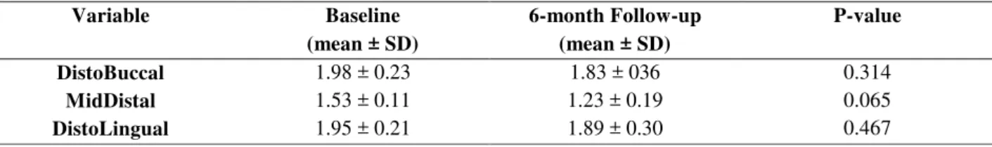
Table 3. Clinical attachment level (mm) in three different sides of second molar tooth at baseline and follow-up
Variable Baseline
(mean ± SD)
6-month Follow-up (mean ± SD)
P-value
DistoBuccal 1.98 ± 0.23 1.83 ± 036 0.314
MidDistal 1.53 ± 0.11 1.23 ± 0.19 0.065
0.467
DistoLingual 1.95 ± 0.21 1.89 ± 0.30
Eshghpour et al. JDMT, Volume 2, Number 2, June 2013 61 Table 4. Height of alveolar bone in PA radiographs in distal part of second molar tooth at baseline and follow-up
Variable Baseline
(mean ± SD)
6-month Follow-up (mean ± SD)
P-value
Alveolar Bone Height 3.1 ± 0.34 2.84 ± 0.48 0.088
Discussion
According to the results of the current study, in the follow-up session, probing depth (PD) and gingival index (GI) were higher than baseline and there were degrees of attenuation in attachment level (CAL) and bone height (BH), but the changes were not statistically significant.
Along with the increase in the incidence of third molar impaction in humans, the number of patients facing complications related tosurgical removal of this impacted tooth is growing.Onecomplication is periodontal problems (7). Results of Kugelberg (3) and Zeigler (4) revealed that extraction of wisdom tooth could lead to significant changes in periodontal condition in distal surface of adjacent second molar tooth.This is in contrast to the results of our study (3,4). However, Grondahl and Lekholm (8) reported no significant difference in BH at the distal part of second molar tooth, whichis in accordance with our findings. They reported the results of a 12-month follow-up. Although we reported changes in BH after 6 months, no significant difference observed between two assessments. In addition, after 2 to 4 years of extraction, Kugelberg et al. (3) found no statistically significant changes in BH.
There exists conflictingresults in previous reports. Peng et al. (9) compared periodontal status of second molar teeth adjacent to the extracted wisdom tooth with the other side second molar. They performed a retrospective study on 57 cases who had their teeth removed at least 5 years before the study. They observed a significant loss in attachment level and bone height in addition to the increased probing depth of experimental sides.
Another retrospective study by Kan et al supported the results of Peng et al study. They performed a similar study on 158 patients who had their wisdom teeth removed 6 months to 3 years prior to the study (10).In contrast, Krausz et al. (7) with similar study design, found significant bone gain in the experimental site after 28 to 58 months. Although Krausz et alstudieshad smaller sample size (25 patients) in comparison to Peng et al. (9) and Kan et al. (10), they had longer follow-up period.
This study was a prospective study in which the periodontal parameters of adjacent second molar tooth 6 months after surgery were compared to the baseline values of the same tooth. According to this difference in
study design, the results of the current study are more valid than mentioned retrospective studies (7,9,10).
Richardson and Dodson (11) performed a review over the effect of removal of wisdom tooth on periodontium of adjacent second molar tooth. They only included prospective RCT studies with more than 6 months follow-up. They included eight studies and concluded that surgical extraction of impacted wisdom tooth had insignificant effect on probing depth and attachment level in distal surface of second molar tooth;the conclusion which is in accordance with the results of the current study.
Conclusion
The results of the current study revealed that well-performed surgical removal of impacted mandibular third molar tooth, does not lead to permanent periodontal problems in distal surface of adjacent second molar tooth.
Acknowledgment
The authors would like to express their gratitude to the staff of Oral and Maxillofacial Surgery Clinic and Dental Research Center for their cooperation.
References
1. Chaparro-Avendaño AV, Perez-Garcia S,
Valmaseda-Castellon E, Berini-Aytes L,
Gay-Escoda C. Morbidity of third molar extraction in
patients between 12 and 18 years of age. Med Oral
Pathol Oral Cir Bucal 2005; 10: 422-31.
2. Kaminishi RM, Lam PS, Kaminishi KS, Marshall
MW, Hochwald DA. A 10-year comparative study
of the incidence of third molar removal in the
aging population. J Oral Maxillofac Surg 2006; 64:
173-4.
3. Kugelberg CF. Periodontal healing two and four
years after impacted lower third molar surgery. A
comparative retrospective study. Int J Oral
Maxillofac Surg 1990; 19: 341–5.
4. Zeigler RS. Preventive dentistry new concepts:
preventing periodontal pockets. Va Dent J 1975;
62
JDMT, Volume 2, Number 2, June 2013 Periodontal Problems 5. Stephens RJ, App GR, Foreman DW. Periodontalevaluation of two mucoperiosteal flaps used in
removing impacted mandibular third molars. J
Oral Maxillofac Surg 1983; 41: 719-24.
6. Knutsson K, Brehmer B, Lysell L, Rohlin M.
Pathoses associated with mandibular third molars
subjected to removal. Oral Surg Oral Med Oral
Pathol Oral Radiol Endod 1996; 82: 10-7.
7. Krausz AA, Machtei EE, Peled M. Effects of
lower third molar extraction on attachment level
and alveolar bone height of the adjacent second
molar. Int J Oral Maxillofac Surg 2005; 34:
756-60.
8. Gröndahl HG, Lekholm U. Influence of
mandibular third molars on related supporting
tissues. Int J Oral Surg 1973; 2: 137-42.
9. Peng KY, Tseng YC, Shen EC, Chiu SC, Fu E,
Huang YW . Mandibular second molar periodontal
status after third molar extraction. J Periodontol
2001; 72: 1647-51
10. Kan KW, Liu JK, Lo EC, Corbet EF, Leung WK.
Residual periodontal defects distal to the
mandibular second molar 6-36 months after
impacted third molar extraction. J Clin Periodontol
2002; 29: 1004-11
11. Richardson DT, Dodson TB. Risk of periodontal
defects after third molar surgery: An exercise in
evidence-based clinical decision making.Oral Surg
Oral Med Oral Pathol Oral Radiol Endod 2005;
100: 133-7.
Corresponding Author: Amirhossein Nejat Faculty of Dentistry
Vakilabad Blvd, Mashhad, Iran Tel: +98-9153148853
Fax: +98-511-8829500