http://dx.doi.org/10.1590/1806-9061-2015-0116
Resonance Spectroscopy
Author(s)
Szeleszczuk ŁI
Pisklak DMI
Wawer II
I Faculty of Pharmacy, Medical University of
Warsaw, Department of Physical Chemistry, Banacha 1, 02-093 Warsaw Poland
Mail Address
Corresponding author e-mail address Łukasz Szeleszczuk
Faculty of Pharmacy, Medical University of Warsaw, Department of Physical Chemistry, Banacha 1, 02-093 Warsaw Poland
Phone: + 48 22 5720950 Email: [email protected]
Keywords
Water, chicken eggshell, NMR, incubation.
Submitted: December/2015 Approved: February/2016
ABSTRACT
The water content of the chicken eggshell has a major influence on gas (CO2 and O2) permeability. Inappropriate water loss during the incubation period increases embryo mortality and decreases chick quality. So far only the procedures that enable to determine the total water content in the eggshell have been described and developed. Our analysis of the 1H MAS NMR spectra of the chicken eggshell samples
revealed three signals, differing significantly in the chemical shift and relaxation times (T1) parameters. In this work we have assigned those signals and described the changes in their intensities that occur during the incubation period. Using 1H MAS NMR it is possible to distinguish
two types of water reservoirs in the chicken eggshell. This approach can be used for more detailed analysis of the water content in the eggshells.
INTRODUCTION
Chicken eggshell (CE) is a complex structure consisting of an organic matrix and minerals, particularly calcium carbonate in the polymorphic form of calcite (Dennis et al. 1996). Studies on the structure of the CE revealed that it is not homogenous and is composed of the shell membranes, mammillary zone, calcium reserve assembly, palisades and cuticle, which is the most external layer of the CE (Hunton, 1995; Hunton, 2005). The role of the CE is not only to protect and provide nutrients to the enclosed embryo but also to allow and control the exchange of water and gas vapor between the inside of the egg and the external environment.
The pores present in the avian eggshell provide the only communicating channels for the exchange of molecules between the developing embryo and the external environment. The number of pores in the CE, their radii and arrangement have been the object of many studies (Tullett, 1978; Board & Scott, 1980; Tullett & Board, 2009). A correlation between the number of pores and egg mass has been found (Ar & Rahn 1985). In general, 7000-17000 funnel-shaped pore channels with diameter of 6-26 µm are distributed on the shell surface and permit the diffusion of metabolic gases and water vapor (La Scala
et al. 2000).
The structure and composition of the CE change during the incubation process (Pizzolante et al., 2009). Recently, we have proven that, during the e
mbryonic development of the chicken inside the
egg,
elution and subsequent absorption of some elements from theshell occur (Szeleszczuk et al., 2015). We have also confirmed that the mechanism of calcium release from insoluble calcium carbonate is based on the formation of calcium bicarbonate Ca(HCO3)2
,
(Pisklak et al., 2012) according to the reaction:2H+ + 2CaCO
The released calcium is then used by the growing chicken embryo for the development of its skeletal system.
During incubation, egg dry matter is metabolized, increasing the water fraction, which amount is additionally increased by metabolic water production (Ar & Rahn, 1980). Water loss during incubation is mandatory if the relative water content is to remain the same. Ar & Rahn (1980) showed that the fraction of the initial mass of the egg lost by evaporation of water across the eggshell during incubation ranges from 10% to 23%, with a mean of 15%.
According to Ackerman & Rahn (1981), O2, CO2 and H2O have common diffusion paths through the eggshell. Since gases are transported across the CE by diffusion (Wangensteen et al., 1970, 1971; Wangensteen and Rahn, 1970, 1971) the gas and water transfer across the shell depends on and is limited by the diffusive properties of gases across the eggshell and shell membrane (Vleck et al., 1983; Rahn and Paganelli, 1982). According to the theoretical treatment of gas flow through pores based on Fick’s law (Tøien et al. 1988; Paganelli 1980), the diffusion coefficient depends, among other factors, on the radii of the pores and the presence of water molecules, as they have a major influence on the adsorption efficiency of the gases inside of the pores.
It is well accepted that water loss through the shell and the permeability of the CE to oxygen and carbon dioxide during the artificial incubation are the key factors that affect hatching success. Inappropriate water loss increases embryo mortality and decreases chick quality. The rate of incubational water loss by the egg affects embryogenesis and total water loss influences the creation an air cell sufficiently large to allow embryonic lung ventilation after internal pipping and successful hatch (van der Pol et al., 2013). Therefore, maintaining a proper water budget during the incubation process ensures high hatchability and hatchling quality (Madeddu et al., 2013). The methods of measuring the eggshell water vapor conductance are currently being improved to allow it to be reliably measured using only fragments of the eggshell (Portugal et al., 2010).
Despite the great number of papers on egg water content, and specifically on its importance for the maintenance of adequate the proper gas exchange and loss during incubation, the authors do not differentiate the types of water present in the CE. Instead, researchers focus mainly on the total egg water content (Ar & Rahn 1980).
Wilson et al. (2006) showed that the use of 1H MAS
NMR (proton Magic Angle Spinning Nuclear Magnetic Resonance spectroscopy) allows differentiating the different types of water present in the human bone. According to their study, water has three structural roles in the bone, and a few types of water reservoirs. Solid state NMR is a highly selective structural technique that can be applied for the study of the nanostructure of different biomaterials, including CE. The magnetic spin properties of the nuclei are very sensitive to the surroundings. The bonding environment, distance and abundance of other magnetic nuclei and order or disorder of the material all have an influence on the NMR proprieties in the solid state.
The aim of this study was to analyze the 1H MAS NMR spectra of chicken eggshells collected on the different incubation days. Previously, using the same technique, we confirmed that the water content of the eggshell decreases during the incubation (Pisklak et al., 2012). In this study, we focused on the characterization of different types of water present in the CE, their T1 (longitudinal, spin-lattice) NMR relaxation parameter values, possible roles and changes in their amounts during the incubation period. There are many potential advantages of using the reported technique in comparison to egg weight loss measurements used in practice to follow water loss rates in incubated eggs (Romão et al. 2008). For instance, 1H MAS NMR provides a molecular view of the structure of CE as it allows to differentiate the water reservoirs and to determine water mobility.
MATERIALS AND METHODS
Twenty four samples of eggshells from commercial broiler breeder (Cobb-500, 30 weeks of age) eggs were collected on the 1st, 14th, 18th and 21st day of
incubation at a hatchery located in Mazovian Province, Poland. The reason for collecting the eggs on the aforementioned days of incubation for sampling is that eggshell resorption significantly increases during the last week of incubation (Crooks & Simkiss, 1974), which may influence CE water content.
from the eggs containing properly developing embryos were analyzed. The samples were stored in an airtight container to prevent water evaporation. Directly before the measurements, the samples were cleaned, the membranes were removed and the eggshells were powdered using a mortar.
Solid-state MAS NMR spectra were recorded at 298 K on a Bruker Avance DSX 400 WB spectrometer in the magnetic field of 9.4 T at 400.13 MHz (1H). The samples were spun at 10 kHz in a 2.5mm zirconia rotor. Spin rate stability was within ± 4 Hz. Chemical shifts were calibrated indirectly through the adamantane signal at 1.78 ppm, relative to TMS. For direct polarization (Bloch decay, BD, DP) experiments, single pulse (a 90° pulse of 3 µs) sequence and a repetition delay of 10 s was employed. Proton T1 relaxation time was determined using the standard inversion recovery T1 pulse sequence. The number of scans was set at 32, with the spectral width of 100 kHz. The collected spectra were deconvoluted and the intensity of the three studied peaks was fitted to the regression line according to the equation:
ln[(M0-Mz(t))/2M0]=-t/T1
where M0 is the equilibrium magnetization (arithmetic mean of the values of the last three intensity values with the longest evolution times) and the Mz is the magnetization after the certain evolution time (t). The total weight of the rotor and sample was measured each time and was in the range of 802 ± 1 mg. Deconvolution of the NMR spectra was performed using the MestReNova program (MestreLab Inc.). The methodology described is commonly applied and was successfully used in a previous CE study (Pisklak et al., 2012).
RESULTS AND DISCUSSION
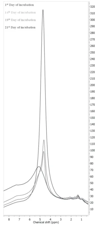
Previously (Pisklak et al., 2012) we have found that the intensity of the main signal in the 1H MAS
NMR spectra, located at 4.8 ppm, significantly decreases during the incubation process due to the water evaporation from the CE. On the other hand, the intensity of the signal at 7.2 increases during incubation as it derives from the bicarbonate anions that are formed during incubation (Fig. 1). Although it was demonstrated that CE moisture content affects its strength (Lott & Reece, 1980), to the best of our knowledge, the changes in the water content of the CE during the incubation have not been studied yet. Instead, researchers focused mainly on the changes in hydration of the whole chicken egg during incubation
(Holub et al. 1994). Furthermore, literature data on the moisture content of the CE are not consistent (Freire & Holanda, 2006; Sugino et al., 1997), which suggest that this topic should be further studied.
Figure 1 – 1H MAS NMR spectra of CE samples collected during incubation.
the CE after the membranes are removed is negligible, the only possible sources of the signals in the 1H NMR
spectra are hydrogen atoms from water molecules and bicarbonate ions.
In order to ensure that both signals (at 1.2 ppm and 4.8 ppm) derived from water hydrogen atoms, we cracked one of the CE samples collected on the first day of incubation and placed half in a the constant temperature chamber (38o C) for 21 days to simulate
the conditions present in the incubation chamber. The spectrum from the other half of the sample was immediately registered. After drying, the sample was removed from the constant temperature chamber and its spectrum was registered (Fig. 2). The superimposition (Fig. 3) of the two spectra revealed that indeed, the intensities of both of those signals decreased, while the intensity and shape of the bicarbonate ion signal (at 7.2 ppm) did not change in the dried sample. The only possible explanation for the changes that occurred under those drying conditions is the evaporation of water.
Because the different water reservoirs of the avian eggshell have not been analyzed yet using the 1H
MAS NMR, we had to use the results of the studies on materials with similar structure and function as a starting point. To the best of our knowledge, this is the first paper dedicated to the study of different water reservoirs in the avian eggshell. According to Vyalikh et al. (2007), who studied 1H MAS NMR spectra of water
in porous inorganic materials, the signal of monomeric water molecules should appear at the 0.8-1.5 ppm, while the signal of the water clusters has a chemical shift of ≈ 5.5 ppm. Wilson et al. (2006) found the signal of isolated water molecules in the carbonated apatite and deproteinated cortical bone at ≈ 2.2 ppm, while the major water clusters had a chemical shift of 5.0-5.5 ppm.
Figure 3 – Superposition of the signals from the deconvoluted 1H MAS NMR spectra of a CE sample before (blue) and after (red) drying.
In addition of the chemical shift, infor-mation on water mobility can be obtained by measuring spin–lattice relaxation times. The spin–lattice relaxation time of bulk water, measured under the same experi-mental conditions, is in the range from 1 s to 3 s (Taylor & Peterson, 2010). Reduced spin–lattice relaxation time is observed when liquid molecules are adsorbed on a solid surface due to a change in molecular mobility (D’Agostion et al., 2014). In or-der obtain the spin–lattice relaxation time values, we performed a series of inversion recovery experiments. The results with the corresponding uncertainties can be found in the Table 1.
No significant differences in the spin– lattice relaxation time values of the three studied signals were found among the collected samples on the same day of incubation. The spin–lattice relaxation time
of the signal at 1.2 ppm was found to be ≈100 ms lower than the spin–lattice relaxation time of the signal at 4.8 ppm. No changes in the spin–lattice relaxation times of the studied signals were observed during incubation.
Both the chemical shifts and the spin–lattice relaxation time values indicate that the major signal in the CE 1H MAS NMR spectra (at 4.8 ppm) originates
from the large reservoirs of water molecules, which are strongly associated by the hydrogen bonds. The other signal (at 1.2 ppm) probably derives from monomeric, adsorbed water molecules that are present mostly in the CE pores. The T1 value of the signal at 1.2 ppm is ≈100 ms lower than that determined at 4.8 ppm, which also confirms this assumption.
During incubation, the intensity of the signal at 4.8 ppm significantly decreased, while the intensity of the signal at 1.2 ppm remained unchanged. Those data suggest that, while the content of the bulk water in CE decreases, the quantity of monomeric water does not change during this period. This monomeric water, probably present in the pores, may be crucial to ensure the proper diffusion of gases through the CE.
Differentiating these two possible types of water has an important practical aspect. The drop of hatchability,
connected with the inappropriate water content of the CE, may be a result of inappropriate ratio (or level) of only one of the described water reservoirs, even if the total amount of water in the shell remains normal.
CONCLUSIONS
The results of the NMR analysis of the CE indicate that two different types or water reservoirs are present. After the deconvolution of the 1H MAS NMR spectra of
the CE, three peaks can be observed. One peak (at 7.2 ppm) originates from the bicarbonate ion (HCO3) and it is characterized by a spin–lattice relaxation time value of 800 ms. Other two peaks (at 1.2 ppm and 4.8 ppm) originate from the different types of water reservoirs. The peak of the monomeric water molecules (at 1.2 ppm) is characterized by the ca.100 ms lower spin–lattice relaxation time value compared with the other reservoir due to the adsorption of water molecules, presumable in the CE pores. During incubation, the intensity of the main water signal (at 4.8 ppm) decreases, the intensity of the bicarbonate ion peak increases, while the intensity of the monomeric (adsorbed) water signal remains unchanged.
REFERENCES
Ackerman RA, Rahn H. In vivo O2 and water vapor permeability of the hen’s eggshell during early development. Respiration Physiology 1981;45(1):1-8.
Ar A, Rahn H. Water in the Avian Egg Overall Budget of Incubation. Integrative and Comparative Biology 1980;20(2):373-384.
Ar A, Rahn H. Pores in avian eggshells: gas conductance, gas exchange and embryonic growth rate. Respiration Physiology 1985;61(1):1-20.
Board RG, Scott VD. Porosity of the Avian Eggshell. American Zoologist 1980;20(2):339-49.
Crooks RJ, Simkiss K. Respiratory acidosis and eggshell resorption by the chick embryo. Journal of Experimental Biology 1974;61(1):197-202.
D’Agostino C, Mitchell J, Mantle MD, Gladden LF. Interpretation of NMR relaxation as a tool for characterising the adsorption strength of liquids inside porous materials. Chemistry -A European Journal 2014;20(40):13009–13015.
Dennis JE, Xiao SQ, Agarwal M, Fink DJ, Heuer AH, Caplan AI. Microstructure of matrix and mineral components of eggshells from white Leghorn chickens (Gallus gallus). Journal of Morphology 1996;228(3):287-306.
Freire MN, Holanda JNF. Characterization of avian eggshell waste aiming its use in a ceramic wall tile paste. Ceramica 2006;52(324):240-244.
Holub A, Baranyiová E, Ponízilová E. Changes in hydration in chicken and duck eggs during incubation. Veterinarni Medicina 1994;39(10):605-14.
Hunton P. Understanding the architecture of the egg shell. World Poultry Science Journal 1995;51:141-7.
Hunton P. Research on eggshell structure and quality: an historical overview. Revista Brasileira de Ciência Avícola 2005;7(2):67-71.
Table 1 – T1 (spin–lattice) relaxation times of the CE samples.
Day of incubation
Sample number
1H NMR chemical shift [ppm]
1.2 4.8 7.2
1. 1. 740 ± 21 ms 849 ± 14 ms 810 ± 16 ms
2. 736 ± 25 ms 850 ± 11 ms 801 ± 18 ms
3. 738 ± 23 ms 836 ± 12 ms 798 ± 17 ms
4. 745 ± 20 ms 849 ± 10 ms 812 ± 15 ms
5. 730 ± 17 ms 841 ± 13 ms 804 ± 14 ms
6. 760 ± 22 ms 845 ± 12 ms 821 ± 18 ms
14. 1. 750 ± 29 ms 825 ± 15 ms 807 ± 21 ms
2. 742 ± 20 ms 839 ± 11 ms 810 ± 19 ms
3. 762 ± 19 ms 842 ± 11 ms 816 ± 18 ms
4. 754 ± 24 ms 841 ± 13 ms 824 ± 22 ms
5. 756 ± 26 ms 838 ± 12 ms 801 ± 17 ms
6. 729 ± 27 ms 845 ± 10 ms 789 ± 16 ms
18. 1. 771 ± 26 ms 824 ± 12 ms 799 ± 19 ms
2. 745 ± 25 ms 831 ± 18 ms 801 ± 17 ms
3. 762 ± 22 ms 840 ± 13 ms 814 ± 22 ms
4. 758 ± 21 ms 851 ± 14 ms 796 ± 24 ms
5. 762 ± 23 ms 846 ± 15 ms 789 ± 21 ms
6. 758 ± 24 ms 841 ± 11 ms 813 ± 20 ms
21. 1. 738 ± 20 ms 843 ± 10 ms 807 ± 22 ms
2. 741 ± 21 ms 838 ± 12 ms 806 ± 24 ms
3. 759 ± 25 ms 851 ± 15 ms 798 ± 21 ms
4. 748 ± 24 ms 842 ± 18 ms 818 ± 20 ms
5. 763 ± 20 ms 838 ± 13 ms 809 ± 24 ms
La Scala Jr N, Boleli IC, Ribeiro LT, Freitas D, Macari M. pore size distribution in chicken eggs as determined by mercury porosimetry. Revista Brasileira de Ciência Avícola 2000;2(2):177-81.
Lott BD, Reece FN. The effect of ambient air moisture and temperature on egg shell breaking strength. Poultry Science 1981;60(1):142-144.
Madeddu M, Zaniboni L, Mangiagalli MG, Cassinelli C, Cerolini S. Egg related parameters affecting fertility and hatchability in the Italian bantam breed Mericanel della Brianza. Animal Reproduction Science 2013;137(3-4):214-219.
Paganelli CV. The Physics of gas exchange across the avian eggshell. American Zoologist 1980;20(2):329-338.
Pisklak DM, Szeleszczuk L, Wawer I. 1H and 13C magic-angle spinning nuclear magnetic resonance studies of the chicken eggshell. Journal of Agricultural and Food Chemistry 2012;60(50):12254–9.
Pizzolante CC, Saldanha ESPB, Laganá C, Kakimoto SK, Togashi CK. Effects of calcium levels and limestone particle size on the egg quality of semi-heavy layers in their second production cycle. Revista Brasileira de Ciência Avícola 2009;11(2):79-86.
Portugal SJ, Maurer G, Cassey P. Eggshell permeability: a standard technique for determining interspecific rates of water vapor conductance. Physiological and Biochemical Zoology 2010;83(6):1023-1031.
Rahn H, Paganelli CV. Role of diffusion in gas exchange of the avian egg. Federation Proceedings 1982;41(6):2134-2136.
Romão JM, Moraes TGV, Teixeira RSC, Cardoso WM, Buxade CC. Effect of egg storage length on hatchability and weight loss in incubation of egg and meat type Japanese quails. Revista Brasileira de Ciencia Avicola 2008;10(3):143-147.
Sugino H, Nitoda T, Juneja LR. General chemical composition of hen eggs. In: Yamamoto T, Juneja LR, Hatta H, Kim M, editors. Hen eggs: their basic and applied. Boca Raton: CRC Press; 1997. p. 13-24.
Szeleszczuk L, Pisklak DM, Kuras M, Wawer I. In vitro dissolution of calcium carbonate from the chicken eggshell: on the study of
calcium bioavailability. International Journal of Food Properties 2015;18(12):2791279.
Taylor RE, Peterson RD. Comparison of spin–lattice relaxation measurements made in the presence of strong radiation damping. Journal of Molecular Structure 2010;970(1-3):155–159.
Tøien O, Paganelli CV, Rahn H, Johnson RR. Diffusive resistance of avian eggshell pores. Respiration Physiology 1988;74(3):345-54.
Tullett SG, Board RG. Determinants of avian eggshell porosity. Journal of Zoology 1977;183(2):203-211.
Tullett SG. Pore size versus pore number in avian eggshells. In: Piiper J, editor. Respiratory function in birds, adults and embryonic. London: Springer-Verlag; 1978. p.219-226.
Van der Pol CW, Van Roovert-Reijrink IAM, Maatjens CM, Van den Brand H, Molenaar. Effect of relative humidity during incubation at a set eggshell temperature and brooding temperature posthatch on embryonic mortality and chick quality. Poultry Science 2013;92(8):2145-2155.
Vleck CM, Vleck D, Rahn H, Paganelli CV. Nest microclimate, water-vapour conductance, and water loss in heron and tern eggs. Auk 1983;100(1):76-83.
Vyalikh A, Emmler T, Grünberg B, Xu Y, Shenderovich I, Findenegg GH, et al. Hydrogen Bonding of Water Confined in Controlled-Pore Glass 10-75 Studied by 1H-Solid State NMR. Zeitschrift für Physikalische Chemie 2007;221(1):155-68.
Wangensteen D, Wilson D, Rahn H. Diffusion of gases across the shell of the hen’s egg. Respiration Physiology 1970/1971;11(1):16-30.
Wangensteen OD, Rahn H. Respiratory exchange by the avian embryo. Respiration Physiology 1970/1971;11(1):31-45

![Table 1 – T1 (spin–lattice) relaxation times of the CE samples. Day of incubation Sample number 1 H NMR chemical shift [ppm] 1.2 4.8 7.2 1](https://thumb-eu.123doks.com/thumbv2/123dok_br/15897981.670784/5.892.49.432.190.648/table-lattice-relaxation-samples-incubation-sample-number-chemical.webp)
