Pesq. agropec. bras., Brasília, v.48, n.3, p.313-319, mar. 2013 DOI: 10.1590/S0100-204X2013000300010
fed n‑3 and n‑6 essential fatty acids
Róberson Sakabe(1), Flávio Ruas de Moraes(1), Marco Antonio de Andrade Belo(1), Fabiana Pilarski(1) and Julieta Rodini Engrácia de Moraes(1)
(1)Universidade Estadual Paulista, Centro de Aquacultura, Departamento de Patologia Veterinária, Via Professor Paulo Donato Castellane, Km 05, CEP 14884900 Jaboticabal, SP, Brazil. E‑mail: [email protected], [email protected], [email protected], [email protected], [email protected]
Abstract – The objective of this work was to investigate the effect of dietary supplementation with essential fatty acids on the kinetics of macrophage accumulation and giant cell formation in Nile tilapia (Oreochromis niloticus). The supplementation sources were soybean oil (SO, source of omega 6, n‑6) and linseed oil (LO, source of omega 3, n‑3), in the following proportions: 100% SO; 75% SO + 25% LO; 50% SO + 50% LO; 25% SO + 75% LO; and 100% LO (four replicates per treatment). After a feeding period of three months, growth performance was evaluated, and glass coverslips were implanted into the subcutaneous connective tissue of
fish, being removed for examination at 2, 4, 6, and 8 days after implantation. Growth performance did not
differ between treatments. Fish fed 100% linseed oil diet had the greatest macrophage accumulation and the
fastest Langhans cell formation on the sixth day. On the eighth day, Langhans cells were predominant on the coverslips implanted in the fish feed 75 and 100% linseed oil. n‑3 fatty acids may contribute to macrophage recruitment and giant cell formation in fish chronic inflammatory response to foreign body.
Index terms: Oreochromis niloticus, foreign body, growth performance, inflammatory response, lipid nutrition.
Cinética da inflamação crônica em tilápia‑do‑nilo
alimentada com ácidos graxos essenciais n‑3 e n‑6
Resumo – O objetivo deste trabalho foi investigar o efeito da suplementação alimentar com ácidos graxos
essenciais sobre a cinética do acúmulo de macrófagos e a formação de células gigantes em tilápia‑do‑nilo (Oreochromis niloticus). As fontes de suplementação foram óleo de soja (OS, fonte de ômega 6, n‑6) e óleo de linhaça (OL, fonte de ômega 3, n‑3), nas seguintes proporções: 100% OS; 75% OS + 25% OL; 50% OS + 50% OL; 25% OS + 75% OL; e 100% OL (quatro repetições por tratamento). Após período de alimentação de três meses, foi avaliado o desempenho produtivo, e lamínulas de vidro foram implantadas no tecido subcutâneo dos
peixes, as quais foram removidas para exame aos 2, 4, 6 e 8 dias após o implante. O desempenho produtivo não diferiu entre os tratamentos. Os peixes alimentados com 100% de óleo de linhaça tiveram maior acúmulo de macrófagos e formação mais rápida de células de Langhans, no sexto dia. No oitavo dia, as células de Langhans foram predominantes nas lamínulas implantadas nos peixes alimentados com 75 e 100% de óleo de linhaça. Os ácidos graxos n‑3 podem contribuir para o recrutamento de macrófagos e a formação de células gigantes, na resposta inflamatória crônica a corpo estranho em peixes.
Termos para indexação: Oreochromis niloticus, corpo estranho, desempenho produtivo, resposta inflamatória, nutrição lipídica.
Introduction
The participation of fatty acids in nonspecific
defense responses is attributed to the production
of eicosanoids with pro‑inflammatory activity
(Balfry & Higgs, 2001). Fish fed diets rich in n‑3 and n‑6 fatty acids present greater B lymphocyte response and survival after challenge with Aeromonas salmonicida and Vibrio anguillarum (Thompson et al., 1996). In addition, they have higher antibody titers when immunized against Edwardsiella ictaluri (Fracalossi et al., 1994).
Multinucleated giant cells (MGC) have been described among fish with infectious or parasitic diseases (Olsen et al., 2006; Hogge et al., 2008; Jacobs et al., 2009), and in experimental in vitro (Couso et al.,
2002) and in vivo (Petric et al., 2003b; Belo et al.,
2005) models. Accumulation of inflammatory cells at the lesion site is the main inflammation event to protect
the organism against foreign elements (Kumar et al.,
2004). MGC formation occurs as an attempt to involve
modulators (Kumar et al., 2004). Petric et al. (2003b)
demonstrated that the implantation of glass coverslips in the subcutaneous tissue of Piaractus mesopotamicus induces the formation of MGC, initially as foreign
body giant cells and, later on, as Langhans type.
Food supplementation with vitamin C increases
the macrophage activity and induces its accumulation
(Petric et al., 2003a). Using the same experimental model, Belo et al. (2005) observed benefic effects of supplementation with vitamin E among teleost fish
kept at a high population density. However, there
is little information about the influence of essential
fatty acids on the activity of macrophages in chronic
inflammation of fish.
The objective of this work was to investigate the effect of dietary supplementation with essential fatty acids on the kinetics of macrophage accumulation and giant cell formation in Nile tilapia (Oreochromis niloticus).
Materials and Methods
Two hundred Nile tilapia juveniles (69.86±5.01 g) were randomly allocated to 20 glass 310 L fiber tanks
(n=10). The tanks were supplied with chlorine‑free running water from an artesian well, with a 1 L min‑1
flow and supplementary aeration.
The fish were weighed and measured at the beginning of the experiment. To do this, they were anesthetized
in an aqueous solution of benzocaine (1 g per 10 L). After this initial biometry, they were acclimatized for 15 days. During this phase, a same baseline feed was provided daily for all groups, corresponding to around 3% of the biomass.
After acclimatization, the fish received the experimental diets containing the essential fatty acids
for 90 days and had their productive performance evaluated. After this, glass coverslips were implanted into the subcutaneous connective tissue, and the
chronic inflammation was evaluated, by quantifying
the number of macrophages, polykaryon cells, and giant cell nuclei and types.
Degummed soybean oil (SO) was used as a source of omega 6 (n‑6), and linseed oil (LO) as a source of omega 3 (n‑3), in the following proportions: 100% SO; 75% SO + 25% LO; 50% SO + 50% LO; 25% SO + 75% LO; and 100% LO. Each treatment consisted of four replicates.
Initially, the baseline diet was standardized such
that it contained 28% digestible protein (DP) and 3,200 kcal (13,397.76 kJ) of digestible energy (DE).
The composition of the baseline diet followed the nutritional requirements of Nile tilapia (National
Research Council, 1993), as shown in Table 1.
After preparing the diets, feed samples were analyzed
to determine their essential fatty acid profile. About 4 g of each sample were homogenized for lipid extraction
using the method of Folch et al. (1957), followed by
esterification of the lipid fraction using the method of
Hartman & Lago (1973).
The fatty acid methyl esters were analyzed using a
GC‑14A Shimadzu gas chromatograph (Shimadzu do Brasil, São Paulo, SP) equipped with a flame ionization
detector, a split injector, and Supelco 2560 fused silica capillary columns of 100 m length and 0.25 mm internal diameter. The following operational parameters were used: detector and injector temperatures of 270 and
250°C, respectively; column temperature programmed at 100°C, increasing up to 180°C at 10ºC min‑1 rate,
and from 180 to 240°C at 1°C min‑1 rate, and holding
at this end temperature for 10 min; nitrogen as carrier
Table 1. Composition and chemical‑bromatological analysis
of the diet.
Ingredient Content (g kg‑1)
Soybean bran 330.00
Corn meal 180.00
Wheat bran 150.00
Rice bran 90.00
Fish meal 165.00
Oil(1) 60.00
Dicalcium phosphate 10.00 Vitamin and mineral supplement(2) 5.00
BHT (antioxidant) 0.20
Calcitic lime 9.80
Composition
Gross protein (%) 29.15
Gross energy (kJ) 12,861.32
Gross fiber (%) 4.72
Ether extract (%) 10.44
Nitrogen free extract (%) 37.10 Dry matter (%) 90.73
(1)The proportions of degummed soybean oil and linseed oil were added
according to each treatment. (2)Vitamin and mineral supplement composi‑ tion: vitamin A, 1,200,000 IU; vitamin B1, 4,800 mg; vitamin B12, 4,800 mg; vitamin B2, 4,800 mg; vitamin B6, 4,800 mg; vitamin D3, 200,000 IU; vitamin K3, 2,400 mg; folic acid, 1,200 mg; biotin, 48 mg; calcium pantothenate, 12,000 mg; choline chloride, 108 g; niacin, 24,000 mg;
Pesq. agropec. bras., Brasília, v.48, n.3, p.313‑319, mar. 2013 DOI: 10.1590/S0100‑204X2013000300010 gas, at 0.6 mL min‑1 and linear velocity of 14 cm s‑1;
split ratio of 1:75 with a total flow of 52 mL min‑1
and column pressure of 167 kPa. Retention time and peak areas were recorded by microcomputer, using
the Class GC 10 software (Shimadzu Corporation, Tokyo, Japan). Dietary fatty acids were expressed in
percentage
Water quality was evaluated daily, at feeding times,
for temperature (28.17±1.0°C); dissolved oxygen concentration (5.15±0.4 mg L‑1); and hydrogen
ion potential (7.45±0.06) and electric conductivity (178.5±5.3 mS cm‑1), determined using an oximeter
YSI‑55, (YSI Incorporated, Yellow Springs, OH, USA) and a pH meter YSI‑63, (YSI Incorporated, Yellow Springs, OH, USA), respectively. Every fortnight, the
ammonia concentration (0.1976±0.095 mg NH3 L‑1)
and alkalinity (28.64±1.12 mg CaCO3 L‑1) were
measured. The results remained within the comfort zone for Nile tilapia (Boyd, 1990).
Daily feed consumption (3% of the biomass),
weight gain, food conversion, and specific growth
rate were evaluated monthly (30, 60, and 90 days).
Food conversion (FC) was estimated with the formula FC = feed consumption/weight gain, and specific growth rate (SGR) with the equation SGR = 100 (ln final weight ‑ ln initial weight/days of experiment).
Ninety days after starting the experiment, ten randomly chosen fish from each treatment were
anesthetized and subjected to implantation of a glass coverslip (Petric et al., 2003b). The coverslips
were removed after euthanasia 2, 4, 6, and 8 days
after the implantation, washed with a 0.65% saline
solution, fixed in Bouin’s solution, and stained with hematoxylin‑eosin. To evaluate the inflammatory
response, seven coverslips were chosen randomly from
the experimental units. All of the macrophages isolated
and the polykaryons formed were counted, as were the number of foreign‑body giant cells and the number of Langhans giant cells. The counting was carried out
under an optical microscope (400 x), in five fields per coverslip, totalizing 35 fields counted per treatment.
All data were statistically analyzed using a completely randomized design, split plotted in time,
with five proportions of fatty acids x four evaluation times. Comparison of the different experimental
groups was carried out by applying the analysis of
variance procedure (SAS Institute, 2001). Significant
differences at 5% probability were estimated with
Tukey’s test, according to Snedecor & Cochran (1974).
Results and Discussion
With the progressive replacement of soybean oil by linseed oil, the supplementation values for linoleic
acid (C18:2n6c) decreased and the linolenic acid values (C18:3n3) increased (Table 2). This result is an evidence that the profile of the fatty acids present in the
feed met the initial proposal of the study.
With the evolution of the inflammation, over the course of the experiment, cell accumulation on the
glass coverslip increased (Table 3). Two days after the implantation, macrophage number was smaller and few
polykaryons with two to five nuclei were formed. On
the fourth day, however, the numbers of polykaryons
with giant cells containing six to ten nuclei increased.
Due to the high rates of cell accumulation on the coverslips, giant cells with more than 20 nuclei and
Langhans cells were already present on the sixth
and eighth days (Figure 1). There was a remarkable increase in the occurrence of these polykaryon cells between these days (Table 3). However, a reduction in the number of macrophages was observed on the eighth day due to the space occupied by the great number of polykaryon cells attached to the coverslips.
Dietary supplementation with essential fatty acids increased the accumulation and dynamics of
macrophages at the inflammation. Greater and faster formation of Langhans MGCs occurred when the diet
contained 100% linseed oil. Macrophage accumulation
and MGC formation increased progressively until the
eighth day after the implantation, corroborating the
findings of Belo et al. (2005).
Table 2. Total fatty acid profile (%) of the five experimental
diets.
Fatty acid Treatments(1) 100%
SO
75% SO + 25% LO
50% SO + 50% LO
25% SO + 75% LO
100% LO C14:0 (myristic acid) 0.59 0.47 0.49 0.93 0.87 C16:0 (palmitic acid) 14.15 15.28 15.74 17.32 18.38 C16:1 (palmitoleic acid) 1.12 1.29 1.88 2.95 3.74 C17:0 (heptadecanoic acid) 0.18 0.14 0.14 0.2 0.19 C18:0 (stearic acid) 3.17 3.14 3.52 3.63 4.1 C18:1n9c (oleic acid) 24.14 25.35 27.53 29.1 31.21 C18:2n6c (linoleic acid) 47.65 37.3 21.36 12.64 3.92 C18:3n3 (linolenic acid) 5.18 14.24 26.4 28.79 33.67 C20:1n9c (eicosenoic acid) 0.34 0.28 0.3 0.36 0.34 C20:5n3 (eicosapentaenoic acid) 0.56 0.19 0.2 0.67 0.45 C22:6n3 (docosahexaenoic acid) 0.81 0.29 0.32 1.01 0.69
Not identified 2.11 2.03 2.12 2.4 2.44
Total 100 100 100 100 100
Newly formed MGC have nuclei distributed
randomly in their cytoplasm, which characterizes
a foreign‑body type of cell. Gradually, the nuclei
become organized at the periphery of the cytoplasm, to
form Langhans cells (Mariano & Spector, 1974). MGC
formation depends on daily recruitment of monocytes from the reserve compartments of the blood. Following diapedesis and transformation into macrophages, these
cells reach the inflammation focus by chemotaxis, in which IL‑4 and IL‑13 induce macrophage fusion
(Brodbeck & Anderson, 2009) and interferon‑gamma participate on Langhans giant cell formation (Anderson
et al., 2008). This evolution was clearly identified in the inflammatory response of Nile tilapia. When in vitro,
macrophages begin the fusion of their membranes after
24 hours in Sparus aurata (Couso et al., 2002), two or three days in Cyprinus carpio (Bayne, 1986), and after a week in Oncorhynchus mykiss (Secombes, 1985). Sado & Matushima (2007, 2008) observed the formation
of giant cells three and seven days after the application
of BCG in Centropomus and Arius, respectively. The
inflammatory response reported for P. mesopotamicus (Petric et al., 2003a, 2003b; Belo et al., 2005) had a predominant formation of Langhans giant cells 15 days after the coverslip implantation, whereas in the present study it occurred eight days after implantation. These results show differences in the evolution of the
macrophage response between different species of fish. Comparative analysis on the cell response between
the different diets clearly showed the effect of diet supplementation with linseed oil on macrophage accumulation and polykaryon formation (Table 3). Two
days after implantation, a significantly greater number
of macrophages and polykaryons with two nuclei was
found among fish fed 75 and 100% linseed oil. On
the fourth day, a greater accumulation of polykaryons
was observed among fish treated with 50 and 75%
linseed oil. Differences were particularly higher when compared with animals treated with 100% soybean oil.
Fish fed 100% linseed oil showed a significant
increase in the formation of Langhans cells on the
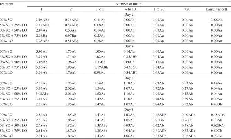
Table 3. Mean values(1) and analysis of variance parameters of cell counts at 2, 4, 6, and 8 days after coverslip implantation
in the different dietary treatments(2).
Treatment Number of nuclei
1 2 3 to 5 6 to 10 11 to 20 >20 Langhans cell Day 2
100% SO 2.16ABa 0.75ABa 0.11Aa 0.00Aa 0.00Aa 0.00Aa 0. 00Aa 75% SO + 25% LO 2.11ABa 0.84ABa 0.08Aa 0.00Aa 0.00Aa 0.00Aa 0.00Aa 50% SO + 50% LO 2.04Aa 0.53Aa 0.14Aa 0.00Aa 0.00Aa 0.00Aa 0.00Aa 25% SO + 75% LO 2.38Ba 0.97Ba 0.25Aa 0.00Aa 0.00Aa 0.00Aa 0.00Aa 100% LO 2.28ABa 0.81ABa 0.28Aa 0.00Aa 0.00Aa 0.00Aa 0.00Aa
Day 4
100% SO 3.01Ab 1.73Ab 1.00Ab 0.14Aa 0.00Aa 0.00Aa 0.00Aa 75% SO + 25% LO 3.09Ab 1.74Ab 1.02Ab 0.25ABb 0.04Aa 0.00Aa 0.00Aa 50% SO + 50% LO 3.08Ac 1.98Ab 1.33Bb 0.60Cb 0.18Aa 0.00Aa 0.00Aa 25% SO + 75% LO 3.06Ab 1.95Ab 1.17ABb 0.43BCb 0.04Aa 0.00Aa 0.00Aa 100% LO 3.09Ab 1.76Ab 0.98Ab 0.34ABb 0.09Aa 0.00Aa 0.00Aa
Day 6
100% SO 2.99Ab 1.95Ab 1.54Ac 1.18Ab 0.69Ab 0.53Ab 0.14Aa 75% SO + 25% LO 3.03Ab 2.02Ab 1.54Ac 1.07Ac 0.72Ab 0.27Ab 0.04Aa 50% SO + 50% LO 3.03Abc 2.01Ab 1.62Ac 1.16Ac 0.90Ac 0.43Ab 0.04Aa 25% SO + 75% LO 3.04Ab 1.90Ab 1.49Ac 1.10Ac 0.78Ab 0.29Ab 0.09Aa 100% LO 2.89Ab 1.95Ab 1.47Ac 1.07Ac 0.84Ab 0.52Ab 0.48Bb
Day 8
100% SO 2.86Ab 1.85Ab 1.43Ac 1.03Ab 0.67ABb 0.60ABb 0.45ABb 75% SO + 25% LO 2.95Ab 1.85Ab 1.41Ac 1.05Ac 0.91Bb 0.76Cc 0.38Ab
50% SO + 50% LO 2.78Ab 1.82Ab 1.38Abc 0.93Ac 0.62Ab 0.48Ab 0.62BCb 25% SO + 75% LO 2.81Ab 1.87Ab 1.35Abc 0.94Ac 0.69ABb 0.63ABc 0.69Cb 100% LO 2.91Ab 1.87Ab 1.43Ac 1.06Ac 0.88ABb 0.62ABb 0.73Cc
(1)Means followed by equal letters, uppercase in the columns and lowercase in the lines, do not differ by Tukey’s test, at 5% probability. (2)SO, degummed
Pesq. agropec. bras., Brasília, v.48, n.3, p.313‑319, mar. 2013 DOI: 10.1590/S0100‑204X2013000300010
sixth day after implantation, in comparison to the other
treatments (Table 3).
After eight days, no significant difference was
observed between treatments in macrophages and
MCG with up to ten nuclei. However, fish fed 75 and
100% linseed oil had greater formation of Langhans
cells, compared to fish fed 75 and 100% soybean oil.
According to Balfry & Higgs (2001), fatty acids in the diet can favor organism defenses, partly by helping to maintain the integrity of the lipid composition of macrophage membranes. The increased macrophage recruitment and polykaryon formation, observed in the present study, with essential fatty acid supplementation
was possibly caused by the favorable action of these compounds on biological membranes, which increased
chronic inflammatory response due to the foreign body.
Belo et al.(2005) reported similar results with regard to vitamin E supplementation.
Moreover, n‑3 polyunsaturated fatty acids (Pufa)
may modulate the expression of inflammatory genes
downregulating some transcription factor activation
(Sethi et al., 2002) and inflammatory cytokines
(Zhao et al., 2005), since they may directly target
the inflammatory gene expression pathways through toll‑like receptor 4 (Lee & Hwang, 2006). This
corroborates the hypothesis that these are active compounds on defense mechanisms among teleost
fish. The increased Langhans giant cell formation on the sixth and eighth days of inflammation observed in
Nile tilapia supplemented with 100% linseed shows the participation of n‑3 fatty acids in the physiopathology of these processes, increasing the macrophage activity. Additionally, fatty acids that modulate immune responses and eicosanoids produced from arachidonic
acid (20:4n‑6) are recognized as inflammatory agents (Abeywardena & Head, 2001; Ganga et al., 2005; Calder, 2006).
Treatments did not differ in weight gain, feed consumption, apparent food conversion, and
specific growth rate (Table 4). Therefore, productive
performance of Nile tilapia supplemented with
Table 4. Productive performance of Nile tilapia (Oreochromis niloticus) as affected by supplementation with different proportions of degummed soybean oil (SO) and linseed oil (LO)(1).
Treatment Weight gain (g) Feed consumption (kg) Feed conversion Specific growth rate 1st month
100% SO 46.61±6.38Aa 0.66±0.02Aab 1.50±0.20Ab 1.27±0.23Aa 75% SO + 25% LO 46.16±4.22Aa 0.59±0.09Aab 1.27±0.11Ab 1.25±0.11Aa 50% SO + 50% LO 45.32±3.02Aa 0.60±0.04Aa 1.31±0.21Ab 1.28±0.14Aa 25% SO + 75% LO 45.09±6.33Aa 0.70±0.01Aab 1.63±0.20Ab 1.13±0.24Aa 100% LO 40.88±7.01Aa 0.59±0.10Aa 1.46±0.21Ab 1.00±0.18Aa
2nd month
100% SO 40.69±2.30Aab 0.81±0.03Aa 2.00±0.04Aa 0.84±0.03Ab 75% SO + 25% LO 38.71±11.55Aa 0.80±0.15Aa 2.09±0.17Aa 0.82±0.17Ab 50% SO + 50% LO 39.37±3.55Ab 0.85±0.10Aa 2.14±0.11Aa 0.84±0.05Ab 25% SO + 75% LO 48.05±11.00Aa 0.88±0.11Aa 2.10±0.42Aa 0.89±0.18Ab 100% LO 37.97±5.12Aa 0.86±0.11Aa 2.27±0.06Aa 0.76±0.08Ab
3rd month
100% SO 26.71±2.77Ab 0.50±0.02Ab 1.92±0.20Aab 0.91±0.08Ab 75% SO + 25% LO 27.83±3.28Aa 0.45±0.03Ab 1.67±0.19Ab 0.84±0.06Ab 50% SO + 50% LO 28.34±3.80Ac 0.52±0.05Ab 1.88±0.14Aab 1.01±0.08Aab 25% SO + 75% LO 25.83±2.72Ab 0.56±0.09Ab 2.10±0.16Aa 0.95±0.14Ab 100% LO 24.48±5.61Ab 0.41±0.03Ab 2.01±0.47Aab 0.85±0.11Ab
(1)Means followed by equal letters, uppercase in the columns and lowercase in the lines, do not differ by Tukey’s test, at 5% probability.
Figure 1. Photomicrograph of mononuclear macrophages
(M), foreign body giant cells (G), and Langhans giant cells (L), six days after the implantation of glass
coverslips in the subcutaneous tissue of the Nile tilapia
(Oreochromis niloticus). Hematoxylin eosin staining.
different essential fatty acids was similar. This result was also obtained in different situations, with different species (Bell et al., 2003; Mourent et al., 2005; Vargas
et al., 2008).
Conclusions
1. Nile tilapia (Oreochromis niloticus) diets supplemented with high levels of linseed oil increase macrophage activity, with greater macrophage
accumulation and faster formation of MGC and
Langhans giant cells at the beginning of the
inflammation.
2. Linolenic acid (C18:3, n‑3) supplementation benefits these nonspecific defense mechanisms.
3. Productive performance of Nile tilapia fed n‑3 or n‑6 fatty acids does not differ.
Acknowledgements
To Fundação de Amparo à Pesquisa do Estado de
São Paulo (Fapesp), for financial support; and to Maria Ines Y. de Campos and Francisca de Assis Ardison, for
assistance in the histological assessments.
References
ABEYWARDENA, M.Y.; HEAD, R.J. Longchain n‑3
polyunsaturated fatty acids and blood vessel function.
Cardiovascular Research, v.52, p.361‑371, 2001. DOI: 10.1016/
S0008‑6363(01)00406‑0.
ANDERSON, J.M.; RODRIGUEZ, A.; CHANG, D.T. Foreign
body reaction to biomaterials. Seminars in Immunology, v.20,
p.86‑100, 2008. DOI: 10.1016/j.smim.2007.11.004.
BALFRY, S.K.; HIGGS, D.A. Influence of dietary lipid composition on the immune system and disease resistance of finfish. In: LIM, C.; WEBSTER, C.D. (Ed.). Nutrition and fish health. New York:
Food Product, 2001. p.213‑234.
BAYNE, C.J. Pronephric leucocytes of Cyprinus carpio: isolation,
separation and characterization. Veterinary Immunology
and Immunopathology, v.12, p.141‑151, 1986. DOI:
10.1016/0165‑2427(86)90118‑2.
BELL, J.G.; MCGHEE, F.; CAMPBELL, P.J.; SARGENT, J.R. Rapeseed oil as an alternative to marine fish oil in diets of
post‑molt Atlantic salmon (Salmo salar): changes in flesh fatty
acid composition and effectiveness of subsequent fish oil ‘wash out’. Aquaculture, v.218, p.515‑528, 2003. DOI: 10.1016/
S0044‑8486(02)00462‑3.
BELO, M.A.A.; SCHALCH, S.H.C.; MORAES, F.R.; SOARES, V.E.; OTOBONI, A.M.; MORAES, J.E.R. Effects of dietary
supplementation with vitamin E and stocking density on
macrophage recruitment and giant cell formation in the teleost fish,
Piaractus mesopotamicus. Journal of Comparative Pathology,
v.133, p.146‑154, 2005. DOI: 10.1016/j.jcpa.2005.04.004. BOYD, C.E. Water quality in ponds for aquaculture. Alabama:
Auburn University, 1990. 482p.
BRODBECK, W.G.; ANDERSON, J.M. Giant cell formation and
function. Current Opinion of Hematology, v.16, p.53‑57, 2009.
DOI: 10.1097/MOH.0b013e32831ac52e.
CALDER, P.C. n‑3 polyunsaturated fatty acids, inflammation, and inflammatory diseases. American Journal of Clinical Nutrition,
v.83, p.1505‑1519, 2006.
COUSO, N.; CASTRO, R.; NOYA, M.; OBACH, A.; LAMAS, J. Formation of short‑lived multinucleated giant cells (MGCS) from
cultured gilthead seabream macrophages. Anatomical Records,
v.267, p.204‑212, 2002. DOI: 10.1002/ar.10103.
FOLCH, J.; LEES, M.; STANLEY, S.P. A simple method for the isolation and purification of total lipids from animal tissues.
Journal of Biological Chemistry, v.226, p.497‑509, 1957.
FRACALOSSI, D.M.; CRAIG‑SCHMIDT, M.C.; LOVELL, R.T.
Effect of dietary lipid sources on production of leukotriene B by
head kidney of channel catfish held at different water temperatures.
Journal of Aquatic Animal Health, v.6, p.242‑250, 1994. DOI:
10.1577/1548‑8667(1994)006<0242:EODLSO>2.3.CO;2. GANGA, R.; BELL, J.G.; MONTERO, D.; ROBAINA, L.; CABALLERO, M.J.; IZQUIERDO, M.S. Effect of dietary lipids on plasma fatty acid profiles and prostaglandin and leptin production
in gilthead seabream (Sparus aurata). Comparative Biochemistry
and Physiology. Part B: Biochemistry and Molecular Biology,
v.142, p.410‑418, 2005. DOI: 10.1016/j.cbpb.2005.09.010. HARTMAN, L.; LAGO, R.C. Rapid preparation of fatty acid
methyl esters from lipids. Laboratory Practice, v.22, p.475‑476,
1973.
HOGGE, C.I.; CAMPBELL, M.R.; JOHNSON, K.A. A new species of myxozoan (Myxosporea) from the brain and spinal cord
of rainbow trout (Oncorhynchus mykiss) from Idaho. Journal of
Parasitology, v.94, p.218‑222, 2008. DOI: 10.1645/GE‑1292.1.
JACOBS, J.M.; RHODES, M.R.; BAYA, A.; REIMSCHUESSEL, R.; TOWNSEND, H.; HARRELL, R.M. Influence of nutritional
state on the progression and severity of mycobacteriosis in striped
bass Morone saxatilis. Disease of Aquatic Organisms, v.87,
p.183‑197, 2009. DOI: 10.3354/dao02114.
KUMAR, V.; ABBAS, A.; FAUSTO, N. Robbins and Cotran:
pathologic basis of diseases. 7th ed. Saunders: Philadelphia, 2004.
1552p.
LEE, J.Y.; HWANG, D.H. The modulation of inflammatory gene expression by lipids: mediation through Toll‑like receptors.
Molecules and Cell, v.21, p.174‑185, 2006.
MARIANO, M.; SPECTOR, W.G. The formation and properties of macrophage polykaryons (inflammatory giant cells). Journal of
Pathology, v.113, p.1‑19, 1974. DOI: 10.1002/path.1711130102.
MOURENT, G.; GOOD, J.E.; BELL, J.G. Partial substitution of fish oil with rapeseed, linseed and olive oils in diets for European
Pesq. agropec. bras., Brasília, v.48, n.3, p.313‑319, mar. 2013 DOI: 10.1590/S0100‑204X2013000300010
composition, plasma prostaglandins E2 and F2µ, immune function
and effectiveness of a fish oil finishing diet. Aquaculture Nutrition, v.11, p.25‑40, 2005. DOI: 10.1111/j.1365‑2095.2004.
00320.x.
NATIONAL RESEARCH COUNCIL. Nutrient requirements of domestic animals: nutrient requirements of warm water fishes and shellfishes. Washington: National Academic, 1993. 102p.
OLSEN, A.B.; MIKALSEN, J.; RODE, M.; ALFJORDEN, A.; HOEL, E.; STRAUM‑LIE, K.; HALDORSEN, R.; COLGUHOUN, D.J. A novel systemic granulomatous inflammatory disease in
farmed Atlantic cod, Gadus morhua L., associated with a bacterium belonging to the genus Francisella. Journal of Fish Disease, v.29,
p.307‑311, 2006. DOI: 10.1111/j.1365‑2761.2006.00714.x. PETRIC, M.C.; MARTINS, M.L.; ONAKA, E.M.; MORAES, J.R.E. de; MORAES, F.R. de; MALHEIROS, E.B. Suplementação alimentar com vitamina C potencializa a formação de macrófagos
policariontes em Piaractus mesopotamicus Holmberg, 1887
(Osteichthyes: Characidae). Boletim do Instituto de Pesca, v.29, p.69‑76, 2003a.
PETRIC, M.C.; MORAES, F.R. de; MORAES, J.R.E. de.
Kinetics of polycarion macrophage formation in granulomatous
inflammatory response of Piaractus mesopotamicus Holmberg,
1887 (Osteichthyes: Characidae). Boletim do Instituto de Pesca, v.29, p.95‑100, 2003b.
SADO, R.Y.; MATUSHIMA, E.R. Avaliação histopatológica,
imuno‑histoquímica e ultra‑estrutural da resposta inflamatória
crônica do robalo (Centropomus spp.) ao BGC. Brazilian Journal
of Veterinary Research and Animal Science, v.44, p.58‑64, 2007.
SADO R.Y.; MATUSHIMA, E.R. Histopathological, immunohistochemical and ultraestructural evaluation of
inflammatory response in Arius genus fish under experimental
inoculation of BCG. Brazilian Archives of Biology
and Technology, v.51, p.929‑935, 2008. DOI: 10.1590/
S1516‑89132008000500009.
SAS INSTITUTE. SAS/STAT software changes and
enhancements through computer program. Release 8.2. Cary:
SAS Institute, 2001.
SECOMBES, C.J. The in vitro formation of teleost multinucleate
giant cells. Journal of Fish Diseases, v.8, p.461‑464, 1985. DOI:
10.1111/j.1365‑2761.1985.tb01279.x.
SETHI, S.; ZIOUZENKOVA, O.; NI, H.; WAGNER, D.D.; PLUTZKY, J.; MAYADAS, T.N. Oxidized omega‑3 fatty acids in fish oil inhibit leukocyte endothelial interactions through activation of PPAR α. Blood, v.100, p.1340‑1346, 2002. DOI: 10.1182/ blood‑2002‑01‑0316.
SNEDECOR, G.W.; COCHRAN, G. Statistical methods. Ames:
Iowa State University, 1974.
THOMPSON, K.D.; TATNER, M.F.; HENDERSON, R.J. Effects
of dietary (n‑3) and (n‑6) polyunsaturated fatty acid ratio on the
immune response of Atlantic salmon, Salmo salar L. Aquaculture
Nutrition, v.2, p.21‑31, 1996. DOI: 10.1111/j.1365‑2095.1996.
tb00004.x.
VARGAS, R.J.; SOUZA, S.M.G. de; KESSLER, A.M.; BAGGIO, S.R. Replacement of fish oil with vegetable oils in diets for jundiá
(Rhamdiaquelen Quoy and Gaimard 1824): effects on performance
and whole body fatty acid composition. Aquaculture Research,
v.39, p.657‑665, 2008. DOI: 10.1111/j.1365‑2109.2008.01946.x. ZHAO, G.X.; ETHERTON, T.D.; MARTIN, K.R.; VANDEN HEUVEL, J.P.; GILLIES, P.J.; WEST, S.G.; KRIS‑ETHERTON, P.M. Anti‑inflammatory effects of polyunsaturated fatty acids
in THP‑1 cells. Biochemical and Biophysical Research
Communications, v.336, p.909‑917, 2005. DOI: 10.1016/j.
bbrc.2005.08.204.