Received: 30 Apr., 2014 Accepted: 15 Aug., 2014 (006373)
1Department of Food Engineering, College of Animal Science and Food Engineering, University of São Paulo – USP, Pirassununga, SP, Brazil, e-mail: carlosaf@usp.br *Corresponding author
Ability of a
Lactobacillus rhamnosus
strain cultured in
milk whey based medium to bind aflatoxin B
1Fernanda BOVO1, Larissa Tuanny FRANCO1, Roice Eliana ROSIM1, Carlos Augusto Fernandes de OLIVEIRA1*
1 Introduction
Lactic acid bacteria (LAB) are genetically diverse bacteria that produce lactic acid as the main product of their metabolism. They are Gram-positive, catalase and oxidase negative bacteria; they do not form spores and move spontaneously; furthermore, they grow anaerobically but are aero-tolerant (Walstra et al., 2006). Lactobacillus, Lactococcus, Leuconostoc, Pediococcus,
and Streptococcus are examples of LAB genus known as starter cultures in the food and beverage, fermented vegetables, cereals, milk, and meat industries. Considering their implication in food processing and their presence in the healthy microbiota of the human gastrointestinal tract, LAB are “generally recognized as safe” (GRAS) (Thipathi et al., 2012).
In addition to the use of LAB as fermentative agents in food products, a new approach that has aroused great interest among researchers is their use as aflatoxin decontaminating agents. Aflatoxins are mycotoxins produced by species of Aspergillus flavus and Aspergillus parasiticus, widely used in food and feed, which have remarkable toxic properties including carcinogenic, mutagenic, teratogenic, immunosuppressive, and hepatotoxic effects in humans and animals (Alberts et al., 2006). Eighteen different types of aflatoxins have been identified so far, and aflatoxin B1 (AFB1) is the most prevalent and toxic metabolite produced by the fungi (Bhat et al., 2010). Several studies have demonstrated the efficiency of different species and strains of LAB in adsorbing aflatoxins from contaminated media (El-Nezami et al., 1998; Pierides et al., 2000; Oatley et al., 2000; Haskard et al., 2001; Peltonen et al., 2001; Azab et al., 2005; Fazeli et al., 2009; Bovo et al., 2013).
LAB necessarily ferment sugars and tend to be nutritionally demanding, requiring specific amino acids and B vitamins
as growth factors (Walstra et al., 2006). Lactose is the major carbon source for LAB, and it is metabolized mainly to lactic acid. Bibal et al. (1988) observed that when the pH decrease resulting from lactic acid accumulation in the medium is not controlled, the cessation of LAB growth occurs concomitantly with the acidification of the culture from pH 6.3 to pH 4.5, which can sometimes cause the incomplete utilization of lactose. Van de Guchte et al. (2002) explained that acids can passively diffuse through the bacterial cell membrane and, after entry into the cytoplasm, rapidly dissociate into protons and charged derivatives to which the cell membrane is impermeable causing an internal acidification that reduces the activity of acid-sensitive enzymes and damages proteins and DNA. Thus, it is important to monitor these conditions during the fermentation in order to favor bacterial growth.
LAB are often cultivated in specific and standardized media, for example, MRS (de Man, Rogosa & Sharpe) broth. However, commercially available culture media with high specificity are generally expensive and are only used in laboratories for small scale production. In the case of large scale production, it is necessary to search for specific low cost culture medium. Currently, there has been an increase in demand for culture media produced with food industry byproducts, such as milk whey.
Resulting from the production of dairy products such as cheese, milk whey is rich in nutrients, for example, lactose, soluble proteins, lipids and minerals, retaining about 55% of total milk nutrients. Although several possibilities of milk whey utilization have been explored, in a major portion of the world, milk whey production is discarded as effluent. Its
Abstract
This study aimed to compare Lactobacillus rhamnosus growth in MRS (de Man, Rogosa and Sharpe) broth and a culture medium containing milk whey (MMW) and to evaluate aflatoxin B1 (AFB1) adsorption capacity by bacterial cells produced in both culture media. L. rhamnosus cells were cultivated in MRS broth and MMW (37 °C, 24 hours), and bacterial cell concentration was determined spectrophotometrically at 600 nm. AFB1 (1 µg/ml) adsorption assays were conducted using 1 x 10¹⁰ non-viable
L. rhamnosus cells (121 °C, 15 minutes) at pHs 3.0 and 6.0 and contact time of 60 minutes. AFB1 quantification was performed by High Performance Liquid Chromatography. Bacterial cell concentration in MMW was higher (9.84 log CFU/ml) than that in MRS broth (9.63 log CFU/ml). There were no significant differences between AFB1 binding results at the same pH value (3.0 or 6.0) for the cells cultivated in MRS broth (46.0% and 35.8%, respectively) and in MMW (43.7% and 25.8%, respectively), showing that MMW can adequately replace the MRS broth. Therefore, it can be concluded that the use of L. rhamnosus cells cultivated in MMW offers advantages such as reduction in large scale production costs, improvement of environmental sustainability, and being a practicable alternative for decontamination of food products susceptible to aflatoxin contamination.
Keywords:aflatoxin B1;decontamination; lactic acid bacteria; milk whey; MRS broth.
OI:
sterilization (121 °C, 15 minutes), proteins were hydrolyzed with a commercial protease, and to prevent Maillard reaction, peptone and yeast crude extract were added subsequently under aseptic conditions.
Another bacterial cell concentration curve was constructed correlating the bacterial growth in MMW and the absorbance measured spectrophotometrically at 600 nm (R2 = 0.999;
Begot et al., 1996). The assays were conducted in 1 L Erlenmeyer flasks at 37 °C for 24 hours. 25 ml samples were withdrawn at the beginning and the end of the experiment to quantify the biomass produced to determine the lactose concentration and to monitor the pH levels (PHS-3B - PHTEK, Curitiba/PR/Brazil) due to the production of lactic acid.
2.3 Lactose determination
Lactose was determined using the DNS Method (dinitrosalicylic acid) proposed by Miller (1959). Lactose PA (Synth, Diadema/SP/Brazil) was used as the standard, and the DNS reagent was prepared by mixing 50 ml of sodium hydroxide 2 N, 2.5 g of 3,5-dinitrosalicylic acid (Vetec Química Fina Ltda., Rio de Janeiro/RJ/Brazil) and 125 ml of distilled water. After complete dissolution, 75 g of sodium potassium tartrate were added (Qhemis, Indaiatuba/SP/Brazil), and the volume was completed to 250 ml with distilled water. DNS reagent was kept protected from light.
The analysis consisted of sample dilution to the range of lactose concentration in the curve constructed (0 to 2 g/L), which correlates lactose concentration with absorbance at 540 nm (R2 = 0.995).
One ml of the diluted sample and 2 ml of the DNS reagent were added in a glass tube and vortexed before heating in water bath at 100 °C for 5 minutes. The sample was cooled to room temperature, and 22 ml of distilled water were added and vortexed again, and, finally, the spectrophotometric absorbance was read at 540 nm.
2.4 AFB1 adsorption assays
AFB1 standard (Supelco™, Bellefonte, PA, USA) was dissolved in toluene and acetonitrile (9:1) and calibrated in spectrophotometer, according to Scott (1990), in order to obtain a 10.0 µg AFB1/ml stock solution. A 1.0 µg AFB1/ml working solution was prepared in a citrate-phosphate buffer solution (pH 3.0 and pH 6.0) using a combination of 0.1 M citric acid (Synth, Diadema, SP, Brazil) and 0.2 M di-basic sodium phosphate (Süd Chemie, Jacareí, SP, Brazil) solutions. The solvent was completely evaporated by direct air injection in a heating bath (TE-019 –Tecnal, Piracicaba, SP, Brazil) at 40 °C.
The L. rhamnosus cells produced in MRS broth or MMW were inactivated by sterilization (121 °C, 15 minutes) because even non-viable cells are able to adsorb the AFB1 (Bovo et al., 2013). The AFB1 binding assays were conducted using the method described by El-Nezami et al. (1998) with some adaptations. Aliquots of the culture broth containing 1 × 10¹⁰ cells were transferred to test tubes and centrifuged
(CT-14 000 - Cientec, Piracicaba, SP, Brazil) at 1800 × g for disposal as waste poses serious pollution problems for the
surrounding environment (Panesar et al., 2007). According to the International Dairy Federation (International Dairy Federation, 2009), milk production raised 24% between 1998 and 2008, with the same 24% increase in cheese production, mostly in Latin America, demonstrating the wide availability of this byproduct for LAB production. Therefore, the aim of this study was to compare the growth of a LAB strain in standard culture medium (MRS broth) and in culture medium containing milk whey and to evaluate the adsorption capacity of AFB1 by bacterial cells produced by both culture media.
2 Materials and methods
2.1 L. rhamnosus and culture conditions in MRS broth
The LAB strain used in this study was L. rhamnosus
HOWARU LYO 40 DCU, kindly donated by Danisco Brazil
Ltda. The lyophilized strain was reactivated in MRS broth (Acumedia, Lansing, MI, USA), composition shown in Table 1,
at 37 °C for 24 hours and grown under these conditions until achieving high cell concentration (> 109 cells/ml). Estimation of
bacterial cell concentration was determined by the turbidimetric method (Begot et al., 1996). A bacterial cell concentration curve was constructed using the absorbance measured at 600 nm (Spectrumlab 22PC - Shanghai Lengguang Technology Co. Ltd, Shanghai, China) and the logarithmic value of bacterial cell concentration obtained by dilution and pour plate counting (Wehr & Frank, 2004) after incubation in MRS agar (Acumedia,
Lansing, MI, USA) at 37 °C for 48 hours under anaerobic conditions. From these data, an equation to calculate bacterial cell concentration in the medium was generated, which adapted perfectly to the data since its coefficient of determination (R2)
was 0.999.
2.2 Culture conditions in medium containing milk whey
L. rhamnosus was cultivated in a medium containing milk whey (MMW) in its composition; Table 1 shows this medium composition. To prevent protein precipitation during
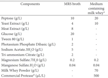
Table 1. Composition of MRS broth and medium containing milk whey.
Components MRS broth Medium
containing milk wheyA
Peptone (g/L) 10 20
Yeast Extract (g/L) 4 10
Meat Extract (g/L) 8
-Glucose (g/L) 20
-Tween 80 (g/L) 1
-Photassium Phosphate Dibasic (g/L) 2
-Sodium Acetate.3H2O (g/L) 5
-Tri-ammonium Citrate (g/L) 2
-Magnesium Sulfate.7H20 (g/L) 0.2 0.2 Manganese Sulfate.H2O (g/L) 0.04 0.04
Milk Whey Powder (g/L) - 70
Commercial ProteaseB (µL/L) - 500
5 minutes. The bacterial pellets were washed with sterile ultra-pure water, re-suspended in 1.5 ml of buffer solutions (pH 3.0 and 6.0) containing AFB1, and incubated under agitation of 180 rpm (TE-140 - Tecnal, Piracicaba, SP, Brazil) for 60 minutes at room temperature. After incubation, the solution was centrifuged at 1800 × g for 5 minutes, and the supernatant was removed and released AFB1 was quantified by high performance liquid chromatography (HPLC). The analyses were performed in triplicate and, for each sample, negative (L. rhamnosus cells suspended in buffer solution) and positive (AFB1 in buffer solution) controls were incubated and analyzed.
2.5 AFB1 quantification by HPLC
AFB1 quantification in the buffer solutions was achieved by injection into a HPLC system equipped with a fluorescence detector RF-10A XL, an autosampler SIL-10AF (Shimadzu,
Tokyo, Japan), and an ODS column 5 µm 4.6 × 150 mm (Phenomenex, Torrance, USA). A flow rate of 1.0 ml/min
was used with a mobile phase containing water, acetonitrile, and methanol (60:20:20). Excitation and emission detection were set at the wavelength of 360 nm and 440 nm, respectively. The limit of detection (LOD) for AFB1 was 0.01 ng/ml, and
its retention time was 10.5 minutes with a retention window of ± 10%. Equation 1 was used to quantify AFB1, where A is the percentage of AFB1 bound by the sample, and B, C, and D are the areas of chromatographic peaks of positive control, sample, and negative control, respectively.
A = [(B-C-D)/B] * 100 (1)
2.6 Statistical analysis
Results were subjected to analysis of variance, in accordance with the procedures established in the General Linear Model of SAS (SAS Institute Inc., 1992), to check for significant
differences between means of variables in the different treatments. The Fisher LSD test (Least Significant Difference) was used to test significant differences between means, adopting α = 0.05 as rejection level.
3 Results and discussion
Greater quantities of L. rhamnosus cells were achieved at lower costs using MMW (9.84 log CFU/ml) when compared with the MRS broth (9.63 log CFU/ml), a medium specifically developed for growing of Lactobacillus bacteria. Thus, MMW can perfectly replace the MRS broth for L. rhamnosus
production, without affecting cell production.
The lactose concentration in MMW decreased approximately by half, from 63.9 g/L to 35.3 g/L, while the pH decreased from 6.1 to 3.5. Lactose fermentation by L. rhamnosus, a homofermentative bacterium, produces lactic acid, which acidifies the medium causing a decrease in the pH. Ostile et al. (2003) also observed that the pH of a medium produced with UHT milk and supplemented with 0.75% of fructose had declined from 6.7 to 4.1 after 24 hours when L. rhamnosus (strain GG) was cultured. The authors also observed that bacterial cell concentration in the medium was 9.2 log CFU/ml after 24 hours.
Narvhus et al. (1998) explained that lactose is the primary substrate for acid production in milk; however, its fermentation is not limited by the amount of lactose available, but by the production of lactic acid and the concomitant lowering of pH, which increasingly inhibits the starter organisms long before the lactose is exhausted. According to Panesar et al. (2007), a complete and rapid fermentation occurs at the optimal pH range of 5.5–6.0, and in some cases, in the range of 6.0–6.5, depending upon the culture used, and it is strongly inhibited at lower pH values, stopping at pH below 4.5. However, LAB acid tolerance gives them a competitive advantage over many other bacteria. These authors added that pH affects some aspects of microbial cells, i.e., the functioning of their enzymes, transport of nutrients into the cell, and RNA and protein synthesis. In the present study, although the pH was below the ideal pH range for optimal cell growth, the growth rates of L. rhamnosus cultivated in MMW was higher than that in MRS broth.
Arauz et al. (2012) explained that LAB production is a meticulous process due to their nutrient demand. In laboratory-scale studies, LAB are usually grown in standard medium such as MRS broth, but the use of this medium in large scale becomes rather expensive. Therefore, complex and expensive culture media should be replaced with simpler and cheaper culture media to improve the LAB commercial production since, according to Rodrigues et al. (2006), the culture medium may represent 30% of the cost of a microbial fermentation. Burns et al. (2008) observed that the major benefits of using milk whey are its nutritional value and reasonable cost. Furthermore, possible environmental problems are avoided since the disposal of this by-product with a high load of organic matter can harm the environment. According to Mizubuti (1994), if 50,000 liters of milk whey were released as effluent, they would be equivalent to the sewage of a town of 25,000 inhabitants.
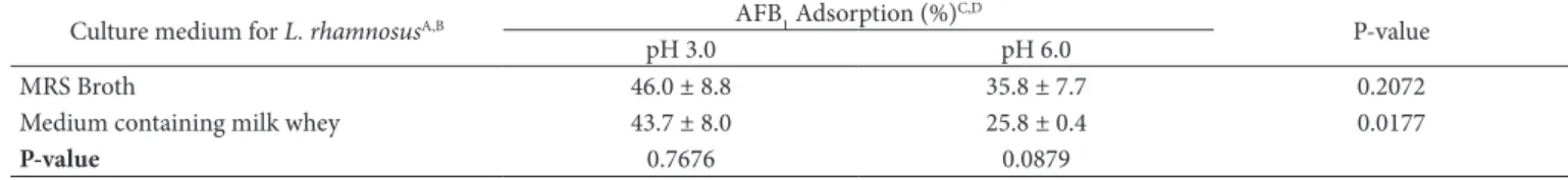
The results obtained for the adsorption of AFB1 by
L. rhamnosus cells produced in MRS broth and in MMW are shown in Table 2. It can be seen that, at the same pH value (3.0 or 6.0), there were no significant differences between the two culture media analyzed. The MMW was the only one that showed significant differences between both pHs, and the AFB1 was better adsorbed by the bacterial cells at pH 3.0 These results show that changing the culture medium did not affect the ability of L. rhamnosus to adsorb aflatoxins at a given pH value, which means that probably there were no changes in the bacterial cell structure.
physical adsorption, ion exchange, and complexation, regardless of the bacterial metabolism. The cell wall of microorganisms is mainly composed by polysaccharides, proteins, and lipids, which contain abundant functional groups such as carboxyl, hydroxyl, phosphate, and amine and hydrophobic adsorption sites, such as aliphatic carbon chains and aromatic rings.
Haskard et al. (2001) and El-Nezami et al. (1998) analyzed viable cells of L. rhamnosus (strains GG and LC-705) and obtained about 80% of AFB1 binding from a contaminated medium. Rahaie et al. (2012) observed that heat treated cells of
L. rhamnosus GG could adsorb 85% of AFB1 (10 ng/ml) from the medium. Peltonen et al. (2001) also studied viable cells of
L. rhamnosus (strains E-97800, LC 1/3 and CSCC 2420) and found AFB1 binding percentages of 22.7%, 54.6% and 33.1%, respectively. It can observed that within a genus or even within a determined species, not all strains are equivalent in terms of toxin removal, on the contrary, aflatoxin binding capacity is a characteristic of only specific strains, and its effectiveness varies markedly (El-Nezami et al., 2004).
The use of heat treated bacterial cells in the AFB1 adsorption assays instead of viable cells was chosen because, as mentioned and proved before, even non-viable bacterial cells can remove the aflatoxin from the medium, and this would allow the drying of bacterial cells and the development of a new product that could be used in two different ways. In the first way, applicable to liquid products, dried bacterial cells would be added to the product for a certain period of time, and then AFB1 adsorption would occur without causing fermentation or damage to the product. Bacterial cells would be removed at the end of this process, resulting in a totally or partially decontaminated product. In the second way, the application would be in solid products, in which dried bacterial cells would be used as food additives, and the AFB1 adsorption process would occur in the gastrointestinal tract, preventing toxin absorption by the organism.
Comparing the results obtained in the present study with those of previous reports is difficult because there are no studies describing the production of bacterial cells in MMW followed by AFB1 adsorption assays in buffer solution. The studies published so far have addressed two different procedures: LAB cells produced in MRS broth and analyzed for aflatoxin adsorption capacity in buffer solution, as shown by Oatley et al. (2000), Lahtinen et al. (2004), Shahin (2007) or Fazeli et al. (2009); or, aflatoxin adsorption assays were performed directly in the dairy product, for example, in milk samples, but in this case, the adsorption of aflatoxin M1, an 4-hydroxylated metabolite derivative from AFB1 biotransformation in the liver of animals that had been fed with contaminated diet, was performed, as
shown in studies of Pierides et al. (2000), Sarimehmetoglu & Küplülü (2004), Elgerbi et al. (2006) and Bovo et al. (2013).
4 Conclusions
The results of this trial showed that the production of
L. rhamnosus cells in MMW did not affect their adsorption capacity for AFB1. The production of greater quantities of L. rhamnosus cells at lower cost was accomplished using MMW, which had a higher cell concentration compared with that of MRS broth, a medium specifically developed for growing of
Lactobacillus bacteria. Therefore, the use of a L. rhamnosus strain which is able to grow in the milk-whey based medium tested and adsorb aflatoxins has a potential application for aflatoxin decontamination in food products.
Acknowledgements
The authors are grateful to Fundação de Amparo à Pesquisa do Estado de São Paulo for the grant and financial support (Grant number 2011/03410-0).
References
Alberts, J. F., Engelbrechta, Y., Steynb, P. S., Holzapfelc, W. H., & Van Zyla, W. H. (2006). Biological degradation of aflatoxin B1 by
Rhodococcus erythropolis cultures. International Journal of Food Microbiology, 109(1-2), 121-126. PMid:16504326. http://dx.doi. org/10.1016/j.ijfoodmicro.2006.01.019
Arauz, L. J., Jozala, A. F., Baruque-Ramos, J., Mazzola, P. G., Pessoa Jr., A., & Penna, T. C. V. (2012). Culture medium of diluted skimmed milk for the production of nisin in batch cultivations. Annals of Microbiology, 62(1), 419-426. http://dx.doi.org/10.1007/s13213-011-0278-6
Azab, R. M., Tawakkol, W. M., Hamad, A. M., Abou-Elmagd, M. K., El-Agrab, H. M., & Refai, M. K. (2005). Detection and estimation of aflatoxin B1 in feeds and its biodegradation by bacteria and fungi.
Egyptian Journal of Natural Toxins, 2, 39-56.
Bata, A., & Lásztity, R. (1999). Detoxification of mycotoxin-contaminated food and feed by microorganisms. Trends Food Science and Technology, 10(6-7), 223-228. http://dx.doi.org/10.1016/ S0924-2244(99)00050-3
Begot, C., Desnier, I., Daudin, J. D., Labadie, J. C., & Lebert, A. (1996). Recommendations for calculating growth parameters by optical density measurements. Journal of Microbiological Methods, 25(3), 225-232. http://dx.doi.org/10.1016/0167-7012(95)00090-9 Bhat, R., Rai, R. V., & Karim, A. A. (2010). Mycotoxins in food and
feed: present status and future concerns. Comprehensive Reviews in Food Science and Food Safety, 9(1), 57-81. http://dx.doi.org/10.1111/ j.1541-4337.2009.00094.x
Table 2. AFB1 adsorption capacity of cells of L. rhamnosus cultured in MRS broth and in a medium containing milk whey.
Culture medium for L. rhamnosusA,B AFB1 Adsorption (%) C,D
P-value
pH 3.0 pH 6.0
MRS Broth 46.0 ± 8.8 35.8 ± 7.7 0.2072
Medium containing milk whey 43.7 ± 8.0 25.8 ± 0.4 0.0177
P-value 0.7676 0.0879
AL. rhamnosus strain HOWARU LYO 40 DCU; BNon-viable cells produced by sterilization at 121 °C for 15 minutes; CAFB
1 removed from a citrate-phosphate buffer containing 1.0 µg
AFB1/ml;
Bibal, B., Goma, G., Vayssier, Y., & Pareilleux, A. (1988). Influence of pH, lactose and lactic acid on the growth of Streptococcus cremoris: a kinetic study. Applied Microbiology and Biotechnology, 28(4-5), 340-344. http://dx.doi.org/10.1007/BF00268192
Bovo, F., Corassin, C. H., Rosim, R. E., & Oliveira, C. A. F. (2013). Efficiency of lactic acid bacteria strains for decontamination of aflatoxin M1 in phosphate buffer saline solution and in skimmed milk. Food and Bioprocess Technology, 6(8), 2230-2234. http://dx.doi. org/10.1007/s11947-011-0770-9
Brinques, G. B. (2009). Otimização da produção de probióticos em biorreatores e suas aplicações em sistemas alimentícios sob a forma imobilizada (Tese de doutorado). Escola de Engenharia, Universidade Federal do Rio Grande do Sul, Porto Alegre. PMid:19936814.
Burns, P., Vinderola, G., Molinari, F., & Reinheimer, J. (2008). Suitability of whey and buttermilk for the growth and frozen storage of probiotic lactobacilli. International Journal of Dairy Technology,
61(2), 156-164. http://dx.doi.org/10.1111/j.1471-0307.2008.00393.x Elgerbi, A. M., Aidoo, K. E., Candlish, A. A. G., & Williams, A. G.
(2006). Effects of lactic acid bacteria and bifidobacteria on levels of aflatoxin M1 in milk and phosphate buffer. Milchwissenschaft,
61(2), 197-199.
El-Nezami, H., Kankaanpaa, P., Salminen, S., & Ahokas, J. (1998). Ability of dairy strains of lactic acid bacteria to bind a common food carcinogen, aflatoxin B1. Food and Chemical Toxicology, 36(4), 321-326. http://dx.doi.org/10.1016/S0278-6915(97)00160-9 El-Nezami, H., Salminen, S., Salminen, E., Haskard, C., & Mykkanen,
H. (2004). Lactic acid bacteria as a tool for enhancing food safety by removal of dietary toxins. In S. Salminen, A. Von Wright & A. Ouwehand, A. (Eds.), Lactic acid bacteria: microbiological and functional aspects (3rd ed., Cap. 12, pp. 397-406). New York: Marcel Dekker.
Fazeli, M. R., Hajimohammadali, M., Moshkani, A., Samadi, N., Jamalifar, H., Khoshayand, M. R., Vaghari, E., & Pouragahi, S. (2009). Aflatoxin B1 binding capacity of autochthonous strains of lactic acid bacteria. Journal of Food Protection, 72(1), 189-192. PMid:19205485.
Haskard, C. A., El-Nezami, H. S., Kankaanpää, P. E., Salminen, S., & Ahokas, J. T. (2001). Surface binding of aflatoxin B1 by lactic acid bacteria. Applied and Environmental Microbiology, 67(7), 3086-3091. PMid:11425726 PMCid:PMC92985. http://dx.doi.org/10.1128/ AEM.67.7.3086-3091.2001
International Dairy Federation. (2009). The World Dairy Situation 2009. In International Dairy Federation. Bulletin of the International Dairy Federation (Vol. 438). Brussels. Retrieved from http://www. lactodata.com/lactodata/docs/lib/fil_ idf_bulletin_438_2009_the_ world_2009.pdf
Lahtinen, S. J., Haskard, C. A., Ouwehand, A. C., Salminen, S. J., & Ahokas, J. T. (2004). Binding of aflatoxin B1 to cell wall components of Lactobacillus rhamnosus strain GG. Food Additives and Contaminants, 21(2), 158-164. PMid:14754638. http://dx.doi. org/10.1080/02652030310001639521
Miller, G. L. (1959). Use of dinitrosalicylic acid reagent for determination of reducing sugar. Analytical Chemistry, 31(3), 426-428. http://dx.doi.org/10.1021/ac60147a030
Mizubuti, I. Y. (1994). Soro de leite: composição, processamento e utilização na alimentação. Semina: Ciências Agrárias, 15(1), 80-94. Narvhus, J. A., Osteraas, K., Mutukumira, T., & Abrahamsen, R. K.
(1998). Production of fermented milk using a malty compound-producing strain of Lactococcus lactis subsp. lactis biovar.
diacetylactis, isolated from Zimbabwean naturally fermented milk.
International Journal of Food Microbiology, 41(1), 73-80. http:// dx.doi.org/10.1016/S0168-1605(98)00036-1
Oatley, J. T., Rarick, M. D., Ji, G. E., & Linz, J. E. (2000). Binding of aflatoxin B1 to bifidobacteria in vitro. Journal of Food Protection,
63(8), 1133-1136. PMid:10945592.
Ostile, H. M., Helland, M. H., & Narvhus, J. A. (2003). Growth and metabolism of selected strains of probiotic bacteria in milk.
International Journal of Food Microbiology, 87(1-2), 17-27. http:// dx.doi.org/10.1016/S0168-1605(03)00044-8
Panesar, P. S., Kennedym, J. F., Gandhi, D. N., & Bunko, K. (2007). Bioutilisation of whey for lactic acid production. Food Chemistry,
105(1), 1-14. http://dx.doi.org/10.1016/j.foodchem.2007.03.035 Peltonen, K., El-Nezami, H., Haskard, C., Ahokas, J., & Salminen, S.
(2001). Aflatoxin B1 binding by dairy strains of lactic acid bacteria and bifidobacteria. Journal of Dairy Science, 84(10), 2152-2156. http://dx.doi.org/10.3168/jds.S0022-0302(01)74660-7
Pierides, M., El-Nezami, H., Peltonen, K., Salminen, S., & Ahokas, J. (2000). Ability of dairy strains of lactic acid bacteria to bind aflatoxin M1 in a food model. Journal of Food Protection, 63(5), 645-650. PMid:10826723.
Rahaie, S., Emam-Djomeh, Z., Razavi, S. H., & Mazaheri, M. (2012). Evaluation of aflatoxin decontaminating by two strains of Saccharomyces cerevisiae and Lactobacillus rhamnosus strain GG in pistachio nuts. International Journal of Food Science and Technology, 47(8), 1647-1653. http://dx.doi.org/10.1111/j.1365-2621.2012.03015.x
Ringot, D., Lerzy, B., Chaplain, K., Bonhoure, J-P., Auclair, E., & Larondelle, Y. (2007). In vitro biosorption of ochratoxin A on the yeast industry by-products: Comparison of isotherm models.
Bioresource Technology, 98(9), 1812-1821. PMid:16919938. http:// dx.doi.org/10.1016/j.biortech.2006.06.015
Rodrigues, L. R., Teixeira, J. A., & Oliveira, R. (2006). Low-cost fermentative medium for biosurfactant production by probiotic bacteria. Biochemical Engineering Journal, 32(3), 135-142. http:// dx.doi.org/10.1016/j.bej.2006.09.012
Sarimehmetoglu, B., & Küplülü, Ö. (2004). Binding ability of aflatoxin M1 to yoghurt bacteria. Ankara Üniversitesi Veteriner Fakültesi Dergisi, 51(3), 195-198.
SAS Institute Inc. (1992). SAS user’s guide: statistics. Cary.
Scott, P. M. (1990). Natural poisons. In K. Helrich (Ed.), Official methods of analysis of the Association of Official Analytical Chemists
(15th ed., pp. 1184-213). Arlington: AOAC.
Shahin, A. A. M. (2007). Removal of aflatoxin B1 from contaminated liquid media by dairy lactic acid bacteria. International Journal of Agriculture & Biology, 9(1), 71-75.
Thipathi, P., Beaussart, A., Andre, G., Rolain, T., Lebeer, S., Vanderleyden, J., Hols, P., & Dufrêne, Y. F. (2012). Towards a nanoscale view of lactic acid bacteria. Micron, 43(12), 1323-1330. PMid:22293169. http://dx.doi.org/10.1016/j.micron.2012.01.001 Van de Guchte, M., Serror, P., Chervaux, C., Smokvina, T., Ehrlich, S.
D., & Maguin, E. (2002). Stress responses in lactic acid bacteria.
Antonie van Leeuwenhoek, 82(1-4), 187-216. http://dx.doi. org/10.1023/A:1020631532202
Walstra, P., Wouters, J. T. M., & Geurts, T. J. (2006). Dairy science and technology (2nd ed.). Boca Raton: CRC Press.