AMP
STUDENT
ARTIGO ORIGINAL
Phenotypic Heterogeneity by Germline Mismatch Repair
Gene Defect in Lynch Syndrome Patients
Heterogeneidade Fenotípica por Mutação Germinativa nos
Genes Mismatch Repair em Doentes com Síndrome de
Lynch
1. Faculty of Medicine. University of Porto. Porto. Portugal.
2. Department of General Surgery. Centro Hospitalar de São João. Porto. Portugal. Autor correspondente: Jorge Hernâni-Eusébio. [email protected]
Recebido: 24 de abril de 2016 - Aceite: 21 de julho de 2016 | Copyright © Ordem dos Médicos 2016
Jorge HERNÂNI-EUSÉBIO1, Elisabete BARBOSA2
Acta Med Port 2016 Oct;29(10):587-596 ▪ http://dx.doi.org/10.20344/amp.7774
RESUMO
Introdução: A síndrome de Lynch é a forma hereditária mais comum de cancro colo-rectal, sendo também responsável por cancro
do endométrio e de outros tipos. Associa-se a mutações germinativas nos genes de mismatch repair do ADN e a instabilidade de mi-crossatélites. As mutações MLH1 e MSH2 têm um fenótipo de síndrome de Lynch ‘clássico’, sendo o MSH2 mais associado a cancro extra-cólico. Mutações do MSH6 e PMS2 têm um fenótipo atípico. A expressão clínica é heterogénea, existindo uma correlação entre o gene mismatch repair mutado e o padrão fenotípico.
Material e Métodos: Análise retrospetiva dos dados clínicos de doentes que cumpriam os critérios de Amesterdão ou que tinha
muta-ções nos genes mismatch repair, entre setembro de 2012 e outubro de 2015.
Resultados: Identificámos 28 doentes. Dezassete tinham cancro colo-rectal sendo a localização no cólon direito predominante. Cinco
tiveram cancro do endométrio (mediana da idade de diagnóstico – 53), sem qualquer mutação no MSH6. Cinco desenvolveram outros cancros. Todos os casos com mutações mismatch repair estudados tinham instabilidade de microssatélites.
Discussão: Na maioria dos casos foi encontrada mutação no MSH2 apesar de o MLH1 ser descrito na literatura como o gene mais
frequentemente mutado. Interessa dizer que os doentes com cancro colo-rectal não evidenciam uma tendência para ter muito infiltrado inflamatório. Na maioria dos casos foi realizada colectomia parcial apesar da incidência elevada de lesões síncronas e metácronas associadas. Histerectomia e anexectomia profilática foi realizada em doentes em menopausa/perimenopausa.
Conclusão: O registo standardizado dos dados dos doentes poderá levar a um melhor acompanhamento e conhecimento desta
sín-drome. O uso das Guidelines de Bethesda poderá identificar novos casos que escapam aos critérios de Amesterdão. A pesquisa de instabilidade de microssatélites deve ser feita em muito maior número. Embora seja descrita na literatura uma correlação genótipo/ fenótipo, o nosso estudo não verificou esta correlação de forma estatisticamente signficativa, talvez por a amostra ser pequena e os registos clínicos insuficientes.
Palavras-chave: Neoplasias Colorretais Hereditárias sem Polipose; Neoplasias do Endométrio; Reparação de Incompatibilidade de
ADN; Síndromes Neoplásicas Hereditárias; Transtornos por Deficiências na Reparação de ADN.
ABSTRACT
Introduction: Lynch syndrome is the most common form of hereditary colorectal cancer, being also responsible for endometrial and
other types of cancers. It is associated with germline mutations in DNA mismatch repair genes and microsatellite instability. MLH1 and MSH2 mutations have a “classical” Lynch syndrome phenotype, with MSH2 having a higher association with extracolonic cancer. MSH6 and PMS2 mutations have an atypical phenotype. Clinical expression is heterogeneous, with correlation between mismatch repair mutated gene and phenotypic patterns.
Material and Methods: We retrospectively analyzed data from patients fulfilling Amsterdam criteria or having mismatch repair gene
mutations, between September 2012 and October 2015.
Results: We identified 28 patients. Seventeen had colorectal cancer with right colon predominance. Five developed endometrial
cancer (median age of diagnosis – 53), with no MSH6 mutations. Five developed other cancers. All mutated mismatch repair cases studied had microsatellite instability.
Discussion: Most cases had MSH2 mutations despite MLH1 being described in the literature as the most frequently mutated.
Interestingly, colorectal cancer patients showed no tendency for high inflammatory infiltrate. Despite the high incidence of synchronous and metachronous tumours, most patients underwent a partial colectomy. Prophylactic hysterectomy and adnexectomy was performed in menopausal/perimenopausal patients.
Conclusion: A standardized registration of patient’s data may lead to better management and knowledge about Lynch syndrome.
Use of Bethesda Guidelines might identify new cases non-identified by Amsterdam criteria. Microsatellite instability analysis must be performed in a much larger scale. The genotypic/phenotypic correlation described in the literature was not verified in our study with statistical significance, perhaps due to small data sample and insufficient clinical registration.
Keywords: Colorectal Neoplasms, Hereditary Nonpolyposis; DNA Mismatch Repair; DNA Repair-Deficiency Disorders; Endometrial
Neoplasms; Neoplastic Syndromes, Hereditary.
INTRODUCTION
Colorectal cancer (CRC) is a very common type of cancer. It has an incidence of 1,200,000 cases annually on a global scale. The mortality rate is of approximately 50%.1
Among these, approximately 20% account for familial forms of CRC.2
non-ARTIGO ORIGINAL
AMP
STUDENT
polyposis colorectal cancer - is the most commonly seen hereditary form of CRC. It is responsible for 3% - 5% of CRCs3 and approximately 3% of endometrial cancers (EC).4
This incidences refers to the general population worldwide.5
A linkage is also shown between LS and other types of cancers.6-8
Therefore, 36,000 – 60,000 of CRCs worldwide are associated with LS,9 with an average age of onset of 45
years.10,11
This is a dominantly inherited disorder with 85% of penetrance,12 featuring germline mutations in at least one of
the DNA mismatch repair (MMR) genes.1,2 Those mutations
are found in more than 80% of patients with LS.13
DNA MMR ‘complex’ is responsible for proofreading and edition posteriorly to the DNA replication phase, ensuring the integrity of the genome.14
Manifesting the mutations in the MMR genes, these patients tend to have a fast process of carcinogenesis15 with
cancers showing microsatellite instability (MSI).16
The knowledge of the impaired activity of the MMR genes in cancers associated with LS led to a common determination of the MMR genes activity by immunohistochemistry (IHQ),17 not only as a marker of a possible cancer correlated
to LS, but also to discover the gene (or genes) that probably underlies the germline mutation.14
Therefore, when we identify a germline mutation in the MMR genes, we have the sine qua non factor that provides the definitive diagnosis of LS.14
Despite that, we should not test every CRC case for these mutations,18 having family history a crucial importance in that
decision19 and in the diagnostic evaluation.1 Amsterdam I20
and II21 criteria have been useful for that purpose. Bethesda
Guidelines for testing CRC for MSI may also be of utility.22,23
Some patients fulfill Amsterdam criteria but have no MMR mutation, which may hamper the diagnostic process and the correct management of those patients at high-risk of developing LS-related tumours.11
In general, it was shown that the lifetime risk of having a cancer in families that are identified with a MMR mutation are the following: 28% - 75% in men and 24% - 52% women for CRC; 27% - 71% for EC; 3% - 13% for ovarian cancer; 2% - 13% for gastric cancer; 1% - 12% for urothelial cancer; 4% - 7% for small-bowel cancer; 1% - 4% for CNS cancer and 2% for bile duct or gallbladder cancer.18,24,25
The wide range of the intervals (specifically for colorectal and endometrial cancers) may be due to the documented phenotypic variation related to germline mutations in different MMR genes.11,12
Those are the MLH1 (mutL homologue 1, located in chromosome 3p21), MSH2 (mutS homologue 2, located in chromosome 2p16), MSH6 (mutS homologue 6, located in chromosome 2p15) and PMS2 (postmeiotic segregation increased 2, located in chromosome 7p22).12,14
Data from the International Society for Gastrointestinal and Hereditary Tumours (InSiGHT) relative to the year of 2012 registered a frequency of LS-associated mutations of 42% for MLH1, 33% for MSH2, 18% for MSH6 and 8%
for PMS2.26 Previous data suggested a frequency of 32%,
38%, 14% and 15%, respectively.27
Despite the typical course of LS (CRC of early onset),14
it is well noticed that there can be several cases of LS with different phenotypic characteristics (like later onset of cancer, ‘rare’ kinds of tumours).6-8,14
With the large-scale implementation of MSI and genetic testing, the specific clinical phenotype of each LS patient is now better understood, according to the MMR mutation.28
Some recent studies give particular emphasis on the possible relevance of epimutations.29
Starting with MLH1 and MSH2 mutations, those tend to have LS of a ‘classical’ phenotype (fulfilling the Amsterdam I criteria)24,30 and cancer displaying high MSI.31
Male patients with MLH1 and/or MSH2 have 27% - 74% of risk for developing CRC; as for female patients that risk is 22% - 61%.32 The mean age for the onset is of 43 - 46
years.24,30
MLH1 patients seem to have high predominance of CRC.11 MSH2 patients have higher probability of developing
extracolonic cancer when compared to MLH1 and are thought to have a higher risk of developing any cancer throughout life.33
Some studies also reported that mutated MSH2 patients are shown to present greater risk of urothelial,34,35 gastric6,36
and ovarian cancer37 when compared to patients with MLH1
mutations.
Also, MSH2 patients have been linked to augmented risk of extracolonic cancers specific of the Muir-Torre syndrome tumours-spectrum.38,39
On the other hand, germline mutations in MSH6 and
PMS2 lead to an atypical phenotype.40,41 Those are patients
expected to have less risk of developing CRC and older age of onset.40,42
Considering particularly MSH6 patients, the penetrance of their mutation is thought to be lower12 and also they do
not show an ubiquitous MSI profile.43
For these patients, the incidence of CRC is lower42
(particularly in women: 10% - 30% risk of CRC; in men it is higher: 22% - 69% risk of CRC) and the age of onset for CRC is higher (most frequently between 50 - 6332 and
with a mean age of diagnosis of 54 years old)12 than that of
patients with the previous enounced mutations.
MSH6-mutated phenotypes are also considered to cause an atypical display of tumours.16
These are the patients with highest risk of developing EC.41,44 That is considered to be the principal clinical
manifestation for female patients with this mutation.42,44
They are usually diagnosed in patients over 50 years of age41,44 and it was observed that at the age of 70, 71% of
female patients with this mutation will have EC.44
Along with MSH2, MSH6 mutation is more related to EC than mutations in MLH1 or PMS2.5
Finally, PMS2 mutations are associated with a special phenotype of LS with large number of colonic polyps and small incidence of CRC. When associated with CRC, this cancer will present MSI. However, it usually develops at an
AMP
STUDENT
ARTIGO ORIGINAL
older age and has an intriguing lack of family history.40
These patients show a risk for developing CRC of 20% for male patients and 15% for female patients, being the diagnosis usually held at the age of 47 - 66 years.32
As accurate as this theoretical LS phenotype according to the MMR gene defect may be, variations can be found even in patients carrying the same germline mutation, giving us a clue that maybe there are other factors correlated to clinical manifestations of LS.30,45,46
According to all of this, we can realize that the clinical expression and manifestations of LS is widely heterogeneous, with particular phenotypic patterns being highly correlated with the MMR gene mutated.
Therefore, we hold as main objective of this study the correlation between different MMR genes affected with mutation and the postulated phenotypic characteristics among them.
With that purpose in mind, we conducted a retrospective study at the Department of General Surgery – Centro Hospitalar de São João (CHSJ), reporting a three year period of time, between September 2012 and October 2015.
MATERIAL AND METHODS
This retrospective study was approved by the Ethics Committee for Health of CHSJ/Faculdade de Medicina da Universidade do Porto (FMUP). There was no conflict of interest reported in the making of this study.
We considered a three year period, between September 2012 and October 2015. According to this period of time, we retrospectively identified 36 patients that were suspected of having LS, as a result of clinical evaluation by a group of physicians from CHSJ and that underwent MMR gene mutation studies. The data was obtained from the Instituto de Patologia e Imunologia Molecular da Universidade do Porto (IPATIMUP) database.
Inclusion criteria were patients with mutations in one or more MMR genes according to the database of IPATIMUP or patients that fulfill the Amsterdam I or II criteria according to a clinical and family history analysis.
Exclusion criteria consisted of incomplete clinical records and patients that have no mutation in MMR genes neither fulfill Amsterdam I or II criteria.
The existing data from every patient were reviewed and additional data was collected from their clinical files. We collected data on the age and gender of the patients as well as MMR genes tested and those that were mutated. Presence of other mutations was assessed. Fulfilling of Amsterdam I and II criteria was also analyzed. In addition, age at CRC diagnosis – if existent – was collected.
As for those with CRC, location of tumor, degree of differentiation, macroscopic characteristics, inflammatory infiltrate, mucinous characteristics, lymph nodes involvement, vascular or perineural invasion, as well as the TNM staging were assessed. MSI status data was also collected.
The surgical treatment and decision following consultation in the Colorectal Oncology Group of CHSJ
were also analyzed.
Further collection of data was performed regarding the existence of EC among women, as well as the age of diagnosis and treatment performed.
We also collected data of other extra-colic cancers, the age of diagnosis and treatment.
Finally, we assessed data regarding the global patient’s follow-up for all included cases.
We mainly sorted the patients by presence of MMR gene mutation (and of these, by different mutated gene) and by fulfillment of Amsterdam I or II criteria.
Then we performed statistical analysis using IBM SPSS® (Statistical Package for Social Sciences) version 23
and conducted a descriptive analysis of all variables. The distribution of continuous variable was performed using the Kolmogorov-Smirnov test that showed that age of patients was normally distributed.
Inferential analysis was performed to evaluate possible associations between categorical variables, using the Chi-square and Fisher’s exact tests.
The evaluation of categorical and continuous variables association was performed by the Independent-samples
t-test.
For evaluating if a relationship existed between variables, the statistical significance was investigated using Chi-square and Fisher’s exact test and we used p = 0.05 as reference.
RESULTS
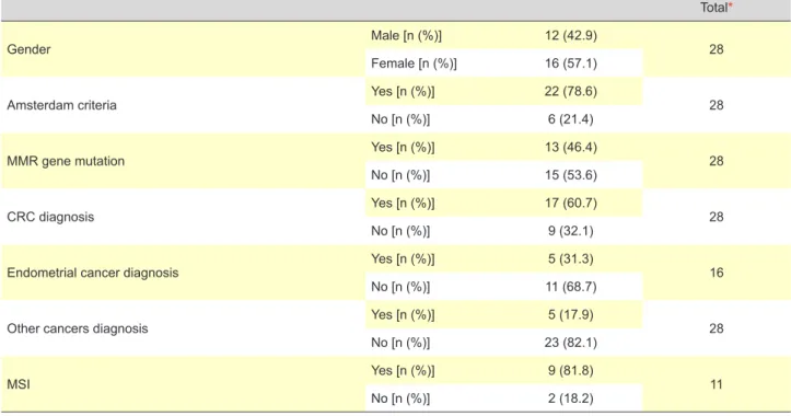
After a first analysis of the 36 identified patients, eight patients were eliminated for not having inclusion criteria. The subject of our analysis was the remaining 28 patients. We started with the analysis of the demographic data of our patients regarding to gender, the fulfilling of Amsterdam I or II criteria, diagnosis of CRC, EC or other cancers of LS spectrum and presence of MSI (Table 1).
We noticed that from the 22 (78.6%) patients fulfilling Amsterdam I or II criteria, only seven (31.8%) had MMR gene mutations. On the other hand, all the patients that did not fulfill Amsterdam I or II criteria – six (21.4%) – had at least one MMR gene mutated.
We also described the CRC, endometrial (Table 2) and other cancers of LS spectrum cases.
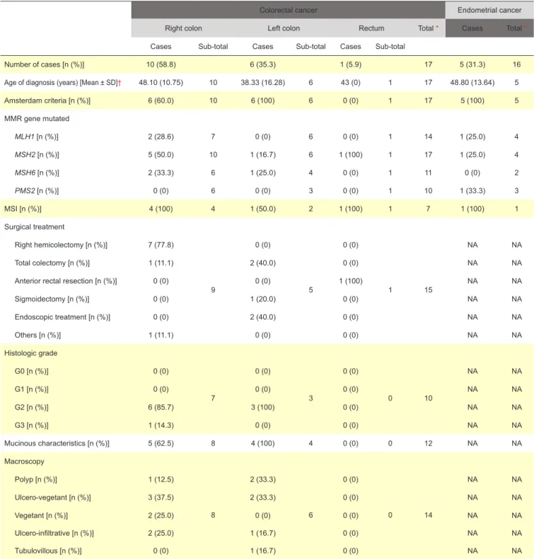
We then sorted the CRC cases by their location (right colon, left colon and rectum).
Median age of diagnosis was of 46 years (min = 36; max = 68) for right colon patients and of 34 years (min = 23; max = 68) for left colon cancers. The only case of cancer of rectum was diagnosed at age of 43 years.
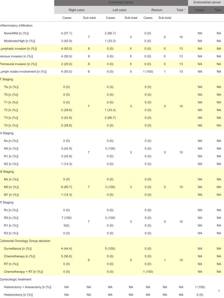
Regarding the CRC tumor characteristics, histologic grading was low in most cases. Also, all cases of CRC in the left colon had mucinous characteristics and so did most of the studied cases in the right colon.
Regarding CRC staging according to AJCC,47 most
patients were stage II or III and only one case (in the right colon) was stage IV.
ARTIGO ORIGINAL
AMP
STUDENT
all patients presented at the group consultation with AJCC Stage III or IV were proposed for adjuvant chemotherapy. The rectum case did chemotherapy + radiotherapy. The chosen chemotherapy scheme was FOLFOX (folinic acid, 5-FU and oxaliplatin).48
Our data showed that the median age of EC diagnosis was 53 years (min = 31; max = 65). None of these cases had CRC. It is important to note that four female patients with CRC performed prophylactic hysterectomy and adnexectomy.
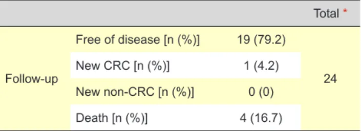
We obtained follow-up data (Table 3) of 24 patients. From these, 19 (79.2%) are free of disease. At the time of last follow-up, one (4.2%) had a metachronous rectal cancer and four (16.7%) patients died. Of those, only one case died of CRC, the other three from endometrial, gastric and CNS cancers.
Concerning MMR genes, 22 patients (78.6%) were tested for MLH1 mutations, 27 (96.4%) for MSH2, 14 (50.0%) for MSH6 and 14 (50.0%) for PMS2.
Finally, we sorted patients by mutated MMR gene (Table 4). We accounted for 13 total cases with mutations and 17 individual mutations among them (with four patients having two concomitant mutations), accounting MLH1 mutations for 17.7% of these individual mutations, MSH2 for 52.9%,
MSH6 for 23.5% and PMS2 for 5.9.
The median age of CRC diagnosis was 49 years (min = 37; max = 42) for MLH1, 43 years (min = 30; max = 57) for MSH2 and 36 years (min = 30; max = 47) for MSH6. No case of CRC was reported as having PMS2 mutation. For EC diagnosis, only one case was described for
MLH1, MSH2 and PMS2 and age of diagnosis was,
respectively, of 65, 39 and 65 years. No case was reported for MSH6.
Only few patients presented other cancers of the LS spectrum and MMR mutations, being those of MSH2 and
MSH6 genes.
When regarding the mutated germline MMR gene, at last follow-up, all patients with MSH2 and MSH6 mutations were free of disease. From the three cases of MLH1 mutations, two (66.7%) resulted in death and one (33.3%) had a metachronous CRC. The PMS2 mutation case also resulted in death. It is of notice that this last patient also had a mutation in MLH1.
As for mutations other than those of the MMR genes, 25 (89.3%) patients did not show any mutation. However, one (3.6%) patient had a mutation in p53 gene and two (7.1%) patients had a mutation in KRAS.
Inferential statistics were performed comparing several variables. However, none of the attempted correlations presented statistical significance.
DISCUSSION
The main objective of this study was to establish a correlation between MMR gene mutation and phenotypic characteristics.
Only seven patients out of 22 that fulfilled the Amsterdam I or II criteria had at least one MMR gene mutation, which could mean that some of these patients may have a MMR gene mutation yet they might not be spotted as it is known that there are mutations still to be discovered in all four of the MMR germline genes.49 We also verified that all
patients not fulfilling Amsterdam I or II criteria had at least one mutated MMR gene, meaning that they could be a first familiar case. All these patients (both having mutation or fulfilling the Amsterdam I or II criteria) were managed as LS patients.
Table 1 - Descriptive demographic table
Total*
Gender Male [n (%)] 12 (42.9) 28
Female [n (%)] 16 (57.1)
Amsterdam criteria Yes [n (%)] 22 (78.6) 28
No [n (%)] 6 (21.4)
MMR gene mutation Yes [n (%)] 13 (46.4) 28
No [n (%)] 15 (53.6)
CRC diagnosis Yes [n (%)] 17 (60.7) 28
No [n (%)] 9 (32.1)
Endometrial cancer diagnosis Yes [n (%)] 5 (31.3) 16
No [n (%)] 11 (68.7)
Other cancers diagnosis Yes [n (%)] 5 (17.9) 28
No [n (%)] 23 (82.1)
MSI Yes [n (%)] 9 (81.8) 11
No [n (%)] 2 (18.2)
AMP
STUDENT
ARTIGO ORIGINAL
cancers should be carefully assessed because they may be also preceding other cancers.
As for MSI, despite being present in most LS patients and being one of the first auxiliary test in LS suspected cases under 50 years (even if they do not fulfill Amsterdam criteria),18 only 11 of our patients underwent this test. This
information may also have repercussions in terms of therapy therefore large scale implementation should be achieved. It is also of notice that Bethesda Guidelines for testing CRC for MSI were only assessed and registered in the history of 1 patient, since they might be of utility as being described
(table continues on the next page)
We realized that CRC is in fact the most frequent type of cancer in our patients, followed by EC and other cancers (two central nervous system – CNS - cancer cases, one case with both gastric and urothelial cancer, one case of gastric cancer and 1 case of lymphoma and another case of breast cancer). It is of capital importance to realize that of the five cases with EC, only one patient had CRC, being of extreme relevance to manage carefully those patients as we know that LS patients may present EC as a first manifestation. Also, the only case with breast cancer was diagnosed nine years before CRC. The remaining four cases of other Table 2 - Descriptive CRC and EC Table
Colorectal cancer Endometrial cancer
Right colon Left colon Rectum Total * Cases Total *
Cases Sub-total Cases Sub-total Cases Sub-total
Number of cases [n (%)] 10 (58.8) 6 (35.3) 1 (5.9) 17 5 (31.3) 16
Age of diagnosis (years) [Mean ± SD]† 48.10 (10.75) 10 38.33 (16.28) 6 43 (0) 1 17 48.80 (13.64) 5
Amsterdam criteria [n (%)] 6 (60.0) 10 6 (100) 6 0 (0) 1 17 5 (100) 5 MMR gene mutated MLH1 [n (%)] 2 (28.6) 7 0 (0) 6 0 (0) 1 14 1 (25.0) 4 MSH2 [n (%)] 5 (50.0) 10 1 (16.7) 6 1 (100) 1 17 1 (25.0) 4 MSH6 [n (%)] 2 (33.3) 6 1 (25.0) 4 0 (0) 1 11 0 (0) 2 PMS2 [n (%)] 0 (0) 6 0 (0) 3 0 (0) 1 10 1 (33.3) 3 MSI [n (%)] 4 (100) 4 1 (50.0) 2 1 (100) 1 7 1 (100) 1 Surgical treatment Right hemicolectomy [n (%)] 7 (77.8) 9 0 (0) 5 0 (0) 1 15 NA NA Total colectomy [n (%)] 1 (11.1) 2 (40.0) 0 (0) NA NA
Anterior rectal resection [n (%)] 0 (0) 0 (0) 1 (100) NA NA
Sigmoidectomy [n (%)] 0 (0) 1 (20.0) 0 (0) NA NA Endoscopic treatment [n (%)] 0 (0) 2 (40.0) 0 (0) NA NA Others [n (%)] 1 (11.1) 0 (0) 0 (0) NA NA Histologic grade G0 [n (%)] 0 (0) 7 0 (0) 3 0 (0) 0 10 NA NA G1 [n (%)] 0 (0) 0 (0) 0 (0) NA NA G2 [n (%)] 6 (85.7) 3 (100) 0 (0) NA NA G3 [n (%)] 1 (14.3) 0 (0) 0 (0) NA NA Mucinous characteristics [n (%)] 5 (62.5) 8 4 (100) 4 0 (0) 0 12 NA NA Macroscopy Polyp [n (%)] 1 (12.5) 8 2 (33.3) 6 0 (0) 0 14 NA NA Ulcero-vegetant [n (%)] 3 (37.5) 2 (33.3) 0 (0) NA NA Vegetant [n (%)] 2 (25.0) 0 (0) 0 (0) NA NA Ulcero-infiltrative [n (%)] 2 (25.0) 1 (16.7) 0 (0) NA NA Tubulovillous [n (%)] 0 (0) 1 (16.7) 0 (0) NA NA
ARTIGO ORIGINAL AMP STUDENT Inflammatory Infiltration None/Mild [n (%)] 4 (57.1) 7 2 (66.7) 3 0 (0) 0 10 NA NA Moderate/High [n (%)] 3 (42.9) 1 (33.3) 0 (0) NA NA Lymphatic invasion [n (%)] 4 (50.0) 8 0 (0) 5 0 (0) 0 13 NA NA Venous invasion [n (%)] 4 (50.0) 8 0 (0) 5 0 (0) 0 13 NA NA Perineural invasion [n (%)] 2 (25.0) 8 0 (0) 5 0 (0) 0 13 NA NA
Table 2 - Descriptive CRC and EC Table (remaining section)
Colorectal cancer Endometrial cancer
Right colon Left colon Rectum Total * Cases Total *
Cases Sub-total Cases Sub-total Cases Sub-total
Lymph nodes involvement [n (%)] 4 (50.0) 8 0 (0) 5 1 (100) 1 13 NA NA
T Staging Tis [n (%)] 0 (0) 7 0 (0) 3 0 (0) 0 10 NA NA T0 [n (%)] 0 (0) 0 (0) 0 (0) NA NA T1 [n (%)] 0 (0) 0 (0) 0 (0) NA NA T2 [n (%)] 2 (28.6) 1 (33.3) 0 (0) NA NA T3 [n (%)] 3 (42.9) 2 (66.7) 0 (0) NA NA T4 [n (%)] 2 (28.6) 0 (0) 0 (0) NA NA N Staging Nx [n (%)] 0 (0) 7 0 (0) 3 0 (0) 0 10 NA NA N0 [n (%)] 3 (42.9) 3 (100) 0 (0) NA NA N1 [n (%)] 3 (42.9) 0 (0) 0 (0) NA NA N2 [n (%)] 1 (14.3) 0 (0) 0 (0) NA NA M Staging Mx [n (%)] 0 (0) 7 0 (0) 3 0 (0) 0 10 NA NA M0 [n (%)] 6 (85.7) 3 (100) 0 (0) NA NA M1 [n (%)] 1 (14.3) 0 (0) 0 (0) NA NA R Staging Rx [n (%)] 0 (0) 7 0 (0) 3 0 (0) 0 10 NA NA R0 [n (%)] 7 (100) 3 (100) 0 (0) NA NA R1 [n (%)] 0(0) 0 (0) 0 (0) NA NA R2 [n (%)] 0 (0) 0 (0) 0 (0) NA NA
Colorectal Oncology Group decision
Surveillance [n (%)] 4 (44.4) 9 5 (100) 5 0 (0) 1 15 NA NA Chemotherapy [n (%)] 5 (55.6) 0 (0) 0 (0) NA NA RT [n (%)] 0 (0) 0 (0) 0 (0) NA NA Chemotherapy + RT [n (%)] 0 (0) 0 (0) 1 (100) NA NA Gynecologic treatment Histerectomy + Anexectomy [n (%)] NA NA NA NA NA NA NA 1 (100) 1 Histerectomy [n (%)] NA NA NA NA NA NA NA 0 (0)
AMP
STUDENT
ARTIGO ORIGINAL
as more sensitive for including more cases that might not be identified only by Amsterdam I or II criteria.22,23
The mean age found in our study for onset of diagnosis of LS does meet the mean age described in the literature.10,11
Analyzing CRC data, we noticed that most cases of CRC were reported to the right colon, which is congruent with what is described for LS.11 Despite that, the cases reported
to the left colon all were studied mainly because of their young age of onset and fulfilling Amsterdam I or II criteria. Otherwise they might have been considered as sporadic CRC. It is also interesting that one of the cases of CRC is present in the rectum, a localization not usually associated with LS.
When considering the mutated MMR gene, we interestingly realize that most cases of CRC had mutations in MSH2. MLH1 is usually described as being the most frequently mutated gene in CRC associated with LS.26
Referring to the surgical treatment performed in our patients, and despite the knowledge that LS patients have high incidence of metachronous or synchronous colorectal tumours,11 most patients underwent segmental colectomy
and only three performed a total colectomy, the surgical treatment preconized in LS patients.50
Discussing the CRC tumor characteristics, most of our patients show a low histological grade of differentiation, despite LS being associated with an unfavorable histological grade.11 Also, all tumours showed mucinous characteristics,
which is congruent with what is described for LS.11 Regarding
macroscopic characteristics, no characteristic was found to be much more frequent than others.
Despite LS cases being mainly described as having high prevalence of inflammatory infiltrate, representing the host-to-tumor response that might give these patients conditions for the described better prognosis when compared to sporadic CRC with no MSI,51,52 our patients did not have
a tendency for the expected moderate/high inflammatory infiltrate.
Now analyzing the decision of the Colorectal Oncology Group of giving the FOLFOX scheme as adjuvant chemotherapy to all AJCC Stage III or IV patients, we have to refer the fact that patients with MSI and context of LS are reported to have a worst response to 5-FU.53
It is interesting to realize that LS first was noticed and diagnosed by onset of EC in five out of 16 female patients. It is not surprising, however, as it may be the first manifestation of LS. It is intriguing that the only gene showing no mutation in this cases was MSH6, a gene usually described as associated with LS with EC phenotype.41,44 It is
also of notice that four female patients with CRC underwent prophylactic hysterectomy and adnexectomy, a valid option for LS female patients in menopause or perimenopausal, after informed consent.18
Analyzing the other cancers of LS spectrum we realized that two patients presented disease related death, accounting for 50.0% of all patients that died. It is an intriguing fact to find out whether those cancers also have a better prognosis than their sporadic equivalent for the same stage (as CRC) or not.
It is of notice that MSH2 was the MMR gene most often studied in our patients, since in our center it is preconized a sequential study, starting with both MLH1 and MSH2 and if both negative, move to analysis of MSH6 and, if negative, proceed to analysis of PMS2.
We realized that most patients have mutations in
MSH2, which is of notice since MLH1 is the gene described
as being the most frequently mutated. MSH6 and PMS2 Table 3 - Descriptive follow-up table
Total * Follow-up Free of disease [n (%)] 19 (79.2) 24 New CRC [n (%)] 1 (4.2) New non-CRC [n (%)] 0 (0) Death [n (%)] 4 (16.7)
* Total number of available data per variable
Table 4 - Descriptive table by MMR gene
MLH1 MSH2 MSH6 PMS2 Total*
Cases Sub-total Cases Sub-total Cases Sub-total Cases Sub-total
Number of cases [n (%)] 3 (37.5) 8 9 (69.2) 13 4 (57.1) 7 1 (12.5) 8 13
Age of CRC diagnosis (years) [Mean ± SD]† 39.5 (3.53) 2 42.4 (7.97) 9 37.67 (8.62) 3 0 (0) 0 14
Age of EC diagnosis (years) [Mean ± SD] 65.0 (0) 1 39.0 (0) 1 0 (0) 0 65.0 (0) 1 3
Gender Male [n (%)] 2 (66.7) 3 2 (22.2) 9 2 (50.0) 4 0 (0) 1 17 Female [n (%)] 1 (33.3) 7 (77.8) 2 (50.0) 1 (100) MSI [n (%)] 2 (100) 2 6 (100) 6 1 (100) 1 1 (100) 1 10 Other cancers [n (%)] 0 (0) 3 1 (11.1) 9 1 (25.0) 4 0 (0) 1 17 Follow-up Free of disease [n (%)] 0 (0) 3 9 (100) 9 4 (100) 4 0 (0) 1 17 New CRC [n (%)] 1 (33.3) 0 (0) 0 (0) 0 (0) Death [n (%)] 2 (66.7) 0 (0) 0 (0) 1 (100)
ARTIGO ORIGINAL
AMP
STUDENT
frequencies are similar to described.26 However, those
results may be biased since each gene was not tested with equal frequencies.
Despite the described association of PMS2 mutations with LS cases without suggestive family history,40 our
case with PMS2 mutation (also mutated for MLH1) fulfilled Amsterdam I or II criteria.
However, as described, the high predominance of CRC in patients with MLH1 or MSH2 mutations and lower with
MSH6 and PMS2 mutations were also noted in our patients.
As the mean ages of onset of CRC are similar to the described for MLH1 and MSH2, the same does not happen to the MSH6, described to have an older age of onset. This also may be due to our small data sample.
No patient with EC had mutations in MSH6, contrary to what is usually described. However, literature suggests that 71% of females with mutation in MSH6 will have EC at age 70,11 therefore emphasizing the previously described role of
prophylactic hysterectomy and adnexectomy.
It is of notice that all patients studied for MSI with MMR germline mutations were positive, even the case mutated for
MSH6, a gene that is described to be most often associated
to MSS status than the others.43
As for other cancers relation to MMR gene mutation, the
MSH6 case had CNS cancer (oligodendroglioma) and the MSH2 case in this situation had breast cancer. The last may
possibly be a Muir-Torre syndrome. Unfortunately, specific data of skin lesions (keratoacanthoma, sebaceous glands tumor) were not registered in our patients.
Besides all of this, it is widely seen that there are phenotypic variations even between patients with the same mutated MMR gene, which points to other factors that can also influence the phenotype other than the MMR gene defect.
It is not surprising that only one case had mutation in p53 as LS associates with a low frequency of this mutation.54
This may be a factor that explains why CRC from LS have a better prognosis than sporadic CRC in the same stage. Finally, regarding inferential statistics performed in order to obtain a relationship between several variables, they were not statistically significant, probably due to the very small data sample. However, some tendencies in the descriptive analysis were of notice, as reported in this discussion.
CONCLUSION
The small size of our data sample along with the insufficient and non-standardized registration of patient’s information may be the main reason for not achieving a statistically significant correlation between variables. Therefore, a multicenter, extended study, perhaps at a national level, might be able to recognize these correlations. Also, the creation of a standardized protocol of evaluation and registration of data for ‘at risk’ patients may be of capital importance for better patient care and management and for better LS understanding.
Family history assessment is of capital importance as
well as considering LS as a potential differential diagnosis in younger patients. Also, the standardized use of Bethesda Guidelines for evaluation of patients might identify new cases that do not fit Amsterdam I or II criteria. As noticed, all our patients not fulfilling Amsterdam criteria had LS diagnostic and at least one mutated MMR gene, which could mean they are a first familial case.
In fact, descriptive analysis of our data shows some tendencies. Globally, most patients have MSI status. However, this should be performed on a much larger scale than it was, since it may influence prognosis and treatment choice. As for CRC, most cases have favorable staging. The information that most female LS patients may have EC in their lifetime is of extreme importance, especially regarding the option of performing prophylactic hysterectomy and adnexectomy in menopause or perimenopausal patients, after informed consent.
Also, further study should be done regarding the outcome of other cancers of LS spectrum when compared to their sporadic equivalents.
Standardized collectio n of cutaneous tumours information should be done, since that information may be of relevance, especially in patients with MSH2 mutations. Also, a protocol for the request of MMR gene testing should be performed. In fact, a large number of mutations in these MMR genes are not yet known or identified, which may result in misdiagnosis and mismanagement. Also, it is suggested in literature that other facts may influence LS phenotypes other than the MMR gene mutation and so further studies should evaluate these factors.
When found, these newly recognized mutations and factors should be spread through best practice guidelines so that clinicians can take advantage of that knowledge in pursuing the best patient care.
ACKNOWLEDGMENTS
The authors would like to thank José Costa Maia, Luís Cyrnes, Daniela Linhares and Teresa Rebello de Andrade for their contributions in this study.
PROTECTION OF HUMANS AND ANIMALS
This retrospective study was approved by the Ethics Committee for Health of CHSJ/Faculdade de Medicina da Universidade do Porto (FMUP).
DATA CONFIDENTIALITY
The authors declare having followed the protocols in use at their working center regarding patients’ data publication.
CONFLICTS OF INTEREST
There was no conflict of interest reported in the making of this study.
FUNDING SOURCES
AMP
STUDENT
ARTIGO ORIGINAL
REFERENCES
1. Lynch HT, Drescher K, Knezetic J, Lanspa S. Genetics, biomarkers, hereditary cancer syndrome diagnosis, heterogeneity and treatment: a review. Curr Treat Options Oncol. 2014;15:429-42.
2. Lynch HT, Lynch PM, Lanspa SJ, Snyder CL, Lynch JF, Boland CR. Review of the Lynch syndrome: history, molecular genetics, screening, differential diagnosis, and medicolegal ramifications. Clin Genet. 2009;76:1-18.
3. Hampel H, Frankel WL, Martin E, Arnold M, Khanduja K, Kuebler P, et al. Feasibility of screening for Lynch syndrome among patients with colorectal cancer. J Clin Oncol. 2008;26:5783-8.
4. Hampel H, Frankel W, Panescu J, Lockman J, Sotamaa K, Fix D, et al. Screening for Lynch syndrome (hereditary nonpolyposis colorectal cancer) among endometrial cancer patients. Cancer Res. 2006;66:7810-7.
5. Weissman SM, Burt R, Church J, Erdman S, Hampel H, Holter S, et al. Identification of individuals at risk for Lynch syndrome using targeted evaluations and genetic testing: National Society of Genetic Counselors and the Collaborative Group of the Americas on Inherited Colorectal Cancer joint practice guideline. J Genet Couns. 2012;21:484-93. 6. Capelle LG, Van Grieken NC, Lingsma HF, Steyerberg EW, Klokman
WJ, Bruno MJ, et al. Risk and epidemiological time trends of gastric cancer in Lynch syndrome carriers in the Netherlands. Gastroenterology. 2010;138:487-92.
7. Watson P, Vasen HF, Mecklin JP, Bernstein I, Aarnio M, Jarvinen HJ, et al. The risk of extra-colonic, extra-endometrial cancer in the Lynch syndrome. Int J Cancer. 2008;123:444-9.
8. Weissman SM, Bellcross C, Bittner CC, Freivogel ME, Haidle JL, Kaurah P, et al. Genetic counseling considerations in the evaluation of families for Lynch syndrome--a review. J Genet Couns. 2011;20:5-19.
9. Evaluation of Genomic Applications in Practice and Prevention (EGAPP) Working Group. Recommendations from the EGAPP Working Group: genetic testing strategies in newly diagnosed individuals with colorectal cancer aimed at reducing morbidity and mortality from Lynch syndrome in relatives. Genet Med. 2009;11:35-41.
10. Hampel H, Stephens JA, Pukkala E, Sankila R, Aaltonen LA, Mecklin JP, et al. Cancer risk in hereditary nonpolyposis colorectal cancer syndrome: later age of onset. Gastroenterology. 2005;129:415-21. 11. Lynch HT, Lynch JF, Lynch PM, Attard T. Hereditary colorectal cancer
syndromes: molecular genetics, genetic counseling, diagnosis and management. Fam Cancer. 2008;7:27-39.
12. Silva FC, Valentin MD, Ferreira Fde O, Carraro DM, Rossi BM. Mismatch repair genes in Lynch syndrome: a review. Sao Paulo Med J. 2009;127:46-51.
13. Peltomaki P. Role of DNA mismatch repair defects in the pathogenesis of human cancer. J Clin Oncol. 2003;21:1174-9.
14. Lynch HT, Snyder CL, Shaw TG, Heinen CD, Hitchins MP. Milestones of Lynch syndrome: 1895-2015. Nat Rev Cancer. 2015;15:181-94. 15. Marra G, Boland CR. Hereditary nonpolyposis colorectal cancer: the
syndrome, the genes, and historical perspectives. J Natl Cancer Inst. 1995;87:1114-25.
16. Peltomaki P, Vasen H. Mutations associated with HNPCC predisposition - Update of ICG-HNPCC/INSiGHT mutation database. Dis Markers. 2004;20:269-76.
17. Giardiello FM, Allen JI, Axilbund JE, Boland CR, Burke CA, Burt RW, et al. Guidelines on genetic evaluation and management of Lynch syndrome: a consensus statement by the US Multi-Society Task Force on Colorectal Cancer. Dis Colon Rectum. 2014;57:1025-48.
18. Vasen HF, Moslein G, Alonso A, Bernstein I, Bertario L, Blanco I, et al. Guidelines for the clinical management of Lynch syndrome (hereditary non-polyposis cancer). J Med Genet. 2007;44:353-62.
19. Lynch HT, Boland CR, Rodriguez-Bigas MA, Amos C, Lynch JF, Lynch PM. Who should be sent for genetic testing in hereditary colorectal cancer syndromes? J Clin Oncol. 2007;25:3534-42.
20. Vasen HF, Mecklin JP, Khan PM, Lynch HT. The International Collaborative Group on Hereditary Non-Polyposis Colorectal Cancer (ICG-HNPCC). Dis Colon Rectum. 1991;34:424-5.
21. Vasen HF, Watson P, Mecklin JP, Lynch HT. New clinical criteria for hereditary nonpolyposis colorectal cancer (HNPCC, Lynch syndrome) proposed by the International Collaborative group on HNPCC. Gastroenterology. 1999;116:1453-6.
22. Laghi L, Bianchi P, Roncalli M, Malesci A. Re: Revised Bethesda guidelines for hereditary nonpolyposis colorectal cancer (Lynch syndrome) and microsatellite instability. J Natl Cancer Inst. 2004;96:1402-3.
23. Umar A, Boland CR, Terdiman JP, Syngal S, de la Chapelle A, Ruschoff J, et al. Revised Bethesda Guidelines for hereditary nonpolyposis colorectal cancer (Lynch syndrome) and microsatellite instability. J Natl Cancer Inst. 2004;96:261-8.
24. Vasen HF, Wijnen JT, Menko FH, Kleibeuker JH, Taal BG, Griffioen G, et al. Cancer risk in families with hereditary nonpolyposis colorectal cancer diagnosed by mutation analysis. Gastroenterology. 1996;110:1020-7. 25. Jenkins MA, Baglietto L, Dowty JG, Van Vliet CM, Smith L, Mead
LJ, et al. Cancer risks for mismatch repair gene mutation carriers: a population-based early onset case-family study. Clin Gastroenterol Hepatol. 2006;4:489-98.
26. Plazzer JP, Sijmons RH, Woods MO, Peltomaki P, Thompson B, Den Dunnen JT, et al. The InSiGHT database: utilizing 100 years of insights into Lynch syndrome. Fam Cancer. 2013;12:175-80.
27. Palomaki GE, McClain MR, Melillo S, Hampel HL, Thibodeau SN. EGAPP supplementary evidence review: DNA testing strategies aimed at reducing morbidity and mortality from Lynch syndrome. Genet Med. 2009;11:42-65.
28. Heald B, Plesec T, Liu X, Pai R, Patil D, Moline J, et al. Implementation of universal microsatellite instability and immunohistochemistry screening for diagnosing lynch syndrome in a large academic medical center. J Clin Oncol. 2013;31:1336-40.
29. Hitchins MP, Ward RL. Constitutional (germline) MLH1 epimutation as an aetiological mechanism for hereditary non-polyposis colorectal cancer. J Med Genet. 2009;46:793-802.
30. Peltomaki P, Gao X, Mecklin JP. Genotype and phenotype in hereditary nonpolyposis colon cancer: a study of families with different vs. shared predisposing mutations. Fam Cancer. 2001;1:9-15.
31. Boland CR, Thibodeau SN, Hamilton SR, Sidransky D, Eshleman JR, Burt RW, et al. A National Cancer Institute Workshop on Microsatellite Instability for cancer detection and familial predisposition: development of international criteria for the determination of microsatellite instability in colorectal cancer. Cancer Res. 1998;58:5248-57.
32. Syngal S, Brand RE, Church JM, Giardiello FM, Hampel HL, Burt RW, et al. ACG clinical guideline: Genetic testing and management of hereditary gastrointestinal cancer syndromes. Am J Gastroenterol. 2015;110:223-62.
33. Vasen HF, Stormorken A, Menko FH, Nagengast FM, Kleibeuker JH, Griffioen G, et al. MSH2 mutation carriers are at higher risk of cancer than MLH1 mutation carriers: a study of hereditary nonpolyposis colorectal cancer families. J Clin Oncol. 2001;19:4074-80.
34. Ehsani L, Osunkoya AO. Expression of MLH1 and MSH2 in urothelial carcinoma of the renal pelvis. Tumour Biol. 2014;35:8743-7.
35. van der Post RS, Kiemeney LA, Ligtenberg MJ, Witjes JA, Hulsbergen-van de Kaa CA, Bodmer D, et al. Risk of urothelial bladder cancer in Lynch syndrome is increased, in particular among MSH2 mutation carriers. J Med Genet. 2010;47:464-70.
36. Wang D, Zhou J, Wang T, Li X, Li S, Chen S, et al. Polymorphisms in MSH2 gene and risk of gastric cancer, and interactions with lifestyle factors in a Chinese population. Cancer Epidemiol. 2012;36:e171-6. 37. Rambau PF, Duggan MA, Ghatage P, Warfa K, Steed H, Perrier R,
et al. Significant frequency of MSH2/MSH6 abnormality in ovarian endometrioid carcinoma supports a histotype-specific Lynch syndrome screening in ovarian carcinomas. Histopathology. 2016;69:288-97 38. Godard V, Coulet F, Bernaudin JF, Housset M, Soubrier F. Mutation du
gène MSH2 dans le syndrome de Muir-Torre. Ann Dermatol Venereol. 1999;126:600-3.
39. Mangold E, Pagenstecher C, Leister M, Mathiak M, Rutten A, Friedl W, et al. A genotype-phenotype correlation in HNPCC: strong predominance of msh2 mutations in 41 patients with Muir-Torre syndrome. J Med Genet. 2004;41:567-72.
40. Senter L, Clendenning M, Sotamaa K, Hampel H, Green J, Potter JD, et al. The clinical phenotype of Lynch syndrome due to germ-line PMS2 mutations. Gastroenterology. 2008;135:419-28.
41. Wagner A, Hendriks Y, Meijers-Heijboer EJ, de Leeuw WJ, Morreau H, Hofstra R, et al. Atypical HNPCC owing to MSH6 germline mutations: analysis of a large Dutch pedigree. J Med Genet. 2001;38:318-22. 42. Plaschke J, Engel C, Kruger S, Holinski-Feder E, Pagenstecher C,
Mangold E, et al. Lower incidence of colorectal cancer and later age of disease onset in 27 families with pathogenic MSH6 germline mutations compared with families with MLH1 or MSH2 mutations: the German Hereditary Nonpolyposis Colorectal Cancer Consortium. J Clin Oncol. 2004;22:4486-94.
ARTIGO ORIGINAL
AMP
STUDENT
Zee AG, et al. Association of hereditary nonpolyposis colorectal cancer-related tumors displaying low microsatellite instability with MSH6 germline mutations. Am J Hum Genet. 1999;65:1291-8.
44. Hendriks YM, Wagner A, Morreau H, Menko F, Stormorken A, Quehenberger F, et al. Cancer risk in hereditary nonpolyposis colorectal cancer due to MSH6 mutations: impact on counseling and surveillance. Gastroenterology. 2004;127:17-25.
45. Gylling AH, Nieminen TT, Abdel-Rahman WM, Nuorva K, Juhola M, Joensuu EI, et al. Differential cancer predisposition in Lynch syndrome: insights from molecular analysis of brain and urinary tract tumors. Carcinogenesis. 2008;29:1351-9.
46. Halvarsson B, Muller W, Planck M, Benoni AC, Mangell P, Ottosson J, et al. Phenotypic heterogeneity in hereditary non-polyposis colorectal cancer: identical germline mutations associated with variable tumour morphology and immunohistochemical expression. J Clin Pathol. 2007;60:781-6.
47. Edge SB, Compton CC. The American Joint Committee on Cancer: the 7th edition of the AJCC cancer staging manual and the future of TNM. Ann Surg Oncol. 2010;17:1471-4.
48. Andre T, Boni C, Navarro M, Tabernero J, Hickish T, Topham C, et al. Improved overall survival with oxaliplatin, fluorouracil, and leucovorin as adjuvant treatment in stage II or III colon cancer in the MOSAIC trial. J
Clin Oncol. 2009;27:3109-16.
49. Hampel H, Frankel WL, Martin E, Arnold M, Khanduja K, Kuebler P, et al. Screening for the Lynch syndrome (hereditary nonpolyposis colorectal cancer). N Engl J Med. 2005;352:1851-60.
50. Parry S, Win AK, Parry B, Macrae FA, Gurrin LC, Church JM, et al. Metachronous colorectal cancer risk for mismatch repair gene mutation carriers: the advantage of more extensive colon surgery. Gut. 2011;60:950-7.
51. Drescher KM, Sharma P, Lynch HT. Current hypotheses on how microsatellite instability leads to enhanced survival of Lynch Syndrome patients. Clin Dev Immunol. 2010;2010:170432.
52. Tougeron D, Maby P, Elie N, Fauquembergue E, Le Pessot F, Cornic M, et al. Regulatory T lymphocytes are associated with less aggressive histologic features in microsatellite-unstable colorectal cancers. PLoS One. 2013;8:e61001.
53. Sargent DJ, Marsoni S, Monges G, Thibodeau SN, Labianca R, Hamilton SR, et al. Defective mismatch repair as a predictive marker for lack of efficacy of fluorouracil-based adjuvant therapy in colon cancer. J Clin Oncol. 2010;28:3219-26.
54. Samowitz WS, Holden JA, Curtin K, Edwards SL, Walker AR, Lin HA, et al. Inverse relationship between microsatellite instability and K-ras and p53 gene alterations in colon cancer. Am J Pathol. 2001;158:1517-24.