Leishmania infantum infection in dogs from maroon
communities in the Eastern Amazon
Infecção por Leishmania infantum em cães de comunidades quilombolas na Amazônia Oriental
Andréia Ferreira da Silva1 Ágatha Rossanni Alves Damasceno1 Welton Seabra Prado1 Rossela
Damasceno Caldeira1 Francisco Dantas Sampaio-Junior1 Diana Maria de Farias1 Laís Cristina Oliveira
da Silva1 Ricardo José de Paula Souza e Guimarães2 Gustavo Góes-Cavalcante1 Alessandra Scofield1* ISSNe 1678-4596
1Laboratório de Parasitologia Animal, Instituto de Medicina Veterinária, Universidade Federal do Pará (UFPA), Campus Castanhal, BR 316,
Km 61, 68740-080, Castanhal, PA, Brasil. E-mail: [email protected]. *Corresponding author. 2Laboratório de Geoprocessamento, Instituto Evandro Chagas (IEC), BR 316, Ananindeua, PA, Brasil.
ABSTRACT: This study was designed to detect L. infantum infection in dogs and to evaluate the factors associated with canine visceral leishmaniasis in the maroon communities of Menino Jesus de Petimandeua and Itaboca in the municipality of Inhangapi, Pará, Brazil. Whole blood and intact skin samples were collected from 143 dogs, and a questionnaire was applied. L. infantum DNA was detected by polymerase chain reaction (PCR) using primers RV1 and RV2. Collection sites were georeferenced to obtain a spatial distribution of the residences visited and infected dogs. L. infantum DNA was detected in 8.4% (12/143) of the skin samples and in 1.4% (2/143) of the blood samples. On the risk map, three clusters were observed in Itaboca and one was observed in Menino Jesus de Petimandeua. We observed that most of the inhabitants in these maroon communities live close to forested areas and do not use protection against insect vectors. The presence of canine reservoirs of L. infantum
associated to environment characteristics (preserved forests and deforested areas) and habits of dog owners (living near forested areas and not using any protection against insects) may favor the transmission of L. infantum in the studied areas.
Key words: Leishmania infantum, maroon communities, dog, PCR, Eastern Amazon.
RESUMO: O presente estudo objetivou detectar a infecção por Leishmania infantum em cães e avaliar os fatores associados com a
leishmaniose visceral canina nas comunidades quilombolas Menino Jesus de Petimandeua e Itaboca, município de Inhangapi, Pará, Brasil. Foram coletadas amostras de sangue e de pele íntegra de 143 cães, sendo aplicado um questionário. A detecção do DNA de L. infantum foi realizada através da PCR com os iniciadores RV1 e RV2. Os locais de coleta foram georreferenciados para realizar a distribuição e análise espacial das residências visitadas e dos cães infectados. DNA de L. infantum foi detectado em 8,4% (12/143) das amostras de pele dos cães e em 1,4% (2/143) das amostras de sangue. No mapa de risco, foram observados três aglomerados em Itaboca e um em Menino Jesus de Petimandeua. Pôde-se constatar que a maioria dos moradores das comunidades quilombolas reside em áreas próximas de mata e não utilizam proteção contra insetos vetores. A presença de reservatórios caninos de L. infantum associada com características ambientais (floresta
preservada e áreas de desmatamento) e os hábitos dos proprietários dos cães (que vivem perto de áreas de floresta e não usam qualquer tipo
de proteção contra insetos) podem favorecer a transmissão de L. infantum nas áreas estudadas.
Palavras-chave:Leishmania infantum, comunidades quilombolas, cão, PCR, Amazônia Oriental.
PARASITOLOGY
INTRODUCTION
Visceral leishmaniasis is a zoonotic
parasitic disease, with an incidence of
200,000-400,000 human cases per year. More than 90%
of visceral leishmaniasis cases occur in India,
Bangladesh, Sudan, South Sudan, Ethiopia, and Brazil (CDC, 2016). According to the World Health Organization (2016), visceral leishmaniasis is a neglected tropical disease, particularly because it
afflicts economically disadvantaged individuals from
developing countries.
In Brazil, visceral leishmaniasis is caused by the protozoan L. infantum (L. chagasi), which is transmitted principally by the phlebotomine species
Lutzomyia longipalpis. Dogs are important domestic
reservoirs of L. infantum, and might serve as sentinels
or indicators of the circulation of this etiologic agent
in risk areas (MORAIS et al., 2013).
In the state of Pará, visceral leishmaniasis has spread to urban areas. Incidence of human
cases has risen, which may be associated with
areas, rampant growth of human populations, and presence of animal reservoirs in urban and rural areas. In addition, other factors, including, the presence of natural conditions for the propagation of phlebotomine flies, adaptation of these insects to
peri-household and household environments, delays
in diagnosis and treatment, and a lack of control measures due to the preliminary nature of regional
epidemiological data, could also be contributing to
the current increase of visceral leishmaniasis cases in
this region (FILHO & SILVEIRA, 2007).
In the municipality of Inhangapi, Pará, there are six maroon communities that are
self-recognized and awaiting land tenure regularization
(INCRA, 2015). Communities of Menino Jesus de Petimandeua and Itaboca are located in a rural area of
the municipality where individuals could be exposed to leishmaniasis vectors and agents. In addition, the Secretaria de Saúde do Estado do Pará (SESPA, 2013)
reported 22 human cases of cutaneous leishmaniasis and six human cases of visceral leishmaniasis in the municipality of Inhangapi between 2009 and 2013; however, data on the occurrence of natural infection by Leishmania spp. in dogs in this region
are lacking. Therefore, the aim of this study was to
detect L. infantum infection in dogs and to evaluate
factors associated with canine visceral leishmaniasis in the maroon communities of Menino Jesus de Petimandeua and Itaboca in the municipality of
Inhangapi, Pará.
MATERIALS AND METHODS
Study area
The study was conducted in the municipality
of Inhangapi (01º25’45”S and 47º55’11”W), in the state of Pará, Brazil. Its population consists of 10,037 inhabitants (IBGE, 2015) spread over an area of 471km². The predominant vegetation is tropical forest, although deforestation has been registered in this
region. The climate is megathermic and humid, with
an average annual temperature of 25°C, an average annual rainfall of 2.350mm, and an average relative humidity of approximately 85% (IDESP, 2014).
Sixty-eight residences were visited in the
maroon communities of Menino Jesus de Petimandeua
(n=30) and Itaboca (n=38), which are localized in a rural
area of this municipality, corresponding to 292 residents. Residences were selected based on information from health officials. Before samples collection, educational
pamphlets were distributed and lectures were given to residents and community health workers to raise
awareness about the importance of leishmaniasis.
Dog sampling
From February to September, 2011, 143
domestic dogs (86 males and 57 females) of different
ages (<6 months, 6 months-2 years, and >2 years)
were examined. Of the 143 dogs, 57 were in Menino Jesus de Petimandeua and 86 were in Itaboca. Animals were classified as symptomatic (showing clinical signs of leishmaniasis, such as weight loss,
cachexia, dermatologic alterations, onychogryphosis,
lymphadenopathy, or kerato conjunctivitis) or
asymptomatic (no clinical signs).
Whole blood and intact skin samples
were aseptically collected from all study animals. Blood samples were collected by puncture of the
cephalic vein using a syringe with a needle, stored in tubes containing ethylene diamine tetraacetic acid
(EDTA), and frozen at -20°C until analysis. To collect skin fragments, animals were tranquilized with
acepromazine (0.1mgkg-1) via intramuscular injection.
Local anesthesia (2% lidocaine hydrochloride; 0.5mg kg-1) was applied without a vasoconstrictor. A 6-mm
(diameter) skin sample was obtained from the withers region of the dogs by punch biopsy. The wound
was sutured with non-absorbable synthetic thread
and needle, and a gentamicin-fibrinolysin ointment
was applied to the skin to aid healing and prevent
secondary infections. Skin fragments were placed in
minitubes containing sterile saline (1mL) and stored
at -20°C until DNA extraction.
Molecular detection of L. infantum
DNA was extracted from blood and skin
samples using the IllustraTM Blood Genomic Prep
Mini Spin Kit (GE Healthcare) and the IllustraTM
Tissue & Cells Genomic Prep Mini Spin Kit (GE Healthcare), respectively, according to the
manufacturer’s instructions.
L. infantum DNA was detected
by polymerase chain reaction (PCR) as previously described by GOMES et al. (2007),
with some modifications. Primers RV1
(5ʹ-CTTTTCTGGTCCCGCGGGTAGG-3ʹ) and RV2
(5ʹ-CCACCTGGCCTATTTTACACCA-3ʹ) were
used to amplify a 145-bp sequence from the LT1
fragment of L. donovani group kDNA mini circles
(LE FICHOUX et al., 1999). The DNA amplification reaction contained 2μL of test DNA (~40ng), 10m
MTris (pH 8.3, 50mMKCl), 1.5mM MgCl2, 200μM
each nucleotide, 50pmol of each primer, and 2.5 units of Taq DNA polymerase (Ludwig Biotec) in a final volume of 25μL. Reactions were performed
conditions: 95°C for 5 minutes, followed by 40 cycles of 95°C for 30 seconds, 61°C for 30 seconds, and 72°C for 1 minute. A final extension was performed at 72°C for 10 minutes.
All reactions were carried with a negative
control (DNA extracted from the blood and lymph node of a dog not infected and PCR negative), a positive control (DNA extracted from the blood and lymph node of a dog naturally infected by L. infantum, positive by imprint and PCR), and a contamination
control (double distilled water instead of DNA). To test the specificity of the primers, reactions containing DNA from other isolated zoonotic
protozoa, including Trypanosoma cruzi (amastigotes
isolated from the cardiac muscle of a dog with acute
Chagas disease), Leishmania amazonensis (MHOM/ BR/2009/26361P promastigotes isolated at Instituto Evandro Chagas), and Leishmania braziliensis
(MHOM/BR/1999/17593P promastigotes isolated at Instituto Evandro Chagas), were included in the experiment as controls.
The PCR products were analyzed by horizontal electrophoresis in a 1.5% agarose gel and then stained with ethidium bromide (0.5mgmL-1). The
amplified products were visualized on a transilluminator
connected to a Quantum-ST4 1000/26M photo
documentation system. The lengths of the amplified
products were estimated by comparison to a 100-bp ladder (Ludwig Biotec).
Collection of residence and canine data
During residence visits, the owners of dogs answered a questionnaire containing information about the proximity of the residence to forested areas, the history of visceral leishmaniasis in family members, utilization of protective measures against arthropod vectors, origin of their dogs, and their approximate age
(<6 months, 6 months-2 years, or >2 years).
Statistical analysis
The Chi-square test (χ2) and Fisher’s
exact test were used to evaluate the association
between the PCR results and the sex and age of dogs. Differences with P values less than or equal to 0.05 were considered statistically significant. Statistical analysis was performed using Bio Estat 5.0.
Spatial distribution and analysis
All the collection sites were georeferenced using GPS MAP equipment (60CSX; Garmin) to
create risk maps using kernel density estimation
(KDE) for animals infected by L. infantum. A
geographical information system (GIS) was used to
convert the obtained coordinates into a base map, edit the obtained graphic elements, process spatial images, establish topological relationships between graphic elements and their respective attributes,
perform spatial analyzes, and visualize the results by means of thematic maps. KDE is a statistical non-parametric interpolation technique that creates a continuous surface (cluster) of calculated densities in all locations; thus, allowing visual identification of
hotspots without changing their local characteristics (BAILEY & GATRELL, 1995).
To evaluate the environmental
characteristics of the study areas (deforestation, forest, “occupations mosaic,” pasture, reforestation,
regeneration with pasture, and secondary vegetation),
images from Land sat satellite (223/61) and the classified images were uploaded to the PRODES
<http://www.obt.inpe.br/prodes/index.php> and
TERRACLASS Project <http://www.inpe.br/cra/ projetos_pesquisas/terraclass2012.php> to make a land use map of the municipality of Inhangapi/PA.
Shapes (limits, roads, hydrography, etc.) were also
obtained from IBGE <http://www.ibge.gov.br/> and
CPRM <http://geobank.cprm.gov.br/>.
RESULTS
In the clinical examination, 5.6% (8/143)
of the animals presented suggestive clinical signs of
canine visceral leishmaniasis, such as cachexia, skin
lesions on the tips of the ears, alopecia, hyperkeratosis,
lymphadenopathy, weight loss (most common clinical sign), and onychogryphosis.
L. infantum DNA was detected in 8.4%
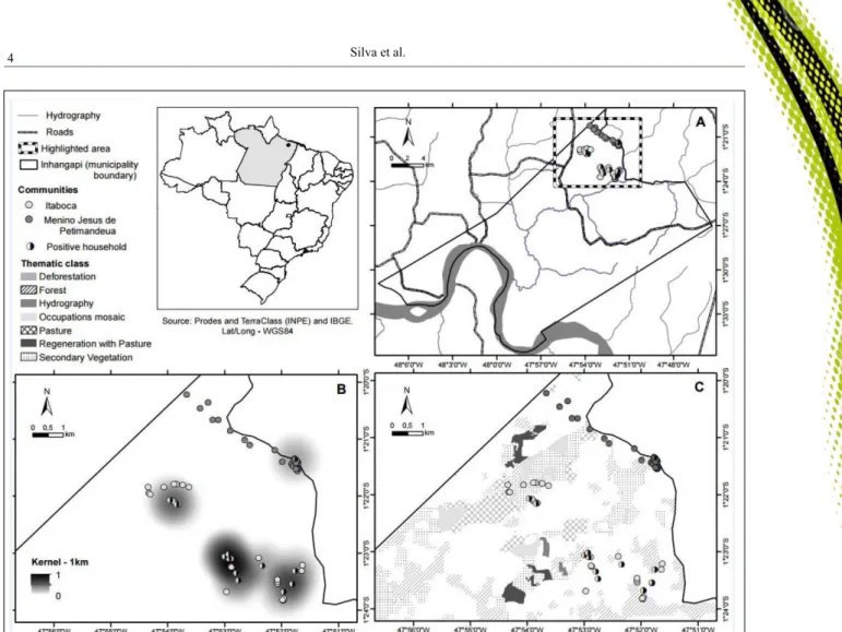
(12/143) of the intact skin samples and 1.4% (2/143) of the blood samples. Eleven positive dogs were from Itaboca, and one positive animal was from Menino Jesus de Petimandeua (Figure 1A). Only one of the
symptomatic animals was positive by PCR. None
of the reactions amplified DNA from the other
trypanosomatids with primers RV1 and RV2.
Most of dogs in the study group were >2 years old (51.05%), followed by 6 months-2 years
old (42.65%) and <6 months (6.30%). Among the PCR positive animals, 50% (6/12) were 6 months-2 years old, and 50% (6/12) were >2 years. In addition, among the PCR-positive dogs, 75% (9/12) were male,
and 25% (3/12) were female. Analyzing the group of
positive animals according to sex and age revealed
that most of the dogs were males >2 years (55.56%)
or males 6 months-2 years (44.44%). There were no
significant differences between the three age ranges
years, P=0.48) and the PCR results. There were also
no significant differences between the sex of the dogs
(P=0.21) and the PCR results.
Origins of 95.1% (136/143) of the animals
examined were known: 68.40% (93/136) were born in the municipality under study, and 31.61% (43/136) were
born in another municipality of Pará. Nine positive dogs
were born in Inhangapi, and three positive dogs were born
in other municipality of Pará.
Regarding the location of residences, 83.83% (57/68) of positive animals were located within 100 meters of a forested area, and the remaining 16.17% (11/68) were located more than 100 meters from a forested area. Most of the positive animals were housed near forested areas,
and L. infantum infection could be associated with their
proximity to forested and rural areas.
With respect to protection against
arthropod vectors, 67.65% (46/68) of dog owners
used no protection, 27.94% (19/68) used mosquito
nets, 1.47% (1/68) used an insecticide, 1.47% (1/68)
used mosquito nets and insecticide, and 1.47% (1/68) used mosquito nets and a fan. During the study, there were no reports of human visceral leishmaniasis cases
in the study communities.
The KDE of the study area, using a 1-km
radius, revealed three clusters in Itaboca and one in
Menino Jesus de Petimandeua, and the cluster with
the highest density was located in Itaboca (Figure
1B). Notably, 100% (38/38) of the studied households
in Itaboca were located in the clusters, and 73.3%
(22/30) of the studied households in Menino Jesus de
Petimandeua (Figure 1B) were located in the cluster.
According to PRODES project data, the extent of deforestation in the municipality of Inhangapi in 2013 was of 68.74%. Figure 1C shows the land use classification from the PRODES and Terra Class
Project data overlapped with clusters obtained from KDE in Itaboca and Menino Jesus de Petimandeua. Three of the clusters were in preserved areas, containing Amazon rainforest and secondary vegetation, while the
remaining cluster, one cluster in Itaboca, was located in an area containing pasture and occupations mosaic.
Areas are represented by an association of various land use arrangements, and due to the spatial resolution of
the satellite images, it is not possible to discriminate the classes (TERRACLASS, 2012).
DISCUSSION
Visceral leishmaniasis is a serious public
health problem in the Northern region of Brazil. During the period from 2009 to 2013, 3,311 human cases were confirmed in this region, and 41% of these cases were registered in the state of Pará (BRASIL, 2014).
The present study reported the first
detection of L. infantum DNA in domestic dogs in
the municipality of Inhangapi. The detection rate for
L. infantum DNA in intact skin samples was higher
than that in blood samples. These data agree with
the findings of QUARESMA et al. (2009), which detected by PCR higher positivity for Leishmania in dogs skin samples from the ear region (80.2%)
than in blood samples (75.3%), as detected by PCR.
QUEIROZ et al. (2010) collected skin samples from 34 naturally infected dogs that were serologically positive for Leishmania sp., and 100% of healthy
skin samples and 95.6% of skin samples with lesions were positive by PCR. The age ranges and the sex of dogs in the maroon communities did not influence the
PCR results, in agreement with the data reported by AMÓRA et al. (2006) and LAURENTI et al. (2013).
In Brazil, different clinical classifications have been used in studies of canine visceral
leishmaniasis (MANCIANTI et al., 1988; GOMES et al., 2007; LAURENTI et al., 2013), and there is no consensus regarding the most appropriate clinical
classification. Factors such as the clinical course of the disease, pathogenicity of the parasite, and the immune and nutritional status of the dogs are directly related to the presence or absence of clinical signs (REY, 2008). The majority of positive dogs in the municipality of
Inhangapi were asymptomatic, and only one positive
animal presented clinical signs suggestive of canine
visceral leishmaniasis. According to LAURENTI et al.
(2013), asymptomatic dogs are highly competent for the transmission of L. infantum to the phlebotomine
vector, and these animals have an important role in the
epidemiology of visceral leishmaniasis, by maintaining the transmission cycle of this agent in endemic areas. In
addition, these authors observed that clinical severity
was inversely related to the competence of dogs to promote infection of the vector.
Continuous deforestation is a serious
environmental problem in Brazilian Amazon. The state
of Pará has one of the highest rates of deforestation (PRODES website), and the municipality of Inhangapi has been registered as having high deforestation levels, mainly due to the replacement of forest for pasture.
In both maroon communities was observed preserved
forest area and deforested areas. In addition, Lu. longipalpis, the main vector of L. infantum in Brazil,
can establish breeding sites in forested areas, and has
also been shown to adapt to degraded environments with anthropic activity (RANGEL & VILELA, 2008; PINHEIRO et al., 2016). In this case, the transmission
of L. infantum to dogs in both maroon communities
may be occurring at the interface between the
peridomestic and wild environments.
The presence of sand fly vectors and numerous risk factors for infection, including the presence of L. infantum reservoir dogs (LAURENTI
et al., 2013), proximity of households to forested areas (ALMEIDA et al., 2009), and the habit of
not using any protection against insect vectors
(QUINTANILHA et al., 2016), may favor the spread
of L. infantum among humans in the studied areas.
CONCLUSION
The presence of canine reservoirs of L.
infantum in addition to the characteristics of the
environment (preserved forests and deforested areas) and habits of dog owners (living near forested areas and not using any protection against insects) may favor the transmission of L. infantum in the studied areas.
ACKNOWLEDGMENTS
The authors are especially thankful to the residents of the maroon communities and health officials for their important collaboration. The authors also thank Instituto Evandro Chagas (IEC) for the Leishmania amazonensis and Leishmania braziliensis isolates, and Fundação Amazônia Paraense (FAPESPA), Pró-Reitoria de Pesquisa e Pós-graduação of Universidade Federal do Pará and Fundação de Amparo e Desenvolvimento da Pesquisa (FADESP) for financial support.
BIOETHICS AND BIOSSECURITY COMMITTEE APPROVAL
REFERENCES
ALMEIDA, A.B.P.F. et al. Seroepidemiological survey of canine leishmaniasis in endemic areas of Cuiabá, State of Mato Grosso. Revista da Sociedade Brasileira de Medicina Tropical, v.42, n.2, p.156-159, 2009. Available from: <http://dx.doi.org/10.1590/ S0037-86822009000200012>. Accessed: 17 Aug. 2015. doi: 10.1590/ S0037-86822009000200012.
AMÓRA, S.S.A. et al. Factors related to positive testing of dogs for visceral leishmaniasis in endemic area in the state of Rio Grande do Norte, Brazil. Ciência Rural, v.36, n.6, p.1854-1859, 2006. Available from: <http://dx.doi. org/10.1590/S0103-84782006000600029>. Accessed: 12 Nov. 2015.
BAILEY, T.C.; GATRELL, A.C. Interactive spatial data analysis. Harlow: Prentice Hall, 1995. 413p.
BRASIL. Situação Epidemiológica – Dados. Ministério da Saúde. Secretaria de Vigilância em Saúde. Departamento de Vigilância Epidemiológica. Brasília: Ministério da Saúde, 20014. Available from: <http://portalsaude.saude.gov.br/index.php/o-ministerio/ principal/leia-mais-o-ministerio/726-secretaria-svs/vigilancia-de- a-a-z/leishmaniose-visceral-lv/11334-situacao-epidemiologica-dados>. Accessed: 12 Jan. 2016.
CDC (CENTERS FOR DISEASE CONTROL AND PREVENTION). Parasites – Leishmaniasis: Epidemiology & Risk Factors. Available: <http://www.cdc.gov/parasites/ leishmaniasis/epi.html>. Accessed 04 Jan. 2016.
FILHO, M.S.R.; SILVEIRA, F.T. Epidemiologic, clinical and immunologic on the human Leishmania (Leishmania) infantum chagasi infection in endemic area of visceral leishmaniasis in Pará. Revista Paraense de Medicina, v.21, n.3, p.7-18, 2007. Available: <http://scielo.iec.pa.gov.br/pdf/rpm/v21n3/v21n3a02. pdf>. Accessed: 05 Jan. 2016.
GOMES, A.H.S. et al. PCR identification of Leishmania in diagnosis and control of canine leishmaniasis. Veterinary Parasitology. v.144, n.3-4, p.234-241, 2007. Available: <http:// www.sciencedirect.com/science/article/pii/S0304401706006030>. Accessed 05 Jan. 2016. doi: 10.1016/j.vetpar.2006.10.008.
IBGE (INSTITUTO BRASILEIRO DE GEOGRAFIA E ESTATÍSTICA). 2015. Available from: <http://www.ibge.gov. br/cidadesat/painel/painel.php?codmun=150340#>. Online. Accessed: 03 Nov. 2015.
IDESP (INSTITUTO DE DESENVOLVIMENTO ECONÔMICO, SOCIAL E AMBIENTAL DO PARÁ). Estatística municipal - Inhangapi. 2014. Available from: <http://www.idesp.pa.gov. br/#>. Online. Accessed: 03 Nov. 2015.
INCRA (INSTITUTO NACIONAL DE COLONIZAÇÃO E REFORMA AGRÁRIA) Quilombolas. Online. 2015. Available from: <http://www.incra.gov.br>. Accessed: 10 nov. 2015.
LAURENTI, M.D. et al. Asymptomatic dogs are highly competent to transmit Leishmania (Leishmania) infantum chagasi to the natural vector. Veterinary Parasitology, v.196, n.3-4, p.296-300, 2013. Available from: <http://www.sciencedirect.com/science/ article/pii/S0304401713001623>. Accessed: 22 Jan. 2016. doi: 10.1016/j.vetpar.2013.03.017.
LE FICHOUX, Y. et al. Occurrence of Leishmania infantum parasitemia in asymptomatic blood donors living in an area of endemicity in Southern France. Journal of Clinical Microbiology, v.37, n.6, p.1953-1957, 1999. Available from: <http://jcm.asm.org/ content/37/6/1953.full.pdf+html>. Accessed: 22 Jan. 2016.
MANCIANTI, F. et al. Studies on canine leishmaniasis control. 1. evolution of infection of different clinical forms of canine leishmaniasis following antimonial treatment. Transactions of the Royal Society of Tropical Medicine and Hygiene, v.82, n.4, p.566-567, 1998. Available from: <http://www.sciencedirect.com/ science/article/pii/003592038890510X>. Accessed: 25 Jan. 2016. doi:10.1016/0035-9203(88)90510-X.
MORAIS, R.C. et al. Detection of Leishmania infantum in animals and their ectoparasites by conventional PCR and real time PCR. Experimental and Applied Acarology, v.59, p.473- 48, 2013. Available from: <http://link.springer.com/article/10.1007 %2Fs10493-012-9611-4>. Accessed: 02 Jan. 2016. doi: 10.1007/ s10493-012-9611-4.
PINHEIRO, M.P.G. et al. Sand flies (Diptera, Psychodidae, Phlebotominae), vectors of Leishmania protozoa, at an Atlantic Forest Conservation Unit in the municipality of Nísia Floresta, Rio Grande do Norte state, Brazil. Parasites & Vectors, v.9, n.83, p.1-8, 2016. Available from: <http://link.springer.com/article/10.1186/ s13071-016-1352-5?view=classic>. Accessed: 06 Feb. 2016. doi: 10.1186/s13071-016-1352-5.
QUARESMA, P.F. et al. Molecular diagnosis of canine visceral leishmaniasis: Identification of Leishamania species by PCR-RFLP and quantification of parasite DNA by real-time PCR. Acta Tropica, v.111, n.3, p.289-294, 2009. Available from: <http://www. sciencedirect.com/science/article/pii/S0001706X09001193>. Accessed: 18 mar. 2015. doi:10.1016/j.actatropica.2009.05.008.
QUEIROZ, N.M.G.P. et al. Canine Visceral Leishmaniasis diagnosis by immunohistochemistry and PCR in skin tissues in association with RIFI and ELISA-test. Revista Brasileira de Parasitologia Veterinária, v.19, n.1, p.32-38, 2010. Available from: <http://dx.doi. org/10.4322/rbpv.01901006>. Accessed: 18 mar. 2015.
QUINTANILHA, M. F. et al. Factors associated with human visceral leishmaniasis: confirmed cases and co-inhabitants in Minas Gerais State, Brazil. Journal of Medical and Biological Science Research, v.2, n.1, p.5-13, 2016. Available from: <http://pearlresearchjournals. org/journals/jmbsr/index.html>. Accessed: 25 Feb. 2016.
RANGEL, E.F.; VILELA, M.L. Lutzomyia longipalpis (Diptera,
Psychodidae, Phlebotominae) and urbanization of visceral
leishmaniasis in Brazil. Cadernos de Saúde Pública, v.24, p.2948-2952, 2008. Available from: <http://dx.doi.org/10.1590/ S0102-311X2008001200025>. Accessed: 15 Aug. 2015.
REY, L. Leishmaníase visceral. In: REY, L. Bases de Parasitologia médica. Rio de Janeiro: Guanabara Koogan, 2008. Cap.6, p.63-70.
SESPA (SECRETARIA DE SAÚDE PÚBLICA DO ESTADO DO PARÁ). Leishmaníase visceral e tegumentar americana no município de Inhangapí. Boletim Epidemiológico, Departamento de Controle de Endemias, Castanhal, Pará. 2013.