Milk-deteriorating exoenzymes from
Pseudomonas fluorescens
041
isolated from refrigerated raw milk
Maurilio L. Martins
1, Uelinton M. Pinto
2, Katharina Riedel
3, Maria C.D. Vanetti
4 1Departamento de Ciência e Tecnologia de Alimentos, Instituto Federal do Sudeste de Minas Gerais, Rio Pomba, MG, Brazil.
2
Departamento de Ciência de Alimentos, Universidade Federal de Ouro Preto, Ouro Preto, MG, Brazil. 3
Institute of Microbiology, Ernst-Moritz-Arndt University of Greifswald, Germany. 4
Departamento de Microbiologia, Universidade Federal deViçosa, Viçosa, MG, Brazil.
Submitted: August 8, 2013; Approved: June 6, 2014.
Abstract
The practice of refrigerating raw milk at the farm has provided a selective advantage for psychro-trophic bacteria that produce heat-stable proteases and lipases causing severe quality problems to the dairy industry. In this work, a protease (AprX) and a lipase (LipM) produced by Pseudomonas fluorescens041, a highly proteolytic and lipolytic strain isolated from raw milk obtained from a Bra-zilian farm, have been purified and characterized. Both enzymes were purified as recombinant pro-teins fromEscherichia coli. The AprX metalloprotease exhibited activity in a broad temperature range, including refrigeration, with a maximum activity at 37 °C. It was active in a pH range of 4.0 to 9.0. This protease had maximum activity with the substrates casein and gelatin in the presence of Ca+2. The LipM lipase had a maximum activity at 25 °C and a broad pH optimum ranging from 7.0 to 10. It exhibited the highest activity, in the presence of Ca+2, on substrates with long-chain fatty acid residues. These results confirm the spoilage potential of strain 041 in milk due to, at least in part, these two enzymes. The work highlights the importance of studies of this kind with strains isolated in Brazil, which has a recent history on the implementation of the cold chain at the dairy farm.
Keywords: raw milk, food deterioration, Pseudomonas fluorescens, extracellular protease, extracellular lipase.
Introduction
In Brazil, the practice of refrigerating raw milk at the dairy farm started in the 90s, was officially instituted by the government in 2002 and it is still being implemented in some areas of the country [Brasil, 2002; Brasil, 2011]. The refrigeration of raw milk in the farm and dairy industries has improved the quality and shelf life of milk and dairy products. However, it does not prevent the growth of psy-chrotrophic microorganisms that produce heat-stable en-zymes such as proteases and lipases (Cousin 1982; Sorhaug and Stepaniak 1997; Decherniet al., 2005; De Jongheet al., 2010; Corrêaet al., 2011; Baglinièreet al., 2013; Quigley et al., 2013).
Many of these enzymes are produced by Pseudomo-nas fluorescens, a frequent psychrotrophic spoilage bacte-rium found in milk (Wiedmann et al., 2000; Dogan and
Boor, 2003; Pintoet al,. 2006; Dufouret al.; 2008; Mar-chandet al., 2009; Baglinièreet al., 2013). As hydrolytic enzymes from this bacterium are generally not inactivated by pasteurization or even by Ultra-High Temperature (UHT) treatment (Griffithset al., 1981; Chenet al., 2003; De Jongheet al., 2010; Baglinière et al., 2013), they can cause severe problems in the dairy industry such as milk protein hydrolysis, development of off-flavors, shelf-life reduction, decrease of yield during cheese production, milk heat-stability loss, and gelation of UHT milk (Fairbairn and Law, 1986; Datta and Deeth, 2001; Chen et al., 2003; Dufouret al., 2008; Baglinièreet al., 2013).
A commom type of protease produced by P. fluorescens is metalloprotease. This class of enzyme contains one zinc atom and up to eight calcium atoms, con-ferring thermostability to the protein (Sorhaug and Ste-paniak, 1997). These authors listed some important
DOI: http://dx.doi.org/10.1590/S1517-838246120130859
Send correspondence to M.C.D. Vanetti. Departamento de Microbiologia,Universidade Federal de Viçosa, 36570-000 Viçosa, MG, Brazil. E-mail: mvanetti@ufv.br.
characteristics of the metalloproteases secreted by strains ofP. fluorescensincluding temperature optimum between 30 and 45 °C, a significant residual activity at 4 °C, and a pH optimum in the neutral range. These authors pointed out that a heat treatment of milk sufficient to fully inacti-vate these enzymes would also create unacceptable changes in the product and it is therefore unpractical for the dairy in-dustry.
Microorganisms that produce lipolytic enzymes, such asP. fluorescens, are important in the dairy industry be-cause they can produce rancid flavors and odors in milk and dairy products that make these foods unacceptable to con-sumers (Cousin 1982). Bacterial lipases generally have mo-lecular masses between 30 to 50 kDa, and the pH optimum is slightly alkaline (in the range of 7 to 9) (Chenet al., 2003; Chakraborty and Paulraj, 2009; Boran and Ugur 2010; Anbu, 2014). Lipase production byP. fluorescensis influ-enced by the type and concentration of carbon and nitrogen sources, iron, pH, dissolved oxygen concentration, and growth temperature (Cousin 1982; Burger et al., 2000; Woodset al., 2001; Rajmohanet al., 2001).
The present work aimed at the molecular character-ization of a protease and a lipase produced by P. fluorescens041, a highly milk deteriorating strain iso-lated from refrigerated raw milk obtained from a Brazilian farm. Both enzymes were overexpressed in Escherichia coli, purified to homogeneity by affinity chromatography and biochemically characterized in order to evaluate their role in the spoilage of milk components.
Material and Methods
Bacterial strains and plasmids
The bacterial strains and plasmids used in this study are listed in Table 1.P. fluorescens041 and 07A strains were isolated from refrigerated raw milk as highly proteolytic and lipolytic psychrotrophic bacteria (Martins et al., 2005; Pintoet al., 2006; Pintoet al., 2010).
Growth conditions
P. fluorescenswas cultured in TYEP (tryptone 1%, yeast extract 0.25%, KH2PO4 0.1%, K2HPO4 0.1%, and CaCl20.25%) broth at 25 °C with aeration or in 12% (w/v) reconstituted skim milk powder.E. coliXL1-Blue was cul-tured in Luria-Bertani (LB) broth or on LB agar plates at 37 °C, as required.
DNA manipulations, PCR reactions and sequencing
DNA manipulations
Cloning, restriction enzyme analysis, and transforma-tion ofE. coliwere performed using established procedures (Sambrooket al., 1989). PCR was performed with TaKaRa Ex Taq polymerase (TaKaRa Shuzo, Shiga, Japan). Plasmid DNA was isolated using the QIAprep Spin Miniprep kit, and chromosomal DNA was purified with the DNeasy tissue kit. DNA fragments were purified from agarose gels by using the QIAquick gel extraction kit (all kits from Qiagen, Hilden, Germany).
Amplification and sequencing of the protease and lipase genes by PCR
The reaction consisted of 2.0 mM MgCl2, 5.0mL of 10X buffer ExTaq, 2.5 mM deoxynucleotide triphosphates (dNTPs), 0.5mM of each primer, 1 U of ExTaqDNA poly-merase, and 40 ng of DNA in a final volume of 50mL. Primers based on the sequences of theaprX(GenBank ac-cession numbers DQ146945, AY298902, AF216700, AY973251) and lip gene (GenBank accession numbers AF216702, AY694785, M86350, S77830, D11455, AB063391, AY304500, AY673674, M74125, AY700013) of otherP. fluorescensstrains were designed (Table 2), and synthesized by Microsynth (Zürich, Switzerland). The re-actions were carried out in a T3 thermocycler (Biometra®, Biolabo Scientific Instruments, Zürich, Switzerland).
The M13 Forward and Reverse Primers were used to sequence theaprX andlipMgenes ofP. fluorescens 041
Table 1- Bacterial strains and plasmids.
Strain or plasmid Description Reference or source
Strains
E. coliXL1-Blue Cloning and subcloning hostsupE44,hsdR17,endA1,recA1, gyrA96,thi1,relA1,lac -F[proAB+,lacIq,lacZ-M15, Tn10 (tetF)]
Bullock et al., 1987
P. fluorescens07A Wild type Martinset al., 2005
P. fluorescens041 Wild type Martinset al., 2005
Plasmids
pCR2.1-TOPO Cloning vector,lacZafragment containing MCS, f1 origin, ColE1, KmrApr Invitrogen
pQE30-Xa Vector for the insertion of a Factor Xa Protease recognition site C-terminal of the 6xHis tag, T5 promoter,lacoperator, ribosome binding site, ATG start codon, His tag sequence, multiple cloning
sites, stop codons in all three reading frames, Col E1 origin of replication, Apr
Qiagen
pQE30-Xa-aprX041 1.43 kb fragment containingaprXfromP. fluorescens041 in pQE30-Xa, Apr This study
cloned into pCR2.1-TOPO according to description of Invitrogen.
Cloning, heterologous expression and purification of
P. fluorescens041 protease and lipase
Once the complete sequences of theaprXandlipM genes were obtained, primers were designed (Table 2) to amplify the open reading frames (ORF) by PCR using the bacterial genomic DNA as a template and TaKaRa Ex Taq as DNA-polymerase. The primers generatedBamHI and SacI sites at the 5’ and 3’ ends of the amplified fragments, respectively.
The amplified DNA fragments of 1,434 bp and 1,422 bp, containing theaprXandlipMstructural genes, re-spectively, were digested withBamHI andSacI and ligated into vector pQE-30Xa (Qiagen) previously cut with the same restriction enzymes. Plasmids harbouring theaprXor lipMORFs inserted downstream of the T5 promoter were named pQE-30Xa-aprX041 or pQE-30Xa-lipM041. The plasmids were subsequently transformed into the expres-sion strainE. coliXL1-Blue.
For overproduction of AprX and LipM,E. coli XL1-Blue cells carrying aprX041 or pQE-30Xa-lipM041 were grown in dYT medium (tryptone 1.6%, yeast extract 1.0%, NaCl 0.5%, and glucose 0.2%) containing ampicillin (100 mg mL-1) at 37 °C under shaking at 300 rpm. At an optical density of 0.5 at 600 nm, isopro-pyl-b-D-thiogalactopyranoside (IPTG) was added to the culture to a final concentration of 1 mM, in order to induce the expression ofaprXandlipM. After 5 h incubation at 37 °C, the cells were collected by centrifugation at 10,000g for 30 min, resuspended in 50 mM Tris-HCl (pH 8.0) and centrifuged at 10,000gfor 30 min, followed by two wash-ing steps with 50 mM Tris-HCl pH 8.0, NaCl 150 mM. The resulting cell pellets were finally resuspended in lysis buffer (8 M urea, 0.1 M NaH2PO4, 0.01 M Tris-HCl, pH 8.0) and the recombinant histidine-tagged enzymes were purified under denaturing conditions using the Ni-NTA Spin Columns (Qiagen) according to the suppliers’ instructions. After purification, the enzymes were
sub-jected to overnight dialysis with Tris-HCl 20 mM, pH 8.0, CaCl25 mM at 4 °C to allow renaturation.
Protein quantification, SDS-PAGE and zymograms
Protein concentration of the purified AprX and LipM enzyme solutions was determined by using the method of Bradford (1976). Proteins were analysed by sodium dodecyl sulfate-polyacrylamide gel electrophoresis [Laem-mli, 1970]. After electrophoresis the gels were stained with Coomassie brilliant blue.
Exoprotease activities of P. fluorescens culture supernatants, resolved proteases after precipitation with ammonium sulfate, and recombinant expressed AprX pro-tease were visualized in SDS-PAGE-gels supplemented with 0.2% (w/v) azocasein (Christensenet al., 2003). After electrophoresis, proteins were renaturated by washing twice in 50 mM Tris-HCl, pH 7.5, 25% (v/v) isopropanol for 15 min at room temperature and once in 50 mM Tris-HCl, pH 7.5. After overnight renaturation at 4 °C, the zymogram was incubated for 4 h in 5 mM CaCl2 and 50 mM Tris-HCl, pH 8.0 at 40 °C. Prior to detection, the gel was washed in 1 M NaOH for 5 min. Protease activity was detected as colourless zones in an orange background.
For the analysis of the lipase pattern after SDS-PAGE, proteins were renaturated as above described. The gels were overlaid with the fluorescent substrate methyl-umbelliferyl-butyrate (0.01 M in dimethylformamide) in order to detect lipolytic activity using UV-light (360 nm) to visualize blue fluorescent bands.
Identification of proteins by mass spectrometry
P. fluorescens041 was grown in TYEP medium at 25 °C for 48 h. The cells were removed from the medium by centrifugation at 10,000gfor 30 min, the supernatant was sterilized by filtration, and the proteins were precipitated with ammonium sulfate. Samples were centrifuged 20 min at 10,000gand the supernatant was discarded. Pellets were washed twice with an 85% ammonium sulfate solution (w/v), and again centrifuged. The pellets were dissolved in 50 Mm Tris-HCl, pH 8.0 and dialysed overnight at 4 °C against Tris-HCl 50 mM, pH 8.0, CaCl25 mM. Aliquots of Table 2- Primers used to amplify theaprXandlipMgene by PCR.
Primer Sequence (5’-3’) Aplication
Apr-F TTATGTCAAAAGTAAAAGAC Amplification ofaprXgene
Apr-R TCAGGCTACGATGTCACTG Amplification ofaprXgene
APRX-F ATTGGATCCAAAGCTATTGTATCTGCCGCG Amplification ofaprXgene and preparation for cloning in pQE-30Xa
APRX-R ATTGAGCTCTCAGGCTACGATGTCACTGGC Amplification ofaprXgene and preparation for cloning in pQE-30Xa
Lip-F ATGGGTRTSTTYGACTATAAAAACC Amplification oflipMgene
Lip-R TTAACCGATCACAATCCCCTCC Amplification oflipMgene
LIPM-F ATTGGATCCAACCTCGGTACCGAGGACTC Amplification oflipMgene and preparation for cloning in pQE-30Xa
LIPM-R ATTGAGCTCTTAACCGATCACAATCCCCTCCC Amplification oflipMgene and preparation for cloning in pQE-30Xa
15 mL of the dialysed samples were separated on SDS-PAGE (12%) gels. Coomassie-stained protein bands were excised, digested with trypsin and analysed by mass spec-trometry (Riedelet al., 2006).
Enzyme assays
Proteolytic activity was investigated on azocasein, according to Christensen et al. (2003), by incubating 250mL of 2% azocasein (w/v) with 150mL sterile filtered culture supernatant or with 75mL of the purified AprX. Lipolytic activity onp-nitrophenyl palmitate was investi-gated by incubating 1 mL of substrate with 100mL super-natant from overnight cultures or with 50mL of the purified lipase LipM.
Characterization of purified enzymes
The proteolytic and lipolytic activities of purified AprX and LipM were determined as described above at various incubation temperatures (4, 25, 30, 37, 40, 45, 50, and 60 °C) and at various pH values using the following buffer systems: sodium succinate (pH 4.0, 5.0, 6.0), Tris-HCl (pH 6.0, 6.5, 7.0, 7.5, 8.0, 8.5, 9.0), and glycine-NaOH (pH 9.0,10.0,11.0,12.0,13.0).
In order to determine heat stability of purified AprX and LipM, they were incubated for 5, 10, 15, 20, 30 and 60 min at 50, 60, 70, 80, 90, and 100 °C. They were also in-cubated at 65 °C for 30 min and 72 °C for 20 s to simulate the milk pasteurization treatments.
To investigate the effect of metal ions on purified AprX and LipM, the reaction mixture was supplemented with 1 mM of each of the following compounds: MnS04, CoCl2, ZnSO4, FeSO4, MgSO4, or FeCl3. The effect of pro-tease inhibitors on the proteolytic activity of purified AprX was determined by supplementing the reaction mixture with 1 mM PMSF, 1 mM EDTA, 1 mM Pefabloc SC, 2% (w/v) SDS, 4 M urea, 0.1% (w/v) DTT, and 0.1% (v/v)
b-mercaptoethanol and subsequent measuring the residual activities on azocasein substrate.
The substrate specificity of purified AprX was deter-mined on casein, elastin, collagen, bovine serum albumin, and gelatin. The reaction mixture consisted of 0.4% (w/v) of each protein in 400mL of 50 mM Tris-HCl, pH 6.5 and 150mL of enzyme solution. After incubation at 37 °C for 1 h, the mixture was withdrawn and the increase in the amount of free amino groups was determined by the ninhydrin method according to Setyoriniet al.(2006).
Activities of purified LipM on differentp-nitrophenyl fatty acid esters (p- nitrophenyl acetate,p-nitrophenyl bu-tyrate, p-nitrophenyl palmitate, and p-nitrophenyl phos-phorylcholine) were also measured according to the assay for lipolytic activity as described above.
Results
Milk-deteriorating hydrolytic activities ofP. fluorescens
P. fluorescens041 showed higher proteolytic (Fig-ure 1A) and lipolytic (Fig(Fig-ure 1B) activities than the strain 07A. Moreover, strain 041 exhibited a higher capacity to hydrolyze milk thanP. fluorescens07A when both strains were inoculated into 12% (w/v) reconstituted skim milk (Figure 1C). Therefore,P. fluorescens041 was selected for further analysis of its hydrolytic extracellular enzymes.
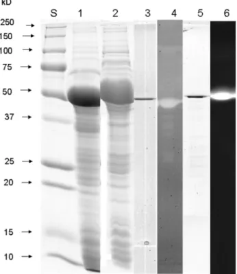
SDS-PAGE analysis of ammonium sulfate precipi-tated protein from supernatants of TYEP cultures of P. fluorescens041 demonstrated the presence of multiple pro-tein bands (Figure 1D). Proteolytic activity of the dominant 50 kDa band was demonstrated by a zymogram incorporat-ing azocasein (Figure 2). Mass spectrometry analysis of the major proteolytic protein band identified this protein as a metalloprotease, which is commonly referred to as AprX. Analysis of the bands with lower molecular weight that showed proteolytic activity (Figure 1D) revealed that these bands were actually degradation products of AprX.
Cloning and sequencing of protease and lipase genes
Primers based on sequences of homologous proteases and lipases from otherP. fluorescensstrains were synthe-sized and used to PCR amplify a segment encoding these enzymes fromP. fluorescens041 genome. Electrophoresis of the PCR products revealed a single product of about 1,500 bp for both genes. These PCR products were se-quenced to reveal their identity asaprX andlipM genes. TheaprXand thelipMgenes ofP. fluorescens041 com-prised open reading frames of 1,434 bp and 1,425 bp and coded for proteins with 477 and 474 amino acids, respec-tively. Based on the amino acid sequences, the molecular mass of both enzymes was predicted to be 49.365 kDa and 49.811 kDa, which was confirmed by SDS-PAGE analysis of the purified enzymes (Figure 2, line 3 and 5). The isoeletric point for AprX was 4.46 and 4.36 for LipM, as determined by using Protean (DNA Star Lasergene 7). They were active on zymograms after renaturation in buffer containing 1 mM of CaCl2(Figure 2, line 4 and 6).
The aprXgene of P. fluorescens041 showed 97% identity with the extracellular alkaline metalloprotease (aprX) gene ofP. fluorescensstrain A506 and with the pro-tease (aprX) gene ofP. fluorescensstrain F. ThelipMgene of P. fluorescens 041 showed 93% identity with polyurethanase lipase A (pulA) gene and 86% with the lipase (lipA) gene ofP. fluorescensstrain A506.
Biochemical characterization of AprX and LipM
refrigera-tion from 4 °C to 7 °C, and low activity in temperatures higher than 45 °C (Figure 3A).
The pH optimum of AprX is between 6.0 and 6.5 (Figure 3B). The protease still exhibits 36% residual activ-ity at pH 4.0 and 62% at pH 9.0.
The protease activity was strongly decreased by pre-incubating the enzyme at different temperatures (Fig-ure 3C); the residual activity of the protease after 60 min at 50, 60, 70, 80, 90 and 100 °C was between 2 and 4%. Inacti-vation of the metalloprotease at temperature and time con-ditions used during the pasteurization process were also evaluated: AprX showed 70% residual activity when it was treated at 75 °C for 20 s (HTST treatment: high temperature and short time) and 4% residual activity when it was incu-bated at 65 °C for 30 min (LTLT: low temperature and long time).
Proteolytic activity of AprX was strongly dependent on the presence of Ca2+. However, other metal ions reduced the proteolytic activity (Table 3). The activity of AprX was decreased when 1 mM EDTA, an inhibitor that specifically acts on metalloproteases, was added to the reaction mix-ture, confirming the type of enzyme. In addition, AprX was
strongly inhibited by denaturing and reducing agents such as SDS, dithiothreitol (DTT),b-mercaptoethanol, and urea (Table 4).
The alkaline metalloprotease was further tested for its capability to hydrolyze different substrates such as casein, bovine serum albumin, collagen, elastin, and gelatin. The highest activities were found on gelatin (100%) and casein (87.6%), followed by collagen (57%), elastin (41.2%), and bovine serum albumin (39.8%).
The temperature optimum of the purified lipase was 25 °C (Figure 4A). LipM showed a residual activity of 3.7% at 4 °C and exhibited low activities at temperatures higher than 37 °C (Figure 4A). Besides, LipM showed the highest lipase activity at pH 7.5 (Figure 4B). At pH values lower than 6.0 and higher than 11.0 only residual lipase ac-tivities could be detected (Figure 4B).
The lipase activity after 60 min of pre-incubation at 50, 60, 70, 80, 90 and 100 °C was nearly undetected (Fig-ure 4C). The treatment of 65 °C for 30 min (LTLT), and 75 °C for 20 s (HTST), reduced the lipolytic activity to 13.2% and 25.4%, respectively.
Lipase activity was dependent on Ca+2 ions in the renaturation buffer. The same was observed for the prote-ase activity. The presence of ions other than Ca+2reduced the lipolytic activity (Table 3). Among the tested sub-strates, LipM exhibited the highest activity for p -nitro-phenyl palmitate (100%), followed byp-nitrophenyl butyr-ate (73%),p-nitrophenyl acetate (20%), andp-nitrophenyl phosphorylcholine (11%). The highest activity on sub-strates with long-chain fatty acid residues such asp -nitro-phenyl palmitate indicates that the enzyme has esterolytic and lipolytic activities.
Discussion
Numerous Pseudomonas spp. have been shown to produce and secrete hydrolytic enzymes (McCarthyet al., 2004; Burgeret al., 2000; Woodset al., 2001; Pintoet al., 2006; Pintoet al., 2010; Liao and McCallus 1998; Koka and Weimer, 2000; Maunsellet al., 2006; Muet al., 2009; Jankiewiczet al., 2010; De Jongheet al., 2011). Interest-ingly, in this study it was verified thataprXencodes for the major, if not the only extracellular protease produced byP. fluorescens041. Mass spectrometry analysis of low molec-ular weight bands that showed proteolytic activity on the azocasein zymogram were identified as degradation
prod-ucts of the metalloprotease AprX. These results are in agreement with the findings of Liao and McCallus (1998)
Figure 2- Coomassie-stained SDS-PAGE and zymogram gel on 12% PAA-gels visualizing recombinant AprX and LipM. Lane S: molecular mass standard (Biorad); lane 1: SDS-PAGE of crude extract ofE. coli
XL1-Blue carrying pQE30-Xa-aprX-041; lane 2: SDS-PAGE of crude ex-tract ofE. coliXL1-Blue carrying pQE30-Xa-lipM-041; lane 3: SDS-PAGE of purified AprX; lane 4: azocasein zymogram of purified AprX; lane 5: SDS-PAGE of purified LipM; lane 6: MU-butyrate zymogram of purified LipM.
who observed thatP. fluorescensCY091 produces a unique extracellular 50 kDa protease, AprX. Our results are also in agreement with several other studies (Koka and Weimer,
2000; Muet al., 2009; Jankiewiczet al., 2010). In contrast, Rajmohanet al.(2002) reported that anotherP. fluorescens isolated from milk produces five distinct proteases when they used the ultrafiltration technique to purify these en-zymes. Nicodemeet al.(2005) observed the presence of more than one protease band for some strains of Pseudomo-Table 3- Effect of metal ions on the activities of alkaline metalloprotease
and lipase.
Metal ion Relative activity (%)
Alkaline metalloproteasea Lipaseb
None 100±2 100±2
Mn2+ 73±1 61±1
Co2+ 48±3 59±8
Zn2+
86±3 49±0
Fe2+
90±5 48±3
Fe3+ 102±1 65±2
Mg2+ 100±1 50±3
a
A reaction mixture containing 250mL of 2% (w/v) azocasein in 50 mM Tris/HCl (pH 8.0), 75mL of AprX, and 1 mM of each metal ion was incu-bated at 37 °C for 12 h. The remaining activity was then measured, as de-scribed in the text. Results show the mean value (n = 3) plus or minus the standard deviation.
bA reaction mixture containing 1 mL of substrate (one volume of 0.3% (w/v)p-nitrophenyl palmitate in isopropanol and nine volumes 0.2% (w/v) sodium desoxycholate and 0.1% (w/v) gummi arabicum in 50 mM sodium phosphate buffer, pH 8.0), 50mL of LipM, and 1 mM of each metal ions was incubated at 25 °C for 20 min. The remaining activity was then mea-sured, as described in the text. Results show the mean value (n = 3) plus or minus the standard deviation.
Table 4- Effect of inhibitors, denaturing and reducing agents on the activ-ity of alkaline metalloprotease.
Compound Relative activity (%)c
Inhibitora
None 100±1
PMSF 95±2
EDTA 51±3
Pefabloc SC 89±1
Denaturing and reducing agentb
None 100±2
SDS 6±1
Urea 38±5
DTT 24±3
b-mercaptoethanol 44±2
a
A reaction mixture containing 250mL of 2% (w/v) azocasein in 50 mM Tris/HCl (pH 8.0), 75mL of AprX, and 1 mM of each inhibitor was incu-bated at 37 °C for 12 h. The remaining activity was then measured, as de-scribed in the text.
b
A reaction mixture containing 250mL of 2% (w/v) azocasein in 50 mM Tris/HCl (pH 8.0), 75mL of AprX, and 2% (w/v) SDS, 4 M urea, 0.1% (w/v) DTT, or 0.1% (v/v)b-mercaptoethanol in 50 mM Tris/HCl (pH 8.0) was incubated at 37 °C for 12 h. The remaining activity was then mea-sured, as described in the text.
c
Results show the mean value (n = 3) plus or minus the standard deviation.
nas,while others produced just one protease, as revealed by a zymogram analyzes.According to Sørhaug and Stepaniak (1997) the number of secreted proteases depends strongly on theP. fluorescens strain. These findings highlight the great diversity ofP. fluorescensisolates and reiterate the importance of studies aiming to elucidate the molecular mechanisms of the hydrolytic enzymes produced by these strains.
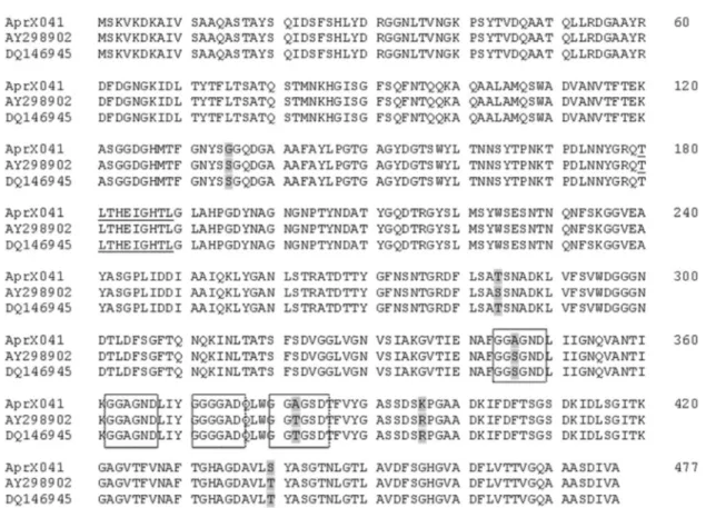
Once the AprX protein produced by strains of P. fluorescensisolated from raw milk showed high similarity with sequences from homologous enzymes in the database (Figure 5), it further confirms the possibility of using the aprXgene as a marker to detectP. fluorescensin milk by using PCR as described by Martinset al.(2005) and Ma-chadoet al.(2013). This approach would reduce the time for detecting these bacteria in raw milk giving flexibility for the dairy manager to choose the best use for a particular milk batch during processing.
Unlike many proteolytic and lipolytic enzymes de-scribed in the literature (Makhzoumet al., 1996; Liao and McCallus, 1998; Rajmohanet al., 2002; Chenet al., 2003; Kojima and Shimizu, 2003; Nornberget al., 2009; Bagli-nièreet al., 2013; Anbu 2014), the protease and lipase eval-uated in this study were relatively more sensitive to heat treatment. This could be attributed due to differences in the
enzymes structures or to differences in experimental proce-dures, as the above mentioned studies have used purified enzymes from culture supernatants and we have purified those from overexpressingE. colistrains. However, some authors (Teoet al., 2003; Jinget al., 2010) verified that His-tag did not affect the metalloprotease activities of some strains, indicating that the recombinant metalloprotease was in an active form. Affinity tags have become essential tools for the production of recombinant proteins in a wide variety of settings (Waugh, 2011).
As the heat treatment and refrigeration processes adopted by the dairy industry during milk processing and storage do not fully inhibit enzymatic activity nor the growth of psychrotrophic bacteria, it is important to pro-duce milk under stringent good manufacturing practices to limit contamination and bacterial spoilage.
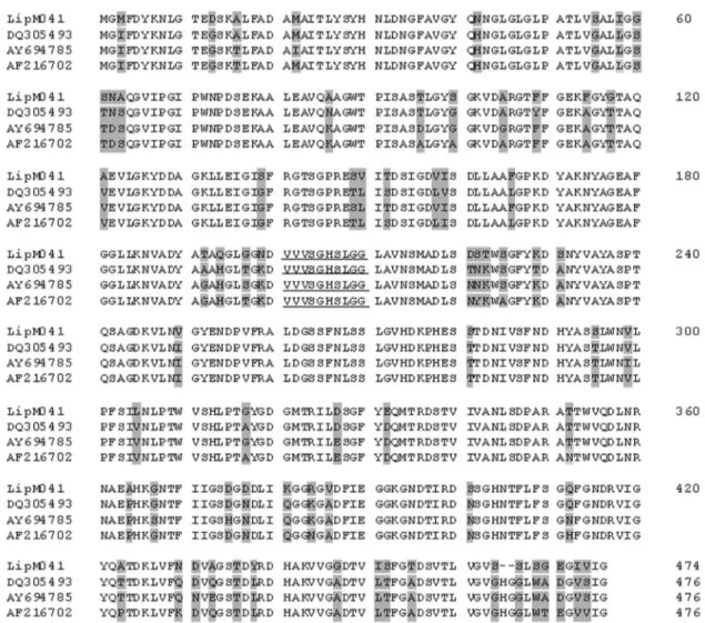
Although LipM exhibits the conserved serine lipase catalytic domain, it presented somewhat lower similarity, as compared to AprX alignment, to the sequences described in the data base (Figure 6). LipM was also less heat stable than some lipases described by other authors (Knaut, 1978; Cousin, 1982; Makhzoumet al., 1996; Boran and Ugur, 2013; Anbu, 2014), although low heat stability has also been observed (Chakraborty and Paulraj, 2009; Dahiyaet al., 2010). According to Cousin (1982), complete
tion of lipases was only obtained by autoclaving milk at 121 °C for 15 min. Knaut (1978) observed that lipases from P. fluorescensspecies were stable even above 100 °C. A heat-treatment of 98 °C for 14 to 25 min was necessary to inactivate lipases from somePseudomonasspecies, includ-ingP. fluorescensandP. fragi(Cousin, 1982).
Overall, the biochemical properties of the purified protease and lipase from this work were similar to those found for proteases and lipases of other P. fluorescens strains isolated from raw milk (Makhzoum et al., 1996; Kimet al., 1997; Schokker and van Boekel, 1997; Liao and McCallus, 1998; Rajmohanet al., 2002; Chenet al., 2003; Kojima and Shimizu, 2003; Dufouret al., 2008; Correaet al., 2011; Baglinièreet al., 2013). It is worth to mention that AprX still exhibited 36% residual activity at pH 4.0, so if present in milk, this enzyme would not only affect the quality of pasteurized milk products but also of fermented products such as yogurt and cheese.
Surprisingly, no lipolytic activity could be detected when the renaturated SDS-PAGE was overlaid with the
lipase substrate methylumbeliferyl-butyrate (results not shown). Probably this occurred due to the degradation of the enzyme by proteases or because Ca+2was not added into the renaturation buffer, and the lipase may need this ion for correct folding.
The purification of AprX and LipM was important for the characterization of these spoilage enzymes and it would be interesting to use them to develop tools for improving their detection in milk. Nowadays, there is a great need for developing fast and reliable methods to detect spoilage en-zymes directly from samples in order to determine the qual-ity of milk that arrives at the dairy industry platform (Datta and Deeth, 2001). Most approaches currently available are time consuming, do not have good sensitivity or have de-tection limits that are too high. Besides characterizing these spoilage enzymes, it is important to estimate the extent of degradation of milk components, and thus further improve enzymatic methods to access milk quality.
In this work a protease and a lipase produced by P. fluorescens041, a highly milk spoilage strain, isolated
from cooled raw milk were purified and characterized. The study showed that both enzymes presented similar bio-chemical properties to other enzymes fromP. fluorescens strains isolated from raw milk. The differences that were observed could be accounted for the experimental proce-dures, especially the use of overexpressed recombinant proteins. The study confirms the spoilage potential of strain 041 in milk due to, at least in part, these two enzymes. The work highlights the importance of studies of this kind with P. fluorescensstrains, the major spoilage bacteria contami-nating milk produced in Brazil (Martinset al., 2005) which has a recent history on the implementation of the cold chain at the dairy farm.
Acknowledgments
The authors thank the Conselho Nacional de Desen-volvimento Científico e Tecnológico (CNPq-Brazil) for a doctoral scholarship provided to M.L. Martins and a post-doctoral scholarship to U.M. Pinto. We are thankful to Leo Eberl from the University of Zurich - Switzerland for his assistance with this work.
References
Anbu P (2014) Characterization of an extracellular lipase by Pseudomonas koreensis BK-l07 isolated from soil. Prep Biochem Biotechnol 44:266-280.
Baglinière et al. (2013) Proteolysis of ultra high temperature-treated casein micelles by AprX enzyme fromPseudomonas fluorescens F induces their destabilization. Int Dairy J 31:55-61.
Boran R, Ugur A (2010) Partial purification and characterization of the organic solvent-tolerant lipase produced by Pseudo-monas fluorescensRB02-3 isolated from milk. Prep Bio-chem Biotechnol 40:229-241.
Bradford MM (1976) A rapid and sensitive method for the quanti-fication of microgram quantities of proteins utilizing the principle of protein dye binding. Anal Biochem 72:248-274. BRASIL (2002) Ministério da Agricultura, Pecuária e
Abasteci-mento. Departamento de Inspeção de Produtos de origem Animal. Instrução Normativa n° 51, de 18 de setembro de 2002. Aprova os Regulamentos Técnicos de Produção, Iden-tidade e Qualidade do Leite tipo A, do Leite tipo B, do Leite tipo C, do Leite Pasteurizado e do Leite Cru Refrigerado e o Regulamento Técnico da Coleta de Leite Cru Refrigerado e seu Transporte a Granel. Diário Oficial da República Fede-rativa do Brasil. Brasília, 20 set. 2002, Seção I, p.13. BRASIL (2011) Ministério da Agricultura, Pecuária e
Abaste-cimento. Departamento de Inspeção de Produtos de origem Animal. Instrução Normativa n°62, de 29 de dezembro de 2011. Aprova o Regulamento Técnico de Produção, Iden-tidade e Qualidade do Leite tipo A, o Regulamento Técnico de Identidade e Qualidade de Leite Cru Refrigerado, Leite Pasteurizado e o Regulamento Técnico da Coleta de Leite Cru Refrigerado e seu Transporte a Granel. Diário Oficial da República Federativa do Brasil. Brasília, 30 dez. 2011, Seção I.
Bullock WO, Fernandez JM, Short, JM (1987) XL-1 Blue: a high efficiency plasmid transforming recA Escherichia coli
strain with b-galactosidase selection. Bio Techniques 5:376-377.
Burger M et al. (2000) Temperature regulation of protease in Pseudomonas fluorescensLD107d2 by an ECF sigma factor and a transmembrane activator. Microbiology 146:3149-3155.
Chakraborty K, Paulraj R (2009) Purification and Biochemical Characterization of an Extracellular Lipase from Pseudomo-nas fluorescensMTCC 2421. J Agri Food Chem 57:3859-3866.
Chen L, Daniel RM, Coolbear, T (2003) Detection and impact of protease and lipase activities in milk and milk powders. Int Dairy J 13:255-275.
Christensenet al. (2003) Quorum-sensing-directed protein ex-pression in Serratia proteamaculans B5a. Microbiology 149:471-483.
Correa APF, Daroit DJ, Velho RV, Brandelli A (2011) Hydrolytic potential of a psychrotrophicPseudomonasisolated from re-frigerated raw milk. Braz J Microbiol 42:1479-1484. Cousin MA (1982) Presence and activity of psychrotrophic
mi-croorganisms in milk and dairy products. J Food Protect 45:172-207.
Datta N, Deeth HC (2001) Age gelation of UHT milk. Trans Inst Chem Eng 79:197-210.
Dahiya P, Arora P, Chaudhury A, Chand S, Dilbaghi D (2010) Characterization of an extracellular alkaline lipase from Pseudomonas mendocinaM-37. J Basic Microbiol 50:420-426.
De Jonghe Vet al.(2011) Influence of storage conditions on the growth ofPseudomonasspecies in refrigerated raw milk. Appl Environ Microbiol 77:460-70.
Decherni S, Benjelloun H, Lebeault JM (2005) Effect of modified atmospheres on the growth and extracellular enzyme activi-ties of psychrotrophs in raw milk. Eng Life Sci 5:350-356. Dogan B, Boor KJ (2003) Genetic diversity and spoilage
poten-tials among Pseudomonas spp. isolated from fluid milk products and dairy processing plants. Appl Environ Micro-biol 69:130-138.
Dufour et al.(2008) Molecular typing of industrial strains of Pseudomonasspp. isolated from milk and genetical and bio-chemical characterization of an extracellular protease pro-duced by one of them. Int J Food Microbiol 125:188-196. Fairbairn DJ, Law BA (1986) Proteinases of psychrotrophic
bac-teria: their production, properties, effects and control. J Dairy Res 53:139-77.
Griffiths MW, Philips JD, Muir DD (1981) Thermostability of proteases and lipases from a number of species of psycho-trophic bacteria of dairy origin. J Appl Bacteriol 50:289-303.
Jankiewicz U, Szawlowska U, Sobanska (2010) Biochemical characterization of an alkaline metallopeptidase secreted by a Pseudomonas fluorescens isolated from soil. J Basic Microbiol 50:125-34.
Jing Y, Toubarro D, Hao Y, Simões N (2010) Cloning, characteri-sation and heterologous expression of an astacin metallo-protease, Sc-AST, from the entomoparasitic nematode Steinernema carpocapsae. Mol Biochem Parasit 174:101-108.
Kim HJ et al. (1997) Purification and characterization of an extracellular metalloprotease from Pseudomonas fluorescens. J Biochem 121:82-88.
Knaut T (1978) Heat resistance of Pseudomonas lipases in milk. XX International Dairy Congress.
Kojima Y, Shimizu S (2003) Purification and characterization of the lipase fromPseudomonas fluorescensHU380.J Biosci Bioeng 96:219-26.
Koka R, Weimer BC (2000) Isolation and characterization of a protease from Pseudomonas fluorescens RO98. J Appl Microbiol 89:280-8.
Laemmli UK (1970) Cleavage of structural proteins during the as-sembly of the head of bacteriophage T4. Nature 227:680-685.
Liao CH. McCallus, DE (1998) Biochemical and genetic charac-terization of an extracellular protease fromPseudomonas fluorescens CY091.Appl Environ Microbiol, 64:914-921. Machado SG, Bazzolli, DMS, Vanetti MCD (2013) Development
of a PCR method for detecting proteolytic psychrotrophic bacteria in raw milk. Int Dairy J 29:8-14.
Makhzoum A, Owusu-Apenten RK, Knapp JS (1996) Purification and properties of lipase from Pseudomonas fluorescens strain 2D.Int Dairy J 6:459-472.
Marchandet al.(2009) Heterogeneity of heat-resistant proteases from milk Pseudomonas species. Int J Food Microbiol 133:68-77.
Martins MLet al.(2005) Detection of the apr gene in proteolytic psychrotrophic bacteria isolated from refrigerated raw milk. Int J Food Microbiol 102:203-211.
Maunsell B, Adams C, O’Gara F (2006) Complex regulation of AprA metalloprotease inPseudomonas fluorescensM114: evidence for the involvement of iron, the ECF sigma factor, PbrA and pseudobactin M114 siderophore. Microbiology 152:29-42.
McCarthy CN, Woods RG, Beacham IR (2004) Regulation of the aprX-lipA operon ofPseudomonas fluorescensB52: differ-ential regulation of the proximal and distal genes, encoding protease and lipase, byompR-envZ.FEMS Microbiol Lett 241:243-248.
Mu Z, Du M, Bai Y (2009) Purification and properties of a heat-stable enzyme of Pseudomonas fluorescens Rm12 from raw milk. Eur Food Res Technol 228:725-734. Nicodemeet al.(2005) Extracellular protease activity of different
Pseudomonasstrains: dependence of proteolytic activity on culture conditions. J Appl Microbiol 99:641-8.
Nornber MFBL, Friedrich RSC, Weiss, RDN, Tondo EC, Bran-delli, A (2009) Proteolytic activity among psychrotrophic
bacteria isolated from refrigerated raw milk. Int J Dairy Technol 63:41-46.
Pinto CLO, Martins ML, Vanetti MCD (2006) Qualidade micro-biológica de leite refrigerado e isolamento de bactérias psi-crotróficas proteolíticas. Cienc Tecnol Aliment 26:1-7. Pinto UM, Costa ED, Mantovani HC, Vanetti MCD (2010) The
proteolytic activity ofPseudomonas fluorescens07A iso-lated from milk is not reguiso-lated by quorum sensing signals. Braz J Microbiol 41:91-96.
Quigleyet al.(2013) The complex microbiota of raw milk. FEMS Microbiol Rev 37:664-698.
Rajmohan S, Dodd CE, Waites, WM (2002) Enzymes from iso-lates ofPseudomonas fluorescensinvolved in food spoilage. J Appl Microbiol 93:205-13.
Riedel K, Carranza P, Gehrig P, Potthast F, Eberl L (2006) To-wards the proteome ofBurkholderia cenocepaciaH111: set-ting up a 2-DE reference map.Proteomics 6:207-216. Sambrook J, Fritsch EF, Maniatis T (1989) Molecular cloning: a
laboratory manual. 2nd ed. Cold Spring Harbor Laboratory, Cold Spring Harbor.
Schokker EP, van Boekel MAJS (1997) Production, purification and partial characterization of the extracellular proteinase fromPseudomonas fluorescens22F. Int Dairy J 7:265-271. Setyorini E, Takenaka S, Murakami S, Aoki K (2006) Purification and characterization of two novel halotolerant extracellular proteases fromBacillus subtilisstrain FP-133. Biosci Bio-technol Biochem 70:433-40.
Sorhaug T, Stepaniak L (1997) Psychrotrophs and their enzymes in milk and dairy products: quality aspects. Trends Food Sci Tech 8:35-37.
Teo JWP, Zhang LH, Poh CL (2003) Cloning and characterization of a metalloprotease fromVibrio harveyistrain AP6. Gene 303:147-56.
Waugh DS (2011) An overview of enzymatic reagents for the re-moval of affinity tags. Protein Expres Purif 80:283-93. Wiedmann M, Weilmeier D, Dineen SS, Ralyea R, Boor KJ
(2000) Molecular and phenotypic characterization of Pseu-domonasspp. isolated from milk. Appl Environ Microbiol 66:2085-95.
Woods RG, Burger M, Beven CA, Beacham IR (2001) The aprX-lipA operon ofPseudomonas fluorescensB52: a mo-lecular analysis of metalloprotease and lipase production. Microbiology 147:345-54.
Associate Editor: Eduardo Cesar Tondo