Type 2 diabetes mellitus in the
pathophysiology of Alzheimer’s disease
Aparecida Marcelino de Nazareth1
ABSTRACT. Both Alzheimer’s disease (AD) and type 2 diabetes mellitus (DM) are two common forms of disease worldwide and many studies indicate that people with diabetes, especially DM, are at higher risk of developing AD. AD is characterized by progressive cognitive decline and accumulation of β-amyloid (Aβ) forming senile plaques. DM is a metabolic disorder characterized by hyperglycemia in the context of insulin resistance and relative lack of insulin. Both diseases also share common characteristics such as loss of cognitive function and inflammation. Inflammation resulting from Aβ further induces production of Aβ1-42 peptides. Inflammation due to overnutrition induces insulin resistance and
consequently DM. Memory deficit and a decrease in GLUT4 and hippocampal insulin signaling have been observed in animal models of insulin resistance. The objective of this review was to show the shared characteristics of AD and DM.
Key words: Alzheimer’s disease, type 2 diabetes mellitus, inflammation.
DIABETES MELLITUS TIPO 2 NA FISIOPATOLOGIA DA DOENÇA DE ALZHEIMER
RESUMO. Tanto a doença de Alzheimer (DA) e diabetes mellitus tipo 2 (DM2) são duas formas comuns de doenças no mundo e muitos estudos indicam que pessoas com diabetes, especialmente DM2, estão em maior risco de desenvolver DA. DA é caracterizada por um declínio cognitivo progressivo e acúmulo de β-amilóide (Aβ) formando placas senis. A DM2 é um distúrbio metabólico caracterizado por hiperglicemia no âmbito da resistência à insulina e falta relativa de insulina. Ambas as doenças também compartilham características comuns tais como a perda da função cognitiva e inflamação. A inflamação resultante de Aβ induz ainda mais a produção de peptídeos Aβ1-42. A inflamação devido
à hipernutrição induz resistência a insulina e consequentemente T2D. Um deficit de memória e uma diminuição de sinalização de insulina hipocampal e GLUT4 foram observados em modelos animais de resistência a insulina. O objetivo desta revisão é mostrar as características comuns das DA e DM2.
Palavras-chave: doença de Alzheimer, diabetes mellitus tipo 2, inflamação.
INTRODUCTION
M
any researchers have struggled tounderstand the molecular basis of the pathophysiology of Alzheimer’s disease (AD) because an exponential number of cases have been predicted for the coming decades and more efective treatments will be required to prevent or halt progression of the disease.
AD is characterized by loss of cognitive function evolving to dementia and death. Despite decades of research, the etiology of the disease is still poorly understood and data have shown that type 2 diabetes mellitus
(DM) is a risk factor for AD.1-4 Moreover, it has
been reported that metabolic disorders result-ing from a high-fat diet and obesity, which can develop to and from DM, result in
cog-nitive decline and AD-like dementia.2,5
Fur-thermore, DM can lead to increased immune system activity and a consequent increase in the secretion of proinlammatory cytokines, which can contribute to brain neuroinlam-mation. Neuroinlammation is one of the
pathophysiological features of AD.6,7
Numer-ous studies have pro posed that inlammatory dysfunctions are associated with neurodegen-erative disorders in both animal mod els and humans. Moreover, AD brains exhibit
defec-This study was conducted at the Federal University of Rio de Janeiro (UFRJ), Rio de Janeiro, RJ, Brazil.
1Physiotherapist, Specialist in Neurofunctional Physical Therapy, Master of Neurosciences from the Federal University of Santa Catarina (UFSC), SC, Brazil, and PhD
in Sciences (Pharmacology and Medicinal Chemistry) from the Federal University of Rio de Janeiro (UFRJ), RJ, Brazil.
Aparecida Marcelino de Nazareth. Federal University of Rio de Janeiro (UFRJ) - Rua Praia de Botafogo, 524/122 - 22250-040 Rio de Janeiro RJ – Brazil. E-mail: [email protected].
Disclosure: The authors report no conflicts of interest.
Received March 28, 2017. Accepted in final form May 11, 2017.
tive insulin signaling, and, more importantly, decreased
responsiveness to insulin.8 Relative insulin deiciency
and insulin resistance are characteristics of DM.9,10 he
aim of this review was to show the shared characteris-tics of AD and DM, for example, insulin resistance and inlammation.
ALZHEIMER’S DISEASE
Alzheimer’s disease (AD) has been studied for decades. First described in 1907 by the German physician,
Alois Alzheimer,11 it is a progressive
neurodegenera-tive disorder12 characterized by β-amyloid plaques and
tangles of hyperphosphorylated tau proteins, besides cholinergic dysfunction. here is a sporadic form of the
disease13,14 and average survival is about eight years.12
he typical AD symptomatology is severe and
pro-gressive impairment of cognitive function,12 which
includes memory loss and language problems15 as well
as non-cognitive dysfunction (executive) often followed by behavioral disorders such as agitation, aggressiveness
and depression12,15 (Table 1).
Neocortex and hippocampus seem to be the areas
most afected by speciicity of the disease12 and the loss
of neurons in these areas is responsible for their atrophy, which is inherent to cognitive dysfunction, especially of
memory, and the disease diagnosis.16-18 Apart from these
areas, the subcortical nuclei that connect the cortex are also afected, including the cholinergic nucleus basalis
of Meynert and medial septum.12,19
β-amyloid protein is found difusely in the brain of
Alzheimer patients.20 It should be highlighted that the
disease onset occurs due to the accumulation of this
pro-tein, which leads to neuronal dysfunction and death.21-23
β-amyloids are peptides of 39-43 amino acid residues24
and, although produced by nearly all cells, there are no
reports about their function.25 It is derived from the
amyloid precursor protein (APP),26 which is cleaved
in two pathways. One of these occurs by the action of
α- and γ-secretase enzymes and is non-amyloidogenic.
his path gives rise to a protein called sAPPα, which
is soluble and seems to be involved in
neuroprotec-tion.27,28 he other is the amyloidogenic path (Figure 1).
In this pathway, APP cleaved by β-secretase generates
sAPPβ and a membrane-bound C-terminal fragment
(C99), which, subsequently cleaved by γ-secretase
pro-duces the Aβ peptide.28,29 Some reports state that one
way of preventing βA formation is by inhibiting these
enzymes.21
APP, whose physiological function is yet not clearly
elucidated,30 has extracellular, membrane and
cytoplas-mic portions. β-amyloid derives from the membrane
portion and is found in both soluble (monomeric and dimeric) and insoluble (aggregate) forms in interstitial
and cerebrospinal luids (CSF).26 he most common
isoforms of β-amyloid are Aβ1-40 and Aβ1-42. his
lat-ter isoform has a hydrophobic nature and aggregates
faster than Aβ1-40.31 he Aβ deposition formation induces
microglia and astrocyte activation, promoting the onset
of neuroinlammation.6,7
Neuroinlammation is another feature that seems to be involved in the pathophysiology of AD. he presence
of microglia and astrocytes around β-amyloid is
associ-ated with supra-regulation of pro-inlammatory
cyto-kines, such as IL-1, IL-6 and TNF-α,32 which mediate the
stimulation of APP production and the amyloidogenic
pathway, inducing the production of Aβ1-42 peptides33
(Figure 1A). All these factors, the activation of the
innate immune response, and the production of Aβ
col-laborate toward the dysfunction and death of neurons.
It is known that the accumulation of Aβ together with
Table 1. Diagnostic criteria for Alzheimer’s disease.
Symptoms
(Initial phase)
Memory: difficulty remembering recent events.
Language: difficulty finding commonly used words.
Performance on ADLs*: difficulty in tasks requiring multiple steps (e.g., preparing dinner).
Spatial orientation: difficulty in orienting for familiar routes (returning home).
Behavior: apathy, depression, agitation and aggression.
Labor
ator
y
tests
Mini-Mental State Examination (MMSE), computed tomography, magnetic resonance imaging, or positron emission tomography, to rule out other possible causes for symptoms.
Differential diagnosis: Pick’s disease; Vascular dementia; Dementia of Lewy Bodies and others.
inlammation induces oxidative stress and decreases
brain insulin signaling.8 One of the functions of insulin
is the regulation of key processes for learning such as synaptic density, dendritic plasticity and the promotion
of neuronal survival.34
According to some reports, the interaction of
envi-ronmental and genetic factors may trigger AD.13 With
regard to genetic factors, data indicate that the incidence of this disease appears to be initiated by the mutation of APP genes and presenilin proteins 1 and 2 (PSEN 1
and PSEN 2),21 which are also involved in β-amyloid13
formation.
In addition to APP and PSEN gene mutations, posi-tional cloning has revealed that the apolipoprotein E
(APOE) is also involved in the onset of the disease.35
APOE is a 299 amino acid lipoprotein encoded by the
APOE gene.36,37 Human ApoE is synthesized in various
organs, especially the liver and brain, playing a key role
in lipid metabolism,36 and can increase β-amyloid
pep-tide aggregation in the brain.38
Besides extracellular deposits of β-amyloid, in AD
there is formation of intracellular neuroibrillary tangles of hyperphosphorylated tau protein and consequent
loss of neuronal synapses.12 Tau is a family of
phospho-proteins39 associated with microtubules, particularly in
neurons40 located mainly in the axonal cone and axonal
endings of these cells.39
Phosphorylation of Tau protein is regulated from
fetal to adult life.41 However, in AD, tau’s microtubule
binding domain is hyperphosphorylated. Tau
hyper-phosphorylation decreases its ainity for microtubules42
promoting neuronal dysfunction due to loss of normal cell morphology, axonal transport, synaptic
dysfunc-tion and neurodegeneradysfunc-tion.43 High levels of
abnor-mal hyperphosphorylated tau protein are observed
in Alzheimer patient neuronal cytosol44 and CSF.45
Evidence indicates that this hyperphosphorylation is
induced by β-amyloid.46
In addition, it has been reported that the hyper-phosphorylation of Tau protein is associated with an
increase in cytokine levels.34,47 Li et al. (2003), upon
plac-ing microglia previously activated with Aβ in co-culture
with neurons, observed an increase in Tau phosphoryla-tion as well as a decrease in the synaptophysin levels of these cells. hese same efects occurred after treatment
of neuronal cells with IL-1β. In contrast, there was an
Figure 1. Inflammatory mediators contribute to increased β-amyloid production and reduced insulin activity. [A] Cytokines released by activated resident macrophages of the CNS (microglia) stimulates the amyloidogenic pathways of APP promoting an increase in β-amyloid levels in AD. Accumulation of Aβ
together with inflammation induces oxidative stress and decreases brain insulin signaling8. [B] High accumulation of glucose and fat in peripheral blood
attenuation of these efects by treatment with the IL-1β
receptor antagonist (IL-1ra), and likewise with an
anti-IL-1 antibody.47
Moreover, other authors have shown that the appli-cation of IL-6 directly to rat hippocampal neurons pro-moted hyperphosphorylation of Tau protein and that this efect was dependent on cdk5/p35 complex, one of the main kinases implicated in tau
hyperphosphoryla-tion in neurodegenerative diseases.48
It is well established that there is degeneration of
cholinergic neurons in Alzheimer’s patients.12 he
remotest AD hypothesis is founded on cholinergic
dys-function.19 Currently, the available drug therapies are
based on this cholinergic hypothesis49 and there are
several therapeutic interventions aimed at improving
cholinergic transmission in these patients.19,49
he neurotransmission promoted by the acetyl-choline neurotransmitter, among other functions, is
involved in cognition, particularly memory.19 However,
the role of the cholinergic system in the pathogenesis of
AD remains unknown.50 here are indications, though,
that picomolar concentrations of β-amyloid induce a
blockade of choline uptake as well as the release of cho-line from the cell, thus promoting the dysfunction of the
cholinergic system.20 Furthermore, there is degeneration
of cholinergic neurons, especially in the basal nucleus of
Meynert, in this disease.12,19
Studies show a reduction in acetylcholinesterase and choline acetyltransferase (ChAT) activity in the brain of Alzheimer’s patients relative to the brains of normal
individuals.51 Furthermore, there are reports of a
reduc-tion in ChAT activity in human postmortem cerebral
cortex52 and of a correlation between cognitive
impair-ment of Alzheimer’s patients and both ChAT activity
and acetylcholine synthesis.12 Also, a reduction in the
levels of muscarinic and nicotinic acetylcholine recep-tors, as well as presynaptic markers of cholinergic neu-rons in postmortem brains of patients with the disease, was observed. he cholinergic deicit in Alzheimer’s
dis-ease has been attributed to the neuronal loss resulting
from deposition of Aβ.53
TYPE 2 DIABETES MELLITUS
DM is implicated as one of the risk factors for AD,1,3 and
data suggest that Alzheimer’s patients have a high risk
of developing type 2 diabetes.54 here appears to be a
bidirectional relationship between the two diseases. Diabetes mellitus is a group of metabolic diseases characterized by chronic hyperglycemia resulting from defects in insulin secretion or action. Types of diabetes include type 1 diabetes mellitus (or insulin-dependent diabetes) that derives from the autoimmune destruction
of pancreatic β cells, responsible for the production of
insulin,55,56 and type 2 diabetes (or insulin-independent
diabetes),57 characterized by high blood glucose levels
in the context of relative insulin deiciency and insulin
resistance9,10 (Table 2).
Insulin is a peptide hormone consisting of 51 amino acids that metabolizes glucose and promotes its uptake
by cells.10,58,59 Insulin is also responsible for the
anab-olism of carbohydrates, proteins and lipids. Its dei-ciency may generate metabolic abnormalities of these
molecules.58,60
Insulin promotes its efect by binding to its recep-tors (IR), which belong to the tyrosine kinase receptor class. In the intramembrane, tyrosine kinase domains are bound to the insulin receptor substrate 1 and 2
(IRS-1 and IRS-2).10,58 hese mediate the response to
insulin via the serine/threonine kinase family known as Protein kinase B (PKB, also known as Akt) and protein kinase C (PKC), which phosphorylate several residues of IRS serine/threonine involved in the metabolic insulin
response.58,60 Akt and PKC kinases are essential in the
development of diabetes and are associated with
hyper-insulinemia, dyslipidemia, and insulin resistance.59 Also,
other non-insulin-dependent kinases can phosphory-late both insulin-dependent substrates. hese kinases include protein kinase activated by cAMP (PKA),
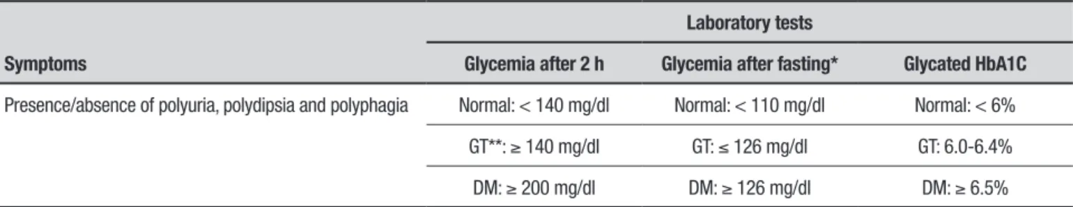
pro-Table 2. Simplified diagnostic criteria for type 2 diabetes mellitus (DM).
Symptoms
Laboratory tests
Glycemia after 2 h Glycemia after fasting* Glycated HbA1C
Presence/absence of polyuria, polydipsia and polyphagia Normal: < 140 mg/dl Normal: < 110 mg/dl Normal: < 6%
GT**: ≥ 140 mg/dl GT: ≤ 126 mg/dl GT: 6.0-6.4%
DM: ≥ 200 mg/dl DM: ≥ 126 mg/dl DM: ≥ 6.5%
tein kinase c-Jun N-terminal (JNK) and kinase 2 of the
G protein-coupled receptor kinase 2 (GRK2).60 Insulin
receptors are densely expressed in the hypothalamus, where they have a role in the regulation of body weight and feeding behavior, as well as in the cerebral cortex, entorhinal cortex and hippocampus, an area involved
with memory.61
Insulin also promotes its efect by binding itself to
insulin-like growth factor 1 (IGF-1R).10 his factor is a
hormone produced mainly in the liver and central ner-vous systems (CNS) that acts as a neurotrophic peptide; it can promote synaptic plasticity through the activa-tion of the IRS-1 signaling pathway phosphatidylinositol
3-kinase (PI3K) and Akt.62,63 IRS-2 receptor also seems
to be involved with neuroplasticity processes such as
learning and memory.64 Insulin improves cognitive
per-formance in humans and animals.4
Both neurons and glial cells express IGF-1R and IR. However, it has been suggested that neurons syn-thesize IGF-1 under physiological conditions, whereas
astrocytes are produced after injuries.65 With regard to
insulin, its synthesis occurs only in neurons and not in
glial cells.10,66
Besides insulin and IGF-1, there is the Glucagon-like peptide 1 (GLP-1) in the periphery and CNS. his is an insulinotropic hormone with neurotransmitter activity whose properties and functions are similar to insulin and IGF-1. GLP-1 is secreted by intestinal cells and neurons, and its receptor, GLP-1R, is widely expressed in the brain, including the cerebral cortex and
hippo-campus.67,68 It has been suggested that glial cells express
GLP-1R and its ligand69 only in pathological conditions,
such as neurodegenerative pathologies.
he main feature of the pathogenesis of DM is
insu-lin resistance.70 Resistance to insulin appears to occur
via genetic factors and/or failure in the recognition of the hormone by IR due to an increase in the levels of
fatty acids, glycerol and glucose.71 It has been shown
that high accumulation of glucose and fat in the blood induces the activation of immune cells, and thereby the
secretion of proinlammatory cytokines, such as IL-1β
and TNF-α. hese cytokines promote the activation,
in neurons, of molecules such as JNK leading to serine phosphorylation of the IRS-1 receptor, inhibiting tyro-sine phosphorylation and triggering insulin resistance
(Figure 1B).72,73
IL-1β in β cells of pancreatic islets and TNF-α is
acti-vated through several transcriptional pathways such as
NF-κB, caspases and inlammasomes. Once activated,
they bind to their respective receptors and recruit sev-eral other proinlammatory mediators. he increase in
proinlammatory cytokines induces a chronic inlam-matory process and reactive oxygen species (ROS)
pro-duction.72,73 Oxidative stress promotes the activation
of molecules such as JNK and thus insulin resistance.74
Insulin resistance and chronic peripheral hyperinsu-linemia implies down-regulation of insulin transport
to the brain, possibly inducing insulin deiciency in it.75
TYPE 2 DIABETES MELLITUS
IN ALZHEIMER’S DISEASE
Research has shown that DM may develop due to a high fat diet and obesity. Also, some studies have indicated that chronic ingestion of high-fat diets and DM are some of the risk factors for decline in cognitive function
and for a dementia similar to AD.5,76,77 In a study using
magnetic resonance imaging, DM patients showed reduced hippocampal volume and accelerated cognitive
decline compared with healthy elderly individuals.78
In addition, another study showed, using a Senes-cence-accelerated mouse prone model (SAMP8), that animals subjected to experimental induction of type 2 diabetes exhibited memory deicit compared to non-diabetic SAMP8 mice. his memory deicit was observed
using the Morris water maze.79 he authors also showed
that these animals exhibited increased β-amyloid
teins in the brain and hyperphosphorylated Tau pro-teins in the hippocampus, indicating changes similar to those observed in Alzheimer’s dementia.
Winocur et al. (2005) showed memory deicit and decrease in expression of insulin-dependent glucose transporter (GLUT4) and hippocampal insulin signal-ing in an animal model of insulin resistance and
obe-sity.4 he authors pointed out that a deicit in insulin
signaling may contribute to injuries in peripheral tissues of diabetes patients and the same can occur with hip-pocampal tissue inducing a deicit in memory in these animals. Also, other researchers showed that decreased sensitivity to insulin has been associated with reduced verbal luency and with cortical volume reduction of
temporal lobes in healthy elderly individuals.80
AD transgenic models submitted to a high-fat diet
showed increases in β-amyloid and tau protein as well as
activated astrocytes in mice brain cortex.81
Senescence-accelerated mice treated with a high-fat diet also showed
an increase in β-amyloid and tau protein levels.79 By
contrast, calorie restriction has been shown to reduce
the deposition of β-amyloid in both elderly mice82 and
transgenic models of AD.83 hese data suggest a close
relationship between type 2 diabetes and AD.
In addition, another study indicated increased
was produced by adipose tissue cells in a concentration
similar to that produced in vivo.84 he authors
sug-gested that adipocytes treated with Aβ reduced IRS-2
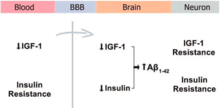
expression, which is involved with memory, and phos-phorylation of Akt-1. A reduction of IRS-2 activation in brains of Alzheimer patients was associated with IGF-1 resistance. Moreover, a reduction in IGF-1 and periph-eral insulin resistance leads to reduced uptake of IGF-1 and insulin into the brain, resulting in accumulation of Aβ85,86 (Figure 2). Neuronal insulin resistance
contrib-utes to Aβ accumulation because the insulin-degrading
enzyme (IDE) also degrades Aβ. he accumulation of
β-amyloid promotes resistance to IGF-1 and insulin
resistance. In contrast, a reduction in Aβ occurs among
rodents with elevated levels of IGF-1.87 his indicates a
role of insulin signaling in the deposition of Aβ in the
brain.
Type 2 diabetes is characterized by hyperglycemia, decreased production of insulin or its availability, and
insulin resistance.57 In this context, intraperitoneal
injections of Aβ1-42 promoted hyperglycemia and
insu-lin resistance in vivo via JAK2 in mice.88 In the study,
insulin resistance appeared to be mediated by the activa-tion of JNK, which induced inhibiactiva-tion of insulin
signal-ing.89 his data was obtained by intracerebroventricular
injection of Aβ oligomers in non-human primates and
through in vitro hippocampal neuron investigation. Studies indicate a reduction in glucose metabolism as well as changes in brain energy metabolism and
impaired neuronal insulin signaling in AD patients.14,90
Insulin signaling, as measured by phosphorylation of
AkT, was also found to be impaired by Aβ1-42 injection in
rat hippocampus91 and in cultured hippocampal neurons
by physiological inhibition of IRS-1pTyr.89 Moreover,
hyperinsulinemia also promotes Aβ1-42 increase in the
CSF of normal elderly.92 However, the mechanisms by
which type 2 diabetes and AD interaction occurs remain unknown.
In both conditions, type 2 diabetes and AD, increased
oxidative stress93,94 and chronic inlammation57,94 occur
(Figure 3). he response to injury in the peripheral ner-vous system (PNS) and CNS by the activation of microg-lia and astrocytes is a normal and beneicial response. Nevertheless, an intense inlammatory response may promote the production of excess cytokines and
oxida-tive stress leading to cell death.94 Neuroinlammation
aggravates insulin resistance through the inhibition of
IR signaling by the activation of the TNF-α receptor
(TNFR).95 he binding of insulin to its receptor induces
tyrosine phosphorylation of IRS, initiating intracellular signaling of insulin. When activated, TNFR activates the
Figure 2. Uptake of 1 and insulin into the brain. A reduction of IGF-1 and peripheral insulin resistance promotes a reduction in uptake of IGF-1 and insulin into the brain, resulting in accumulation of Aβ. Neuro-nal insulin resistance contributes to Aβ accumulation because the insu-lin degrading enzyme (IDE) also degrades Aβ. BBB: Blood-brain barrier; IGF-1: Insulin-like growth factor-1; Aβ: β-amyloid.
Figure 3. Distinct and shared symptoms of Type 2 Diabetes and
Al-zheimer's disease. Metabolic changes that occur in type 2 diabetes mellitus, such as hyperglycemia and obesity, can induce the presence of Aβ plaques, hyperphosphorylated Tau protein and loss of neurons. Aβ plaques can induce insulin resistance. The pathophysiology of both pathologies includes inflammation, increased oxidative stress, cognitive deficit and insulin resistance.
JNK pathway, one of which blocks insulin signaling by
serine phosphorylation of IRS-1.96 his infers an insulin
signaling deicit in CNS and PNS.
Cytokines and chemokines produced by both adipose tissue resident macrophages and by adipocytes of obese patients can cross the blood-brain barrier and contribute
to the onset of brain inlammation.97,98 Also, change in
the immune system accompanies physiological aging
(inlamm-aging).99 In this context, it has been suggested
that peripheral inlammatory mediators (TNF-α, IL6,
IL-1β) due to inlammation and/or infection, or
inlammation in the CNS leading to neurodegeneration. he binding of these cytokines to their receptors located in neuronal cells induces the activation of kinases such as JNK and protein kinase double-stranded RNA-dependent (PKR), which phosphorylate IRS-1 at serine residues thereby inhibiting tyrosine phosphorylation. hus, there is blocking of the action of insulin in
neuro-nal cells.34 All these factors indicate that peripheral and
central inlammatory mediators contribute to neuroin-lammation and resistance to neuronal insulin, which afects. among other functions, the cognitive function of diabetes and AD patients.
CONCLUSION
Alzheimer’s disease (AD) has been studied for decades. Several types of studies, including epidemiological and genetic investigations, indicate a relationship among obesity, insulin resistance, type 2 diabetes and
neurode-generative disorders such as AD.2,76 Some of these studies
have also found a link between severe chronic inlam-mation and cognitive impairments in AD. Knowledge and understanding of the pathophysiology as well as inlammatory and type 2 diabetes mechanisms involved in AD are important for early diagnosis of the disease (e.g. through biomarkers) and for treatment of AD.
REFERENCES
1. Luchsinger JA, Tang MX, Shea S, et al. Caloric intake and the risk of Alzheimer disease. Arch Neurol. 2002;59:1258-63.
2. Mittal K, Katare DP. Shared links between type 2 diabetes mellitus and Alzheimer’s disease: A review. Diabetes Metab Syndr. 2016;10:S144-9. 3. Vagelatos NT, Eslick GD. Type 2 diabetes as a risk factor for Alzheimer’s
disease: the confounders, interactions, and neuropathology associated with this relationship. Epidemiol Rev. 2013;35:152-60.
4. Winocur G, Greenwood CE, Piroli GG, et al. Memory impairment in obese Zucker rats: an investigation of cognitive function in an animal model of insulin resistance and obesity. Behav Neurosci. 2005;119:1389-95. 5. Beydoun MA, Beydoun HA, Wang Y. Obesity and central obesity as risk
factors for incident dementia and its subtypes: a systematic review and meta-analysis. Obes Rev. 2008;9:204-18.
6. Eikelenboom P, Zhan SS, van Gool WA, et al. Inflammatory mechanisms in Alzheimer’s disease. Trends Pharmacol Sci. 1994;15:447-50. 7. McGeer PL, McGeer EG. The inflammatory response system of brain:
implications for therapy of Alzheimer and other neurodegenerative diseases. Brain Res Brain Res Rev. 1995;21:195-218.
8. Verdile G, Keane KN, Cruzat VF, et al. Inflammation and Oxidative Stress: The Molecular Connectivity between Insulin Resistance, Obesity, and Alzheimer’s Disease. Mediators Inflamm. 2015;2015:105828. 9. Chamberlain JJ, Rhinehart AS, Shaefer CF, Jr. et al. Diagnosis and
Management of Diabetes: Synopsis of the 2016 American Diabetes Association Standards of Medical Care in Diabetes. Ann Intern Med. 2016;164:542-52.
10. Duarte AI, Moreira PI, Oliveira CR. Insulin in central nervous system: more than just a peripheral hormone. J Aging Res. 2012;2012:384017. 11. Alzheimer A, Stelzmann RA, Schnitzlein HN, et al. An English transla-tion of Alzheimer’s 1907 paper, “Uber eine eigenartige Erkankung der Hirnrinde”. Clin Anat. 1995;8:429-31.
12. Francis PT, Palmer AM, Snape M, et al. The cholinergic hypothesis of Alzheimer’s disease: a review of progress. J Neurol Neurosurg Psychiatry. 1999;66:137-47.
13. Blennow K, de Leon MJ, Zetterberg H. Alzheimer’s disease. Lancet. 2006;368:387-403.
14. Chen Y, Zhao Y, Dai CL , et al. Intranasal insulin restores insulin signaling, increases synaptic proteins, and reduces Abeta level and microglia acti-vation in the brains of 3xTg-AD mice. Exp Neurol. 2014;261:610-9. 15. Lopez OL, McDade E, Riverol M, et al. Evolution of the diagnostic
criteria for degenerative and cognitive disorders. Curr Opin Neurol. 2011;24:532-41.
16. Braak H, Braak E. Morphological criteria for the recognition of Alzheim-er’s disease and the distribution pattern of cortical changes related to this disorder. Neurobiol Aging. 1994;15:355-6.
17. Terry RD, Peck A, DeTeresa R, et al. Some morphometric aspects of the brain in senile dementia of the Alzheimer type. Ann Neurol. 1981; 10:184-92.
18. Terry RD, Masliah E, Salmon DP, et al. Physical basis of cognitive altera-tions in Alzheimer’s disease: synapse loss is the major correlate of cogni-tive impairment. Ann Neurol. 1991;30:572-80.
19. Contestabile A. The history of the cholinergic hypothesis. Behav Brain Res. 2011;221:334-40.
20. Auld DS, Kar S, Quirion R. Beta-amyloid peptides as direct cholinergic neuromodulators: a missing link? Trends Neurosci. 1998;21:43-9. 21. Allsop D, Howlett D, Christie G, et al. Fibrillogenesis of beta-amyloid.
Biochem Soc Trans. 1998;26:459-63.
22. Evin G, Weidemann A. Biogenesis and metabolism of Alzheimer’s disease Abeta amyloid peptides. Peptides. 2002;23:1285-97. 23. Hardy JA, Higgins GA. Alzheimer’s disease: the amyloid cascade
hypoth-esis. Science. 1992;256:184-5.
24. Coulson EJ, Paliga K, Beyreuther K, et al. What the evolution of the amyloid protein precursor supergene family tells us about its function. Neurochem Int. 2000;36:175-84.
25. Bates KA, Verdile G, Li QX, et al. Clearance mechanisms of Alzheimer’s amyloid-beta peptide: implications for therapeutic design and diagnostic tests. Mol Psychiatry. 2009;14:469-86.
26. Kummer MP, Heneka MT. Truncated and modified amyloid-beta species. Alzheimers Res Ther. 2014;6:28.
27. Kojro E, Gimpl G, Lammich S, et al. Low cholesterol stimulates the nonamyloidogenic pathway by its effect on the alpha -secretase ADAM 10. Proc Natl Acad Sci U S A. 2001;98:5815-20.
28. Selkoe DJ, Schenk D. Alzheimer’s disease: molecular understanding predicts amyloid-based therapeutics. Annu Rev Pharmacol Toxicol. 2003;43:545-84.
29. Nunan J, Small DH. Regulation of APP cleavage by alpha-, beta- and gamma-secretases. FEBS Lett. 2000;483:6-10.
30. Muller UC, Zheng H. Physiological functions of APP family proteins. Cold Spring Harb Perspect Med. 2012;2:a006288.
31. Walsh DM, Selkoe DJ. A beta oligomers - a decade of discovery. J Neurochem. 2007;101:1172-84.
32. Takeda S, Sato N, Morishita R. Systemic inflammation, blood-brain barrier vulnerability and cognitive/non-cognitive symptoms in Alzheimer disease: relevance to pathogenesis and therapy. Front Aging Neurosci. 2014;6:171.
33. Serpente M, Bonsi R, Scarpini E, et al. Innate immune system and inflammation in Alzheimer’s disease: from pathogenesis to treatment. Neuroimmunomodulation. 2014;21:79-87.
34. Ferreira ST, Clarke JR, Bomfim TR, et al. Inflammation, defective insulin signaling, and neuronal dysfunction in Alzheimer’s disease. Alzheimers Dement. 2014;10:S76-83.
35. Bertram L, Lill CM, Tanzi RE. The genetics of Alzheimer disease: back to the future. Neuron. 2010;68:270-81.
36. Mahley RW. Apolipoprotein E: cholesterol transport protein with expanding role in cell biology. Science. 1988;240:622-30.
37. Zannis VI, Breslow JL, Utermann G, et al. Proposed nomenclature of apoE isoproteins, apoE genotypes, and phenotypes. J Lipid Res. 1982; 23:911-4. 38. Ma J, Yee A, Brewer HB, Jr. et al. Amyloid-associated proteins alpha
1-antichymotrypsin and apolipoprotein E promote assembly of Alzheimer beta-protein into filaments. Nature. 1994;372:92-4.
39. Brandt R, Leger J, Lee G. Interaction of tau with the neural plasma membrane mediated by tau’s amino-terminal projection domain. J Cell Biol. 1995;131:1327-40.
41. Butler M, Shelanski ML. Microheterogeneity of microtubule-associated tau proteins is due to differences in phosphorylation. J Neurochem. 1986;47:1517-22.
42. Mazanetz MP, Fischer PM. Untangling tau hyperphosphorylation in drug design for neurodegenerative diseases. Nat Rev Drug Discov. 2007; 6:464-79.
43. Roy S, Zhang B, Lee VM, et al. Axonal transport defects: a common theme in neurodegenerative diseases. Acta Neuropathol. 2005;109:5-13. 44. Kuret J, Congdon EE, Li G, et al. Evaluating triggers and enhancers of
tau fibrillization. Microsc Res Tech. 2005;67:141-55.
45. Blennow K. Cerebrospinal fluid protein biomarkers for Alzheimer’s disease. NeuroRx. 2004;1:213-25.
46. Rapoport M, Ferreira A. PD98059 prevents neurite degeneration induced by fibrillar beta-amyloid in mature hippocampal neurons. J Neurochem. 2000;74:125-33.
47. Li Y, Liu L, Barger SW, et al. Interleukin-1 mediates pathological effects of microglia on tau phosphorylation and on synaptophysin synthesis in cortical neurons through a p38-MAPK pathway. J Neurosci. 2003;23: 1605-11.
48. Quintanilla RA, Orellana DI, Gonzalez-Billault C, et al. Interleukin-6 induces Alzheimer-type phosphorylation of tau protein by deregulating the cdk5/p35 pathway. Exp Cell Res. 2004;295:245-57.
49. Lleo A, Greenberg SM, Growdon JH. Current pharmacotherapy for Alzheimer’s disease. Annu Rev Med. 2006;57:513-33.
50. Rylett RJ, Williams LR. Role of neurotrophins in cholinergic-neurone function in the adult and aged CNS. Trends Neurosci. 1994;17:486-90. 51. Davies P, Maloney AJ. Selective loss of central cholinergic neurons in
Alzheimer’s disease. Lancet. 1976;2:1403.
52. Bowen DM, Smith CB, White P, et al. Neurotransmitter-related enzymes and indices of hypoxia in senile dementia and other abiotrophies. Brain. 1976;99:459-96.
53. Grimaldi M, Marino SD, Florenzano F, et al. beta-Amyloid-acetylcholine molecular interaction: new role of cholinergic mediators in anti-Alzheimer therapy? Future Med Chem. 2016;8:1179-89.
54. Jansen R, Dzwolak W, Winter R. Amyloidogenic self-assembly of insulin aggregates probed by high resolution atomic force microscopy. Biophys J. 2005;88:1344-53.
55. Daneman D. Type 1 diabetes. Lancet. 2006;367:847-58.
56. Devendra D, Liu E, Eisenbarth GS. Type 1 diabetes: recent develop-ments. BMJ. 2004;328:750-4.
57. Spranger J, Kroke A, Mohlig M, et al. Inflammatory cytokines and the risk to develop type 2 diabetes: results of the prospective population-based European Prospective Investigation into Cancer and Nutrition (EPIC)-Potsdam Study. Diabetes. 2003;52:812-7.
58. Copps KD, White MF. Regulation of insulin sensitivity by serine/threonine phosphorylation of insulin receptor substrate proteins IRS1 and IRS2. Diabetologia. 2012;55:2565-82.
59. Farese RV, Sajan MP, Standaert ML. Insulin-sensitive protein kinases (atypical protein kinase C and protein kinase B/Akt): actions and defects in obesity and type II diabetes. Exp Biol Med (Maywood ). 2005;230:593-605.
60. Boura-Halfon S, Zick Y. Phosphorylation of IRS proteins, insulin action, and insulin resistance. Am J Physiol Endocrinol Metab. 2009;296:E581-91.
61. Marks JL, King MG, Baskin DG. Localization of insulin and type 1 IGF receptors in rat brain by in vitro autoradiography and in situ hybridization. Adv Exp Med Biol. 1991;293:459-70.
62. Cardona-Gomez GP, Mendez P, DonCarlos LL, et al. Interactions of estrogens and insulin-like growth factor-I in the brain: implications for neuroprotection. Brain Res Brain Res Rev. 2001;37:320-34.
63. Dudek H, Datta SR, Franke TF, et al. Regulation of neuronal survival by the serine-threonine protein kinase Akt. Science. 1997;275:661-5. 64. Martin ED, Sanchez-Perez A, Trejo JL, et al. IRS-2 Deficiency impairs
NMDA receptor-dependent long-term potentiation. Cereb Cortex. 2012;22:1717-27.
65. Torres-Aleman I. Toward a comprehensive neurobiology of IGF-I. Dev Neurobiol. 2010;70:384-96.
66. Unger JW, Moss AM, Livingston JN. Immunohistochemical localization of insulin receptors and phosphotyrosine in the brainstem of the adult rat. Neuroscience. 1991;42:853-61.
67. Brubaker PL, Drucker DJ. Minireview: Glucagon-like peptides regulate cell proliferation and apoptosis in the pancreas, gut, and central nervous system. Endocrinology. 2004;145:2653-9.
68. Sarkar S, Fekete C, Legradi G, et al. Glucagon like peptide-1 (7-36) amide (GLP-1) nerve terminals densely innervate corticotropin-releasing hormone neurons in the hypothalamic paraventricular nucleus. Brain Res. 2003;985:163-8.
69. Kappe C, Tracy LM, Patrone C, et al. GLP-1 secretion by microglial cells and decreased CNS expression in obesity. J Neuroinflammation. 2012;9:276.
70. Rehman K, Akash MS. Mechanisms of inflammatory responses and development of insulin resistance: how are they interlinked? J Biomed Sci. 2016;23:87.
71. Meigs JB. Epidemiology of cardiovascular complications in type 2 diabetes mellitus. Acta Diabetol. 2003;40 Suppl 2:S358-61.
72. Rehman K, Akash MS. Mechanisms of inflammatory responses and development of insulin resistance: how are they interlinked? J Biomed Sci. 2016;23:87.
73. Roman-Pintos LM, Villegas-Rivera G, Rodriguez-Carrizalez AD, et al. Diabetic Polyneuropathy in Type 2 Diabetes Mellitus: Inflammation, Oxidative Stress, and Mitochondrial Function. J Diabetes Res. 2016; 2016:3425617.
74. Rehman K, Akash MS. Mechanisms of inflammatory responses and development of insulin resistance: how are they interlinked? J Biomed Sci. 2016;23:87.
75. Stein LJ, Dorsa DM, Baskin DG, et al. Reduced effect of experimental peripheral hyperinsulinemia to elevate cerebrospinal fluid insulin concen-trations of obese Zucker rats. Endocrinology. 1987;121:1611-5. 76. Arrieta-Cruz I, Gutierrez-Juarez R. The Role of Insulin Resistance and
Glucose Metabolism Dysregulation in the Development of Alzheimer s Disease. Rev Invest Clin. 2016;68:53-8.
77. Biessels GJ, Reagan LP. Hippocampal insulin resistance and cognitive dysfunction. Nat Rev Neurosci. 2015;16:660-71.
78. Bruehl H, Wolf OT, Sweat V, et al. Modifiers of cognitive function and brain structure in middle-aged and elderly individuals with type 2 diabetes mellitus. Brain Res. 2009;1280:186-94.
79. Mehla J, Chauhan BC, Chauhan NB. Experimental induction of type 2 diabetes in aging-accelerated mice triggered Alzheimer-like pathology and memory deficits. J Alzheimers Dis. 2014;39:145-62.
80. Benedict C, Brooks SJ, Kullberg J, et al. Impaired insulin sensitivity as indexed by the HOMA score is associated with deficits in verbal fluency and temporal lobe gray matter volume in the elderly. Diabetes Care. 2012;35:488-94.
81. Julien C, Tremblay C, Phivilay A, et al. High-fat diet aggravates amyloid-beta and tau pathologies in the 3xTg-AD mouse model. Neurobiol Aging. 2010;31:1516-31.
82. Mouton PR, Chachich ME, Quigley C, et al. Caloric restriction attenuates amyloid deposition in middle-aged dtg APP/PS1 mice. Neurosci Lett. 2009;464:184-7.
83. Patel NV, Gordon MN, Connor KE, et al. Caloric restriction attenuates Abeta-deposition in Alzheimer transgenic models. Neurobiol Aging. 2005;26:995-1000.
84. Tharp WG, Gupta D, Smith J, et al. Effects of glucose and insulin on secretion of amyloid-beta by human adipose tissue cells. Obesity (Silver Spring). 2016;24:1471-9.
85. Adlerz L, Holback S, Multhaup G, et al. IGF-1-induced processing of the amyloid precursor protein family is mediated by different signaling pathways. J Biol Chem. 2007;282:10203-9.
86. Bosco D, Fava A, Plastino M, et al. Possible implications of insulin resis-tance and glucose metabolism in Alzheimer’s disease pathogenesis. J Cell Mol Med. 2011;15:1807-21.
87. Carro E, Trejo JL, Spuch C, et al. Blockade of the insulin-like growth factor I receptor in the choroid plexus originates Alzheimer’s-like neuro-pathology in rodents: new cues into the human disease? Neurobiol Aging. 2006;27:1618-31.
88. Zhang Y, Zhou B, Deng B, et al. Amyloid-beta induces hepatic insulin resistance in vivo via JAK2. Diabetes. 2013;62:1159-66.
89. Bomfim TR, Forny-Germano L, Sathler LB, et al. An anti-diabetes agent protects the mouse brain from defective insulin signaling caused by Alzheimer’s disease- associated Abeta oligomers. J Clin Invest. 2012;122:1339-53.
90. Haley AP, Knight-Scott J, Simnad VI, et al. Increased glucose concen-tration in the hippocampus in early Alzheimer’s disease following oral glucose ingestion. Magn Reson Imaging. 2006;24:715-20.
insulin signaling, and hippocampal metabolism. J Alzheimers Dis. 2012;30:413-22.
92. Watson GS, Peskind ER, Asthana S, et al. Insulin increases CSF Abeta42 levels in normal older adults. Neurology. 2003;60:1899-903.
93. Anderson EJ, Lustig ME, Boyle KE, et al. Mitochondrial H2O2 emission and cellular redox state link excess fat intake to insulin resistance in both rodents and humans. J Clin Invest. 2009;119:573-81.
94. Heneka MT, Carson MJ, El KJ, et al. Neuroinflammation in Alzheimer’s disease. Lancet Neurol. 2015;14:388-405.
95. Hotamisligil GS. Inflammation and metabolic disorders. Nature. 2006; 444:860-7.
96. Hirosumi J, Tuncman G, Chang L, et al. A central role for JNK in obesity and insulin resistance. Nature. 2002;420:333-6.
97. Banks WA. Blood-brain barrier transport of cytokines: a mechanism for neuropathology. Curr Pharm Des. 2005;11:973-84.
98. Lumeng CN, Bodzin JL, Saltiel AR. Obesity induces a phenotypic switch in adipose tissue macrophage polarization. J Clin Invest. 2007;117: 175-84.