Variability and Action Mechanism of a Family of
Anticomplement Proteins in
Ixodes ricinus
Bernard Couvreur1., Je´roˆme Beaufays1., Ce´dric Charon1., Kathia Lahaye1
, Franc¸ois Gensale1, Vale´rie Denis1, Benoıˆt Charloteaux2, Yves Decrem1, Pierre-Paul Pre´voˆt1, Michel Brossard3, Luc Vanhamme1,4, Edmond Godfroid1
*
1Laboratory for Molecular Biology of Ectoparasites, Institut de Biologie et de Me´decine Mole´culaires (IBMM), Universite´ Libre de Bruxelles, Gosselies, Belgium,2Centre de Biophysique Mole´culaire Nume´rique, Gembloux Agricultural University, Gembloux, Belgium,3Institute of Zoology, University of Neuchaˆtel, Neuchaˆtel, Switzerland,4Laboratory of Molecular Parasitology, Institut de Biologie et de Me´decine Mole´culaires (IBMM), Universite´ Libre de Bruxelles, Gosselies, Belgium
Background.Ticks are blood feeding arachnids that characteristically take a long blood meal. They must therefore counteract
host defence mechanisms such as hemostasis, inflammation and the immune response. This is achieved by expressing batteries of salivary proteins coded by multigene families.Methodology/Principal Findings.We report the in-depth analysis of a tick multigene family and describe five new anticomplement proteins inIxodes ricinus. Compared to previously described Ixodesanticomplement proteins, these segregated into a new phylogenetic group or subfamily. These proteins have a novel action mechanism as they specifically bind to properdin, leading to the inhibition of C3 convertase and the alternative complement pathway. An excess of non-synonymous over synonymous changes indicated that coding sequences had undergone diversifying selection. Diversification was not associated with structural, biochemical or functional diversity, adaptation to host species or stage specificity but rather to differences in antigenicity. Conclusions/Significance. Anticomplement proteins fromI. ricinusare the first inhibitors that specifically target a positive regulator of complement, properdin. They may provide new tools for the investigation of role of properdin in physiological and pathophysiological mechanisms. They may also be useful in disorders affecting the alternative complement pathway. Looking for and detecting the different selection pressures involved will help in understanding the evolution of multigene families and hematophagy in arthropods.
Citation: Couvreur B, Beaufays J, Charon C, Lahaye K, Gensale F, et al (2008) Variability and Action Mechanism of a Family of Anticomplement Proteins inIxodes ricinus. PLoS ONE 3(1): e1400. doi:10.1371/journal.pone.0001400
INTRODUCTION
Parasites probably affect every living organism and it may reasonably be estimated that at least half the animals on earth are parasites [1]. By definition, parasites live at the expense of their host but hosts defend themselves and, in turn, parasites evolve counter-measures. Parasitism is probably therefore a major driving force in evolution [1]. Bloodfeeding arthropods such as ticks constitute a very good example of the evolutionary arms race between hosts and parasites.
Ticks are obligate blood feeding arachnids. They infest many species of mammals, birds, reptiles and amphibians worldwide. They are the vectors of protozoan, bacterial and viral pathogens of prime medical and veterinary importance. Examples of such important pathogens are Borrelia burgdorferi, Tick-borne Encepha-litis Virus (TBEV), Babesia bovisor Theileria parva, the respective agents of Lyme disease and viral encephalitis in humans, and babesiosis and theileriosis (East Coast Fever) in cattle [2]. Blood losses due to heavy infestation may weaken the animal, render it more susceptible to other diseases or cause death by exsanguina-tion [3]. In addiexsanguina-tion, the occurrence of tick toxicosis including tick paralysis is associated with the presence of toxins in the saliva [4]. There are two main families of ticks,Ixodidaeor hard ticks and
Argasidaeor soft ticks. TheIxodidaefamily is further divided into two
subdivisions: Prostriata, which contains only the subfamilyIxodinae, and Metastriata, which includes the subfamilies Bothriocrotinae,
Amblyomminae,HaemaphysalinaeandRhipicephalinae[3]. Argasid ticks
typically feed for short periods of time (up to 2 hours) whereas Ixodid ticks remain attached to and feed on their vertebrate host for up to two weeks [3]. The feeding time of an adultIxodes ricinus
female is typically 7–10 days [3]. Such a long blood meal is only possible because these parasites have developed ways to circum-vent host defense mechanisms including hemostasis (coagulation,
platelet aggregation and vasoconstriction), the inflammatory response and innate and adaptive immunity [reviewed in 5,6,7,8]. Furthermore, pain or itching caused by the inflammatory response stimulates hosts to scratch and dislodge the parasite.
The complement system is a first line of defence against invading pathogens and it links the innate and adaptive responses of the vertebrate immune system [reviewed in 9]. It consists of a cascade of plasma enzymes leading to activation of three effector mechanisms: (i) generation of the short potent pro-inflammatory peptides C3a and C5a, ii) deposition of opsonizing C3b proteins on cell surfaces, (iii) formation of the membrane attack complex (MAC). MACs create pores in the membrane, leading to cell death. Complement is activatedvia three main pathways. The classical pathway (CP) is
Academic Editor:Jeffrey Gold, Oregon Health & Science University, United States of America
ReceivedSeptember 25, 2007;AcceptedDecember 11, 2007;PublishedJanuary 2, 2008
Copyright:ß2008 Couvreur et al. This is an open-access article distributed under the terms of the Creative Commons Attribution License, which permits unrestricted use, distribution, and reproduction in any medium, provided the original author and source are credited.
Funding:LVH is a Senior Research Associate at the Belgian National Fund for Scientific Research. CC is supported by a PhD studentship from the Fonds pour la Formation a` la Recherche dans l’Industrie et dans l’Agriculture of the Belgian Federal Governement. This work was supported by contracts 215107 and 415702 from the Ministe`re de la Re´gion Wallonne in Belgium.
Competing Interests:The activities of IxAC-B1 to B5 are being patented.
* To whom correspondence should be addressed.E-mail: edmond.godfroid@ ulb.ac.be
initiated mainly when the C1 complex binds to the Fc region of certain antibody isotypes in immune complexes. The lectin-mediated pathway is activated by mannose-binding lectin interacting with mannose residues on microbial surfaces.
The alternative pathway (AP) is spontaneously activated by hydrolysis of plasma C3 into C3 (H2O). C3 (H2O) binds soluble factor B (fB). Bound fB is cleaved by serine protease factor D into soluble Ba peptide and the larger Bb fragment. The resulting C3 (H2O)Bb complex is the initial C3 convertase. It cleaves fluid-phase C3 into C3a peptide and metastable C3b. C3b binds covalently to a pathogen or cell surface via a short-lived thioester bond. Factor B interacts with C3b, leading to its cleavage by factor D and the formation of the C3 convertase (C3bBb). This complex generates new C3b molecules and amplifies the complement cascade by forming new C3 convertases or C5 convertases (C3b2Bb). C5 convertase cleaves C5 into C5a and C5b. C5b initiates the formation of MAC [9].
Host cells are protected from attack by the complement system by plasma and membrane-bound regulatory molecules that inactivate complement proteins. C3 convertases are deactivated by dissociation mediated by surface proteins such as Decay-Accelerating Factor (DAF) and Complement Receptor-1 (CR1), as well as soluble factor H. These proteins bind to C3b and displace Bb [9]. They also act as co-factors for serine protease factor I which cleaves C3b [10]. On the other hand, the half-life of C3 convertase is increased at least 10-fold by properdin [11]. It is present in the plasma in oligomer form (dimer, trimer or tetramer) [12,13]. Each monomer is a 53 kDa protein composed of six repetitive thrombospondin domains (TSP), flanked with an N-terminal and C-N-terminal region [14,15,16]. Properdin binds to surface-bound C3b and increases its ability to interact with factor B [17]. It also binds to pre-formed C3 convertases leading to increased stability and preventing inactivation by regulators such as factor H and factor I [9]. Moreover, properdin oligomers attached to C3b on cell surfaces interact with preformed fluid-phase C3b or C3bBb through its other subunits [18]. The essential role of properdin in complement activation was demonstrated by the capacity of an anti-properdin monoclonal antibody to inhibit activation of the alternative pathway. This monoclonal antibody prevents the interaction between properdin and C3b [19].
The AP is the major line of defense against invading pathogens such as bacteria [20]. It is also involved in guinea pig resistance to the hard tickDermacentor andersoni[21,22]. Saliva or salivary gland extracts fromIxodes dammini [23],I. hexagonusand I. uriae[24],I.
scapularis[25] andI. ricinus[24,26] have been found to have an
inhibitory activity on the alternative complement pathway. Valenzuela et al. [25] purified the active anticomplement
component from the saliva of adult I. scapularis. N-terminal
sequencing combined with the screening of a cDNA library led to the description of the coding sequence of a tick anticomplement protein named ISAC (I. scapularisanti-complement). Recombinant ISAC mimics the anticomplement activity of tick saliva. It interferes with the formation of C3 convertase from C3 and fB and destabilizes pre-formed C3 convertase. Sequences closely
related to ISAC were then cloned by RT/PCR fromI. scapularis
nymphs [27], found by screening a cDNA library with sera from repeatedly infested guinea pigs [28] or by PCR-screening of a
nymph cDNA library [29]. In I. pacificus, sequencing large
numbers of cDNA clones from adult salivary glands led to the discovery of ISAC-I [30]. Finally, using degenerate primers designed from the published ISAC sequence, Daix et al. [31] recently cloned the related IRAC I and IRAC II fromI. ricinus.
In soft ticks too, anticomplement activity is present in saliva and salivary gland extracts [32]. InOrnithodoros moubata, this activity is
due to protein OmCI which inhibits both the alternative and
classical pathways. Its sequence is unrelated to the Ixodes
anticomplement molecules mentioned above (,15% amino-acid
identity). OmCI binds to C5 component of the complement cascade and belongs to the lipocalin superfamily [33].
The recent characterization of large numbers of cDNA sequences from salivary glands of Ixodid ticks includingI. scapularis[34,35] and
I. pacificus[30] indicated that most salivary proteins are expressed as
large clusters of related proteins, probably coded by multigene families. Moreover, genome size and organization were examined in
Ixodes scapularis, Boophilus microplus [36] and Amblyomma americanum
[37]. These genomes are large: 2.16109, 7.16109bp and 1.046109 bp respectively. Reassociation rates of genomic DNA indicate that they are composed mainly of moderately repetitive elements, which include transposable elements and members of multigene families. This organization in multigene families is therefore probably a major feature of hard tick genome organization and perhaps an adaptation to bloodfeeding.
In the work described here we completed an inventory of sequences related toI. scapularisanticomplement protein ISAC in the salivary glands ofI. ricinus. We were able to detect five new sequences that we used to study diversification mechanisms possibly at work in a family of tick salivary proteins (hereby referred to as the IxAC family) and we investigated their action mechanism. The results showed thatI. ricinusanticomplement proteins specifically bind to properdin, leading to the inhibition of the formation of C3 convertase and inhibition of the alternative complement activation pathway. Sequence diversification is associated with antigenic diversity rather than major divergence in molecular characteristics or activity, host specificity or stage specificity.
In this study, we investigated the general significance of multigene families in the context of a host-parasite relationship. This is a specific in-depth analysis of a tick multigene family. It highlights the value of performing dedicated gene-targeted inventories when studying specific aspects of tick adaptation to a bloodfeeding lifestyle. Remarkably, IxACs fromI. ricinusare also the first inhibitors that specifically target a positive regulator of complement.
RESULTS
A large family of anticomplement proteins in the
hard tick
Ixodes ricinus
The fragments of two genes related to the prototypical Ixodes
scapularisanticomplement protein ISAC of Valenzuela et al. [25]
were found by serendipity in cDNA from pooled salivary glands. The complete coding sequences as well as parts of the 39and 59
UTR’s were then reconstituted by RACE. The new genes (accession numbers: AM407396 and AM407397) coded for two new proteins showing,40 % identity with ISAC and the recently
described IRAC I and IRAC II fromI. ricinus[31], but over 65 % identity with each other. This led us to suspect the existence of a much larger family of anticomplement proteins inI. ricinus.
In order to make as complete an inventory of this family as possible, a total of 2 different reverse transcription experiments, 6 different PCR amplifications and 12 ligations were performed on polyA+RNA from salivary glands ofI. ricinusfemales (Table S1).
122 clones with inserts of the expected size ($600 bp) were
sequenced. A few were disregarded as they coded for proteins unrelated to anticomplement proteins (e.g. ribosomal proteins). A few additional clones with inserts smaller than expected (400– 500 bp) were sequenced too. They were found to code for homologs of uncharacterized ‘‘putative salivary proteins’’ fromI.
found, most could be assigned to previously described IRAC I, IRAC II, AM407396 and AM407397 on the basis of sequence identity. AM407396 was the most frequent (46.6 %) followed by AM407397 (23.3 %), IRAC I (15.5 %) and IRAC II (0.9%). Three additional new sequences were also identified. They were assigned accession numbers AM407398, AM407399 and AM407400 respectively. They accounted for 3.4 %, 8.6 % and 1.7% of
clones, respectively. Sequences found to be identical in $3
independent clones were considered genuine. AM407400 was only represented by two clones which showed a difference of two nucleotides. Therefore, the latter sequence was confirmed independently by amplifying internal fragments with gene-specific primers from salivary gland cDNA. Finally, the same set of sequences was found from the various independent RT-PCR experiments (Table S1). Overall, the results suggested that a complete or near-complete inventory of IxAC anticomplement messengers from theIxodes ricinussalivary gland was achieved at least for the population investigated.
PCR experiments using the primers listed in Table 1 were also performed on cDNA from pooled salivary glands of 3 day-fed male and femaleR. appendiculatus. No PCR products were observed (not shown). We were nevertheless able to amplify the coding sequence for the known lipocalin RaHBP-2 from the same cDNAs using specific primers. Interrogation of databases including the
prelim-inary releases of sequences from the non-Ixodes hard tick R.
appendiculatus, B. microplus, A. americanum and A. variegatum genome
projects yielded no or only insignificant hits.
Phylogenetic analysis of tick anticomplement
sequences
Nucleotide and peptide sequences of ISAC, IRAC I and AM407396 were used to interrogate databases with the same results. A total of 48 entries were recovered (Table S2). These were fromI. scapularis(45 entries),I. ricinus(2 entries) andI. pacificus(1 entry). They had been cloned from salivary glands (30), whole fed nymphs and nymph salivary glands (15) or unspecified tick material (3).
Two sequences containing ambiguous positions and ten sequences with incomplete coding sequences for the mature protein, including Salp9 (AF278574) and Isac-like clone 113 (AY956386), were initially discarded. These 12 entries were from
I. scapularis. The remaining 36 entries were aligned with our five
new sequences fromI. ricinus(AM407396 to AM407400). Distance dendrograms were constructed from alignments of nucleotide sequences or predicted amino-acid sequences of putative mature proteins translated from the open reading frames (Figure 1). All sequences clustered into two main groups or subfamilies, IxAC-A and IxAC-B, which were strongly supported by bootstrap analysis (1000/1000). IxAC-A could be further divided into two clusters. A first, large, group contained onlyI. scapularissequences closely related to prototypical ISAC. It was strongly supported by bootstrap analysis (.900/1000). A second, smaller, cluster contained Isac-1 from I.
pacificus, IRAC II fromI. ricinus, Salp20-like protein 2 and EST nu
DN970085 fromI. scapularis. Bootstrap support was lower ($830/ 1000). IRAC I could not be joined robustly (bootstrap value,750/ 1000) to any of the previous two clusters. The IxAC-B subfamily
contained our five new sequences AM407396 to AM407400 fromI.
ricinusbut none from other tick species. No robust cluster emerged
within this subfamily.
The maximum-likelihood method was also applied to the initial nucleotide and amino-acid alignment of putative mature proteins. It supported the same topology as the distance method with slightly different bootstrap values (Figure 1). The two subfamilies were strongly supported (1000/1000). The two clusters within IxAC-A were also recovered but bootstrap support was lower than with the distance method. Again, IRAC I could not be placed robustly in any of the two clusters within IxAC-A (bootstrap support,650/1000).
The overall topology of the distance trees was not altered after including the leader peptide sequences in the alignments (not shown) or the 12 discarded sequences (not shown). Most of the latter clustered with ISAC within the ISAC-like cluster (not shown). Only Salp9 grouped with Isac-I, IRAC II and EST nuDN970085 within the second cluster in IxAC-A. Salp9, a 79 residues peptide, aligned to the C-terminal half of DN970085 to which it showed 90% identity. Because ISAC and Salp9 were the earliest tick anticomplement sequences published, we decided to name the large and small clusters within IxAC-A ‘‘ISAC-like’’ and ‘‘Salp9-like’’, respectively.
We therefore decided to rename the five new sequences (AM407396 to AM407400) IxAC-B1 to IxAC-B5 to indicate the fact that they clustered into the new group or subfamily IxAC-B and away from the previously described IRAC I and IRAC II which belong to the IxAC-A subfamily.
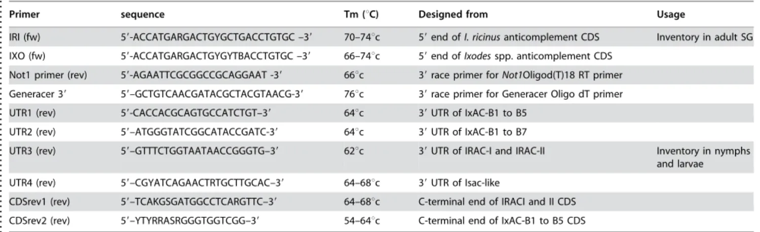
Table 1.PCR primers for RT-PCR inventory ofI. ricinusgenes coding for anticomplement proteins.
. . . .
Primer sequence Tm (uC) Designed from Usage
IRI (fw) 59-ACCATGARGACTGYGCTGACCTGTGC –39 70–74uc 59end ofI. ricinusanticomplement CDS Inventory in adult SG
IXO (fw) 59-ACCATGARGACTGYGYTBACCTGTGC –39 66–74uc 59end ofIxodesspp. anticomplement CDS
Not1 primer (rev) 59-AGAATTCGCGGCCGCAGGAAT -39 66uc 39race primer forNot1Oligod(T)18 RT primer
Generacer 39 59–GCTGTCAACGATACGCTACGTAACG-39 76uc 39race primer for Generacer Oligo dT primer
UTR1 (rev) 59-CACCACGCAGTGCCATCTGT–39 64uc 39UTR of IxAC-B1 to B5
UTR2 (rev) 59–ATGGGTATCGGCATACCGATC-39 64uc 39UTR of IxAC-B1 to B7
UTR3 (rev) 59–GTTTCTGGTAATAACCGGGTG–39 62uc 39UTR of IRAC-I and IRAC-II Inventory in nymphs
and larvae
UTR4 (rev) 59–CGYATCAGAACTRTGCTTGCAC–39 64–68uc 39UTR of Isac-like
CDSrev1 (rev) 59–TCAKGSGATGGCCTCARGTTC–39 64–68uc C-terminal end of IRACI and II CDS
CDSrev2 (rev) 59–YTYRRASRGGGTGGTCGG–39 54–64uc C-terminal end of IxAC-B1 to B5 CDS
Forward primers IRI and IXO were designed manually from the 59end of coding sequences of anticomplement proteins available at the start of this project fromI. ricinusandIxodesspp. respectively. The trinucleotide ACC was added 59to the start codon to improve eukaryotic expression. Commercial reverse primers Generacer 39 and Not1 are available from Invitrogen and Amersham Biosciences, respectively. Family-specific reverse primers were designed from UTR or coding sequences of the different subfamilies of tick anticomplement proteins as indicated. Calculated mean melting temperatures (Tm) are also indicated. CDS, coding sequences; UTR, untranslated region; SG, salivary gland.
doi:10.1371/journal.pone.0001400.t001
....
...
....
...
...
....
...
...
....
...
...
....
...
...
....
...
...
....
...
...
..
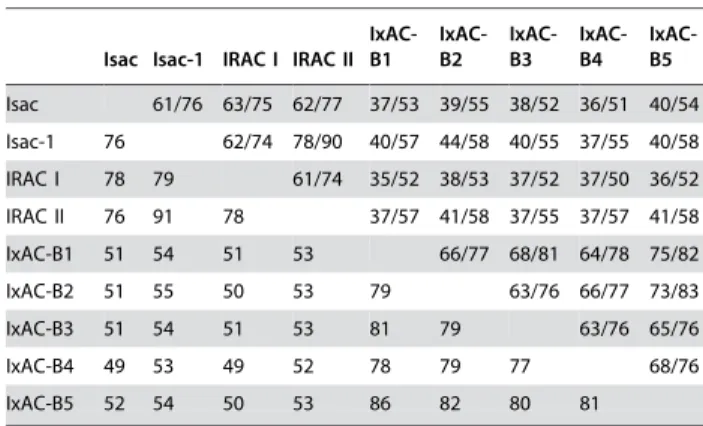
Percentages of identity and similarity were calculated for representative IxACs (Table 2). Within a subfamily, amino-acid sequences were over 60 % identical whereas identity dropped to
,40 % between the two subfamilies. The two subfamilies could
also be differentiated by an indel of 4 amino-acids (position 74 to 77 in Figure 2). Within the Isac-like cluster or within the Salp9-like cluster, amino-acid sequences were at least 70% identical.
According to information provided on the entry files or in the original publications, the sequences had been obtained from adults (12), nymphs (34) or unspecified stages (2). However, no robust ‘‘adult-only’’ or ‘‘nymph-only’’ clusters could be discerned
(Figure 1). Moreover, ‘‘adult’’ EST nu DN968378 was found to
be 100% identical to ‘‘nymphal’’ Salp20 (AF209917) (Figure 1). To summarize, phylogenetic analysis of all available tick anticomplement sequences indicated that they robustly segregated into two distinct groups or subfamilies, which we termed IxAC-A
and IxAC-B. Within-group amino-acid identity was .60%
whereas between-group identity dropped to ,40%. The larger
IxAC-A contained sequences from I. scapularis, I. ricinus and I.
pacificus. It could be subdivided into two or possibly three clusters.
Our new sequences fromI. ricinus constituted a completely new group which we named IxAC-B. No stage-specific group of sequences was identified at this point of the research.
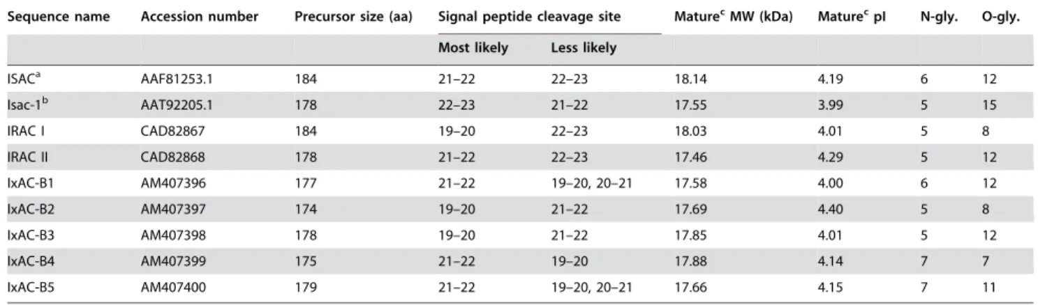
Protein properties
The properties of these newly discovered proteins were predicted from their amino-acid sequences and compared to prototypical ISAC from I. scapularis and related Isac-1 from I. pacificus. Calculated PM and pI ranged from 17.46 to 18.03 and 4.01 to 4.29 respectively (Table 3).
All anticomplement proteins presented four conserved cysteine residues predicted to make two disulphide bridges (Figure 2).
Most likely signal peptide cleavage sites for representative IxACs are indicated in Table 3 and Figure 2. In each individual peptide sequence, SignalP predicted a second or even a third probable, Figure 1. Phylogenetic analysis ofIxodesanticomplement proteins. A distance dendrogram was constructed from an alignment of 41 tick mature anticomplement proteins using programs in the Phylip 3.65 package (see text). Branch length is proportional to distances between peptide sequences. The bootstrap values are indicated near major nodes, calculated from 1000 replicates of the peptide and nucleotide sequence alignments, respectively. Bold characters:I. ricinusentries; *:I. pacificussequence; all others are fromI. scapularis. Prototypical ISAC is boxed. Sequences are identified by their accession number in databases or by descriptive names when available. Ad., isolated from adults; Ny., isolated from nymphs; NS, not specified.
though less likely, cleavage site. These were at position 19 (after C residue), 21 (after SS residues) or 22 (after SSN/E). Altogether, cleavage at any 3 locations within the CqSSq(S)qE/N motif is theoretically possible.
The presence of a signal peptide and the absence of any hydrophobic transmembrane region suggests that these proteins are secreted. This was supported by a TargetP program analysis and confirmed experimentally as recombinant IxACs were recovered in the culture medium after transfection of COS7 or 293T cells.
In a western blot analysis using an anti-V5 antibody, all recombinant IxACs from I. ricinus appeared as a series of thin bands at 50–70 kDa (Figure 3a). The apparent molecular weights are consistent with reported values for purified native anticom-plement proteins fromI. scapularis (,65 kDa) [25] andI. damini
(,49 kDa) [23]. They contrast with the predicted MW of
,18 kDa. This difference and the appearance of the bands were
in agreement with extensive glycosylation. Indeed, several consensus sites for N- and O- glycosylation were found in the sequences (Table 3). Furthermore, the presence of N-linked glycosylation was experimentally confirmed by treatment with N-glycosidase F leading to a fall in observed MW to 35–45 kDa (Figure 3b). Recombinant Salp20 expressed in insect cells also appears as a smear possibly representing differentially glycosylated forms of the protein [29]. The authors experimentally confirmed the presence of N-linked and O-linked sugars.
Finally, hydrophobic cluster analysis (HCA) showed that the distribution of clusters of hydrophobic amino-acids had a nearly identical distribution in all 7 IxACs fromI. ricinus as well as in ISAC and ISAC-1 (Figure 4). This suggested that these 9 proteins had identical folds or tertiary structures.
The excess of non-synonymous over synonymous
changes indicates that the coding sequences had
undergone diversifying selection
Calculation of the percentage identity between 7 I. ricinus
anticomplement sequences indicated that they were more closely related at the nucleotide level than at the amino-acid level (Table 2). A theoretical ancestral sequence was also re-constructed using the Ancescon program. It was aligned to the 7 actual sequences. The numbers of synonymous changes per synonymous sites (dS) and non-synonymous changes per non-non-synonymous sites (dN) were calculated using the Nei-Gojobori method. Values for dN/dS were consistently
.1 for pairwise comparisons of actual sequences with one another and with the putative ancestral sequence (Table 4, higher-right triangle). The ratio from overall means of dN and dS values was 2.44. Fisher’s exact test for positive selection did not reject the hypothesis of dN.dS except in the case of IRAC I compared to IRAC II (P value,0.05) (Table 4, lower left triangle).
All the data therefore show that diversifying selection had taken place within the IxAC family inI. ricinus.
Table 2.Nucleotide and amino-acid identity/similarity of mature anticomplement proteins.
. . . .
Isac Isac-1 IRAC I IRAC II IxAC-B1
IxAC-B2
IxAC-B3
IxAC-B4
IxAC-B5
Isac 61/76 63/75 62/77 37/53 39/55 38/52 36/51 40/54
Isac-1 76 62/74 78/90 40/57 44/58 40/55 37/55 40/58
IRAC I 78 79 61/74 35/52 38/53 37/52 37/50 36/52
IRAC II 76 91 78 37/57 41/58 37/55 37/57 41/58
IxAC-B1 51 54 51 53 66/77 68/81 64/78 75/82
IxAC-B2 51 55 50 53 79 63/76 66/77 73/83
IxAC-B3 51 54 51 53 81 79 63/76 65/76
IxAC-B4 49 53 49 52 78 79 77 68/76
IxAC-B5 52 54 50 53 86 82 80 81
Percent identity/similarity of amino acid sequences are indicated in the upper right triangle. Percent identities of nucleotide sequences are indicated in the lower left triangle.
doi:10.1371/journal.pone.0001400.t002
..
...
...
....
...
...
....
...
...
....
...
...
....
...
...
....
...
...
....
.
Figure 2. Alignment ofIxodesanticomplement proteins. The 7 anticomplement proteins fromI. ricinus(IRAC I and II; IxAC-B1 to B5) were aligned with the prototypical anticomplement protein fromI. scapularis(ISAC) and the homolog fromI. pacificus(ISAC I). Individual residues printed in white on a black background are conserved in all 9 aligned sequences; white residues on a grey background are conserved in 7 or 8 of 9 entries; black residues on a grey background are conserved in 5 or 6 entries; black residues on a white background are conserved in less than 5 entries. -, gap; ! !, region of predicted signal peptide cleavage; C1 to C4, conserved cysteine residues.
doi:10.1371/journal.pone.0001400.g002
IxACs from
I. ricinus
inhibit the alternative
complement pathway (AP) but not the classical
pathway (CP)
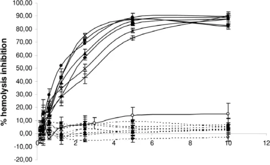
The effect of similar amounts of the seven I. ricinus IxACs
transiently expressed in 293T cells (Figure 3) were assessed in hemolytic assays of both the classical (CP) and alternative complement pathways (AP). A clear dose-dependent inhibition of the AP was observed for all seven recombinant proteins tested as they inhibited the lysis of rabbit erythrocytes by normal human serum. The shape of the curves and plateau values for hemolysis inhibition were identical for the seven proteins (Figure 5).
Addition of recombinant IxACs in the AP assay 15 minutes after adding red blood cells to human serum in the AP assay showed that they were able to inhibit ongoing hemolysis of rabbit erythrocytes. (Figure S1).
The same dilutions of the 7 recombinant proteins were retested in the CP assay in the presence of normal human serum. As shown in Figure 5, there was no inhibition of lysis of antibody-sensitized sheep erythrocytes.
In both assays, no inhibition was observed after addition of recombinant RaHBP2.
We therefore concluded that the 7 IxAC fromI. ricinusall had a similar inhibitory effect on the alternative but not the classical complement activation pathway.
IxACs inhibit the cleavage of human C3 and factor B
During complement activation by the alternative pathway, plasma protein C3 is cleaved into the large opsonizing factor C3b and the small pro-inflammatory peptide C3a and precursor B is cleaved into a large Bb fragment and a small Ba peptide. We investigated the effect ofI. ricinusIxAC proteins on the cleavage of factor B and on the production of C3a in the AP assay. Supernatants of completed AP hemolytic assays were analyzed by Western blot using antisera to fB or C3a.
As shown in Figure 6A, anti-C3a antibody recognized major
bands at 116 kDa, 77kDa and,10 kDa. Arrows indicate clearly
identifiable bands; they corresponded to the a-chain of C3
(115 kDa) and the small C3a peptide (9 kDa) [38,39]. The latter was almost completely suppressed in samples from assays run in the presence of recombinant IxACs as compared to sample
Table 3.Calculated properties of anticomplement proteins.
. . . .
Sequence name Accession number Precursor size (aa) Signal peptide cleavage site MaturecMW (kDa) MaturecpI N-gly. O-gly.
Most likely Less likely
ISACa AAF81253.1 184 21–22 22–23 18.14 4.19 6 12
Isac-1b AAT92205.1 178 22–23 21–22 17.55 3.99 5 15
IRAC I CAD82867 184 19–20 22–23 18.03 4.01 5 8
IRAC II CAD82868 178 21–22 22–23 17.46 4.29 5 12
IxAC-B1 AM407396 177 21–22 19–20, 20–21 17.58 4.00 6 12
IxAC-B2 AM407397 174 19–20 21–22 17.69 4.40 5 8
IxAC-B3 AM407398 178 19–20 21–22 17.85 4.01 5 12
IxAC-B4 AM407399 175 21–22 19–20 17.88 4.14 7 7
IxAC-B5 AM407400 179 21–22 19–20, 20–21 17.66 4.15 7 11
Amino-acid sequences were deduced from the sequenced open reading frames. Indicated values were calculated from deduced amino-acid sequences using online programs at CBS and EBI (see text). Signal peptide cleavage sites are indicated by the position of the residues between which the cleavages were predicted to occur. (a) I. scapularissequence [25]. (b)I. pacificussequence [30]. (c) after removal of the predicted signal peptide at the most likely cleavage position (see text). aa, amino-acids; MW, molecular weight; kDa, kiloDaltons; pI, isoelectric point; N-gly., number of predicted N-glycosylation sites; O-gly., number of predicted O-glycosylation sites. doi:10.1371/journal.pone.0001400.t003
....
....
...
...
....
...
...
....
...
...
....
...
...
....
...
...
....
...
...
.
Figure 3. Western blot analysis of recombinant IxAC-V5His proteins fromI. ricinus.Standardised amounts of recombinant IxAC-V5His proteins from supernatants of transfected 293T cells were analysed by SDS/PAGE and detected by western blotting using an anti-V5 monoclonal antibody. A) Parallel analysis of IxACs, B) N-deglycosylation of IxAC-B1-V5His.1, untreated, 2, incubated with PNGase (New England Biolabs).
from assays run in the presence of RaHBP2 or without added protein.
Antiserum to factor B recognized purified factor B as a single band on a non-denaturating western blot (Figure 6B). A second band was recognized in samples from control AP assays run in the absence of added protein or in the presence of unrelated RaHBP2. It resolved into two distinct bands presumably corresponding to differently charged forms of Bb [39,40]. It was absent in a sample from AP assays run in the presence of recombinant IxACs.
We concluded thatI. ricinus IxAC inhibited the formation of C3a and cleavage of fB. Moreover, there were no detectable
differences in the degree of this inhibition between different members of the two IxAC sub-families.
I. ricinus
IxACs specifically interact with properdin
We then attempted to identify the target(s) of IxACs using ELISA methodology. Components of C3 convertase (i.e. C3, C3b, fB, fD or properdin) were coated on microtiter plates and incubated with recombinant IxAC_V5His. Binding of IxACs was monitored using an anti-V5 antibody. We first tested one member of the IxAC-A subfamily and one from the B subfamily. IRAC II and IxAC-B1 purified from the baculovirus/Sf9 expression system, but not Figure 4. Comparison ofIxodesanticomplement protein tertiary structure.Aligned IxAC amino-acid sequences fromI. ricinuswere submitted to hydrophobic cluster analysis (HCA). Groups of adjacent hydrophobic residues are outlined and shaded. Proline (asterisk), glycine (open rectangle), serine (dotted square) and threonine (open square) are highlighted. The overall distribution of hydrophobic clusters and their size, shape and orientation are very similar.
doi:10.1371/journal.pone.0001400.g004
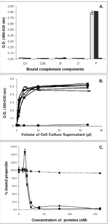
unrelated protein Iris, strongly bound to properdin. They did not bind to C3, C3b, fB or fD (Figure 7A). In addition, standardized
amounts of the sevenI. ricinusIxACs but not RaHBP2 bound to
properdin in a dose-dependent manner (Figure 7B).
We also tested the binding of properdin to C3b-coated plates in the presence of increasing amounts of IRAC II, IxAC-B1 and control Iris. Binding was revealed by a monoclonal antibody to properdin (Figure 7C). Increasing amounts of IRAC II, IxAC-B1 but not Iris lead to a decrease in the amount of bound properdin. We concluded that I. ricinus IxACs specifically interacted with properdin and prevented its binding to C3b. Again, no difference could be discerned amongst IxACs.
IxAC proteins inhibit the formation of the C3
convertase complex by interacting with properdin
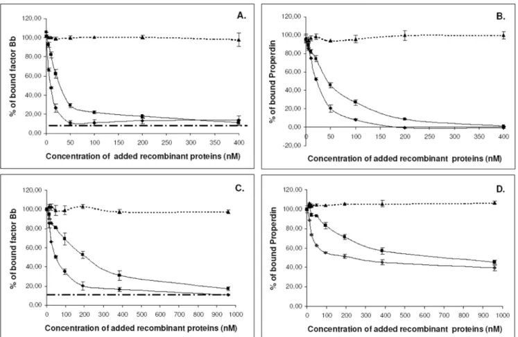
We also studied the effect ofI. ricinusIxACs on the formation and stability of the alternative pathway C3 convertase (C3bBbP). This was reconstitutedin vitroby adding purified components fB, fD and properdin to C3b-coated plates. Bound Bb and properdin were detected using specific antibodies. Approximately 10 times less bound Bb was detected in the absence of properdin than in its presence (Figure 8A). We tested the effect of one member of each of the two IxAC subfamilies on the formation and stability of this complex.
Figure 5. Effect of recombinantI. ricinusIxAC proteins on the alternative and classical pathways of complement activation. Assays of the alternative (AP, solid lines) and classical (CP, dashed lines) complement activation pathway were conducted in the presence of normalized amounts of recombinantI. ricinusIxACs produced in the supernatant of transfected 293T cells. Values for the percent inhibition of rabbit red blood cell lysis in the presence of human serum are indicated. The values are means6standard deviation of triplicates. RaHBP2 was used as negative control. Black diamond :IRAC I; black square:IRAC II; black triangle:IxAC-B1; cross:IxAC-B2; star:IxAC-B3; closed circle:IxAC-B4; Plus:IxAC-B5; open circle:RaHBP2. doi:10.1371/journal.pone.0001400.g005
Table 4.Evidence for positive selection inI. ricinusIxAC coding sequences.
. . . .
Irac I Irac II Ancestral IxAC-B1 IxAC-B2 IxAC-B3 IxAC-B4 IxAC-B5
Irac I 5.92 2.06 2.30 2.18 2.30 2.41 2.08
Irac II 0.028 1.77 2.38 2.03 2.30 2.38 2.05
Ancestral 1.000 1.000 2.82 1.92 2.54 2.46 2.07
IxAC-B1 1.000 1.000 1.000 4.13 2.63 3.39 3.28
IxAC-B2 1.000 1.000 1.000 0.223 3.63 3.28 3.17
IxAC-B3 1.000 1.000 1.000 1.000 0.435 3.25 3.57
IxAC-B4 1.000 1.000 1.000 1.000 1.000 1.000 2.51
IxAC-B5 1.000 1.000 1.000 0.491 1.000 0.441 1.000
Pairwise dN and dS values were calculated using the Nei-Gojobori method as implemented in the Mega3 package. dN/dS ratios are indicated in the upper right triangle. Fisher’s test P values are indicated in the lower left triangle. A putative ancestral coding sequence forI. ricinusanticomplement proteins was inferred from an alignment of the IxAC fromI. ricinuscoding sequences using the Ancescon package.
doi:10.1371/journal.pone.0001400.t004
....
....
...
...
....
...
...
....
...
...
....
...
...
....
...
In a first series of experiments, increasing amounts of purified IRAC II and IxAC-B1 were added together with the individual convertase components to C3b-coated plates. We observed a dose-dependent decrease in the amount of bound Bb (Figure 8A) or properdin (Figure 8B), indicating inhibition of complex formation. At the highest protein concentrations ($200 mM), the amount of bound Bb dropped to values observed when reconstituting the C3 convertase without properdin (Figure 8A). No such effect was observed with the unrelated Iris protein (Figure 8).
In a second series of experiments, C3bBbP was pre-formed on ELISA plates and then incubated with increasing amounts of IRAC II and IxAC-B1 proteins and the unrelated control Iris protein (Figure 8C and D). The results indicated that IxAC
proteins induced the displacement of all pre-bound factor Bb (Figure 8C) and about 50 % of pre-bound properdin.
We also performed time-course experiments of C3 convertase formation with (C3bBbP) or without properdin (C3bBb) in the presence of 200 mM IRAC II, IxAC-B1 or Iris. The amount of bound Bb was much lower in the absence of properdin than in its presence. In this case, the presence of IxACs or Iris had no effect, indicating that the proteins had no direct effect on the interaction between C3b and Bb. On the contrary, the formation of C3 convertase in the presence of properdin was strongly affected by IxACs. Values of bound Bb dropped to values observed without properdin (Figure S2).
Overall, these results show thatI. ricinusIxAC proteins inhibit the formation of the C3 convertase complex by interacting specifically Figure 6. Inhibition of C3a formation and factor B cleavage.Aliquots of supernatant from AP hemolysis assays conducted in the presence of standardized amounts of IxACs fromI. ricinusand unrelated control RaHBP2 were analyzed by western blotting. Panel A: Blots from gels run under denaturing conditions were probed with monospecific anti-C3a serum. Thea-chain of precursor C3 (116 kDa) and the C3a peptide (,10 kDa) are indicated by arrows. Panel B: Blots from gels run under non-denaturing conditions were probed with a antiserum to factor B. Purified factor B was used as a positive control.
doi:10.1371/journal.pone.0001400.g006
with properdin. They also induce the displacement of pre-bound properdin, and indirectly, Bb, in a dose-dependent manner.
IxAC proteins inhibit complement activation on
agarose-coated ELISA plates
We also tested the ability of IxACs to inhibit activation of the AP on agarose-coated ELISA plates using human serum as a source of complement factors. This experimental set-up is closer to physiological activation of complement than protein-protein interactions conducted on plastic surfaces. Recombinant IRAC II and IxAC-B1 were added in the assay at various time-points after addition of human serum (Figure 9).
The results indicated that IxAC proteins prevented C3b and factor B deposition in agarose-coated wells when added together with serum (not shown). They also stopped further C3 and factor B deposition when they were added at various times (30, 45 and 60 min) after initiation of the reaction as shown by the immediate plateauing of the curve. Nevertheless, they did not displace bound C3 or Bb as the measured amounts of bound factors did not drop (Figure 9). The results also showed that members of the two IxAC subfamilies are able to inhibit the formation of C3 convertase (Figure 9A–B and Figure 9C–D) in a similar manner. Although they were able to stop the ongoing formation of C3 convertase they could not undo previously formed complexes.
An additional experiment was also performed. After 60 min. of reaction, the reaction medium was replaced with fresh buffer containing the recombinant proteins but no human serum. A drop in the amount of bound Bb was observed in the presence of IRAC II or IxAC-B1 but not Iris (not shown).
These results therefore confirmed that IxAC proteins are able to inhibit the activation of the AP on a surface by preventing deposition of C3b and Bb.
Class specificity is observed within Vertebrates
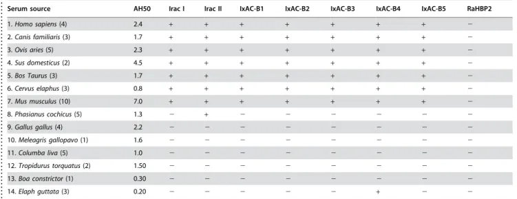
We next tested the hypothesis that diversification of anticomple-ment proteins helpsI. ricinuscounteract the complement activity of its diverse host organisms. Freshly prepared sera from different vertebrate species were first titrated in the AP assay in order to define the volume causing 50% hemolysis (AH50). A wide range of
AH50 values were observed, from the equivalent of 0.25ml per
microwell test (50ml final volume) forBoa constrictorto 7.0ml per test for Balb/c mice (Table 5). Heat-inactivated samples completely lost their hemolytic activity, confirming that this activity was indeed due to complement (not shown).
Identical amounts of normalized IxAC proteins fromI. ricinusand control RaHBP2 were then added to the AP hemolysis assay in the presence of AH50 volumes of serum. The 7 anticomplement proteins reproducibly inhibited all mammalian sera in a dose-dependent manner (Table 5 and Figure S3). In some species, such as humans (Figure 3) orB. TaurusandM. musculus(Figure S3) the dose-response curves were similar for the seven IxACs fromI. ricinus. In other mammals such as,C. familiaris, O. aries, S. domesticusand C.
elaphus (Figure S3) lower doses of the proteins had different
efficiencies. They all reached similar plateau values at higher doses of protein, though. We also observed that most species were not equally sensitive to IxAC inhibition of the AP. Thus hemolysis of rabbit red blood cells by mouse serum was inhibited by not more
than,30% whereas the hemolytic activity of human serum was
inhibited at,85 %. Intermediate plateau values were observed for
the other species tested (Figure S3). On the contrary, IxACs fromI.
ricinusdid not affect most bird and squamate reptile sera, with the
exception of IRAC II and IxAC-B4, which inhibited AP activity of one bird (Phasianus colchicus) and one snake (Elaph guttata), respectively. Figure 7. ELISA analysis of the binding of IxAC proteins to
immobilized C3 convertase components.Panel A: Binding of IxACs to AP components. Purified recombinant IRAC II, IxAC-B1 or unrelated protein Iris were added to microtiter wells previously coated with purified factors C3, C3b, fB, fD or properdin (P). Bound proteins were detected with an anti-V5 monoclonal antibody using an ELISA format. Light dotted histogram: IRAC II; dark dotted histogram: IxAC-B1; black histogram: Iris. Panel B: Increasing amounts of normalized supernatant from transfected culture 293T cells were added to immobilized properdin. Bound IxACs were detected with an anti-V5 antibody. Black diamond: Iris; black square: IRAC I; black triangle: IRAC II; cross: IxAC-B1; star: IxAC-B2; closed circle: IxAC-B3; plus: IxAC-B4; minus : IxAC-B5. Panel C. Competition between properdin and IxACs for C3b binding. Purified properdin and increasing amounts of IRAC II, IxAC-B1 or unrelated control IRIS were added simultaneously to C3b-precoated microtiter wells. Bound properdin was detected with an anti-properdin monoclo-nal antibody. Black diamond: IRAC II; black square: IxAC-B1; black triangle: Iris.
To summarize, all 7 IxAC fromI. ricinusinhibited the AP in all mammal species tested. Inhibition of the AP in only one bird by only IRAC II and only one squamate species by only IxAC-B4 was also observed.
Expression patterns of individual
I. ricinus
IxACs
Primer pairs designed to specifically detect each IxAC messenger (Table S3) were used to analyse their pattern of expression by RT/ PCR in individual ticks, at various stages of their life cycle and during the bloodmeal.
We first investigated IxAC expression during the life cycle. All 7
IxAC sequences were detected in polyA+ RNA from the original
pool of 70 salivary glands from adult females. No PCR product was
observed from poly A+ RNA that had not undergone reverse
transcription, indicating that we did not amplify fragments of genomic DNA (Figure 10a). Younger stages of the life cycle (larvae and nymphs) expressed IRAC I, IxAC-B1, B3 and B5 (Figure 10b). We also investigated whether they expressed additional anticomple-ment proteins not found in adults by searching for the full coding sequences in 10 pooled nymph and 10 pooled larvae cDNA in high-fidelity conditions using primer pairs designed from the 59 and 39
ends of IxAC-A or IxAC-B coding sequences (Table 1). Primers amplified the expected,600 bp products which were then inserted
into pCRII and sequenced. Only IRAC I was retrieved from
nymphs (7 independent clones), and IxAC-B1 and IxAC-B5 from larvae (2 and 1 clones respectively). The ‘‘nymphal’’ or ‘‘larvae’’ nucleotide sequences were 100% identical to ‘‘adult’’ ones.
We then investigated the expression of the IxAC repertoire during the bloodmeal. Messengers for IRAC I, IRAC II, IxAC-B1, IxAC-B3 and IxAC-B5 were detected in unfed females and the complete repertoire was detected in salivary glands from females on day 3 of the bloodmeal and on day 5 (Figure 10b).
When analysing the pattern of IxAC expression in 10 individual adult females we found that no single specimen expressed the whole range of anticomplement IxAC proteins. Each individual tick expressed one (1 tick), two (6 ticks) or three proteins (3 ticks). IxAC-B1 was expressed in 8/10 ticks tested, followed by IxAC-B3 and B4 (3/10), then IRAC I, IRAC II, IxAC-B2 and B5 (2/10). Four individual ticks expressed members of both IxAC-A and IxAC-B and 6 individual ticks expressed only members of IxAC-B. None expressed solely members of IxAC-A.
Taken together, the results indicated that IxAC are expressed in all ticks tested, throughout the bloodmeal and at all the development stages examined. There was no evidence for stage-specific variants although some members of the family may be induced or up regulated during the first days of the bloodmeal and in the adult. Individual females expressed individual members of the family and no individual tick expressed the complete series ofI. ricinusIxACs. Figure 8. Effect of IxAC proteins on the formation and stability of C3 convertase.Panel A and B: The effect of IxAC proteins on the formation of C3 convertase was evaluated by incubating simultaneously purified factors B, D and properdin with increasing amounts of IRAC II or IxAC-B1 on C3b-coated wells. Panel C and D: The effect of IxAC proteins on the stability of C3 convertase was assessed by incubating preformed C3 convertase (fB, fD and properdin pre-incubated for 1 hour) on C3b-coated plates with increasing amounts of recombinant IxACs. Bound factor B or properdin was detected with an anti-factor B antibody (A–C) or anti-properdin antibody (B–D), respectively. Recombinant IRIS was used as negative control. Black diamond: IRAC II; black square: IxAC-B1; black triangle: IRIS.
doi:10.1371/journal.pone.0001400.g008
Figure 9. Effect of IxAC proteins on the deposition of C3b and factor B on agarose-coated plates.Loading human serum on agarose-coated microplate wells activates the alternative complement pathway. Purified recombinant IRAC II (A–B) or IxAC-B1 (C–D) were added after 30, 45 or 60 minutes. The reactions were stopped at various times. C3b and factor B deposition was detected using anti-C3 antibody (A–C) or anti-factor B antibody (B–D), respectively. Black diamond: no added protein; black square: 30 min.; black triangle: 45 min.; cross: 60 min.
doi:10.1371/journal.pone.0001400.g009
Table 5.Host specificity of anticomplement activity by recombinant IxACs fromI. ricinus.
. . . .
Serum source AH50 Irac I Irac II IxAC-B1 IxAC-B2 IxAC-B3 IxAC-B4 IxAC-B5 RaHBP2
1.Homo sapiens(4) 2.4 + + + + + + + 2
2.Canis familiaris(3) 1.7 + + + + + + + 2
3.Ovis aries(5) 2.3 + + + + + + + 2
4.Sus domesticus(2) 4.5 + + + + + + + 2
5.Bos Taurus(3) 1.7 + + + + + + + 2
6.Cervus elaphus(3) 0.8 + + + + + + + 2
7.Mus musculus(10) 7.0 + + + + + + + 2
8.Phasianus cochicus(5) 1.3 2 + 2 2 2 2 2 2
9.Gallus gallus(4) 2.2 2 2 2 2 2 2 2 2
10.Meleagris gallopavo(1) 1.6 2 2 2 2 2 2 2 2
11.Columba liva(5) 1.0 2 2 2 2 2 2 2 2
12.Tropidurus torquatus(2) 1.50 2 2 2 2 2 2 2 2
13.Boa constrictor(1) 0.30 2 2 2 2 2 2 2 2
14.Elaph guttata(3) 0.20 2 2 2 2 2 + 2 2
We assessed the ability of individual recombinant IxACs proteins to inhibit the alternative pathway (AP) of complement in sera from various vertebrate species as indicated. AP activation was assessed by hemolysis of added rabbit erythrocytes. (+) and (2) indicate the existence or absence of a dose-response relationship between added recombinant proteins and percent inhibition of hemolysis. AH50 value, serum amount (ml) that causes 50 % hemolysis of rabbit erythrocytes in 50ml of the AP assay. Host N-u1–7: mammals; 8–11: birds; 12–14: Squamates. Values between brackets indicates the nuindividuals from which the serum pools were constituted. doi:10.1371/journal.pone.0001400.t005
....
...
....
...
...
....
...
...
....
...
...
....
...
...
....
...
...
....
...
...
....
...
...
Antigenic diversification in the IxAC family in
I.
ricinus
A monospecific mouse antiserum to IxAC-B1 was produced in mice by DNA immunization followed by a booster with purified recombinant IxAC-B1. It was used to perform western blot analysis of standardized amounts of IRAC I to IxAC-B5 (see Figure 3a) in parallel with the anti-V5 commercial antibody. As shown in Figure 11A, mouse anti-IxAC-B1 serum only recognized IxAC-B1 and none of the other IxACs fromI. ricinus. This shows that epitopes recognized on IxAC-B1 in this assay were not present on any other member of the family.
The neutralizing potential of these antibodies was also assessed. Standardized amounts of 7 recombinant IxACs fromI. ricinuswere pre-incubated with heat-inactivated anti-IxAC-B1 serum before assessing their ability to interfere with complement activity. Neutralization of AP inhibition activity as indicated by recovery of RBC lysis was observed only against IxAC-B1 (Figure 11B). Seroneutralization of AP inhibition by IxAC-B1 was not observed with pre-immune sera or with antisera directed to the unrelated protein Iris (not shown).
These data show that an antiserum raised against one member of the IxAC family is able to recognize and functionally inhibit this member alone and no other member of the family. It suggests that immunodominant epitopes of a member of the family are not shared by other members.
DISCUSSION
The interaction between hard ticks and their hosts is a good example of an ongoing ‘‘arms race’’ between a parasite and its host. As the tick feeds on its vertebrate host for periods of several days it must counteract all the host’s defense mechanisms including hemostasis, inflammation and the immune response. This is accomplished by injecting batteries of active proteins in the saliva. Most are coded by multigene families only a few of which have been fully characterized. In this work we have attempted to provide an exhaustive analysis of one such family, the family of salivary inhibitors of the alternative complement pathway inIxodes
ricinus. Our analysis includes: i) a complete or nearly complete
inventory of members of the family, ii) a detailed study of its action mechanism: these proteins bind to properdin, a positive activator of complement, thereby preventing early rejection of the tick by the innate response, and iii) investigation of the possible selective pressure causing diversification: sequence diversification comes with antigenic diversity and helps circumvent the host’s adaptive immune response.
A large family of complement inhibitors in the hard
tick
I. ricinus
We describe five new anticomplement sequences (AM407396 to AM 407400) from the hard tickIxodes ricinus. These share less than Figure 10. Expression patterns of individual IxACs.PolyA+RNA was extracted from variousI. ricinusmaterial as indicated and reverse transcribed. The resulting cDNAs were submitted to PCR analysis using pairs of primers specific for the indicated IxACs. Non- reverse transcribed polyA+RNA from the pool was included as negative control (Ctrl). PCR products were run on 1.2 % agarose gels. M, DNA size markers. B1 to B5, IxAC-B1 to IxAC-B5. Sizes in bp are indicated. Panel A: Analysis of salivary glands of a tick population (70 specimens, pool) and from individual female ticks at day 5 of the bloodmeal (A to J). Panel B: Analysis of pooled salivary glands of tick female populations at day 0 (25 specimens), day 3 (25 specimens) and day 5 (70 specimens) of the bloodmeal as well as from pooled gorged nymphs (25 specimens) and larvae (25 specimens).
doi:10.1371/journal.pone.0001400.g010
40 % amino-acid identity with previously described ISAC of I.
scapularis[25] and with IRAC I and IRAC II fromI. ricinus[31].
The new sequences clustered in a distinct group of anticomple-ment sequences as indicated by phylogenetic analysis (Figure 1).
We also proposed the acronym ‘‘IxAC’’ for Ixodes
Anti-Complement to the larger family of Ixodes anticomplement
proteins related to prototypical ISAC.
Our analysis provides a reasonably exhaustive and accurate inventory for the tick population under study. This is supported by several lines of evidence: i) 12 RT/PCR inventories performed independently on two different cDNA using 6 different primers pairs consistently yielded the same set of sequences; ii) Sequences identical to previously described IRAC I and IRAC II were recovered; iii) IxAC-B1 and IxAC-B2 were first discovered by serendipidity and then repeatedly found again by RT/PCR analysis of salivary gland cDNA. The existence of additional variants (either alleles or paralogs) in the larger I. ricinusspecies may be investigated on specimens captured in the field or on specimens from other laboratory colonies.
We also noticed consistent differences in the frequency of the 7
I. ricinusIxACs in RT/PCR inventories (Table S1). This may be
due to the differential expression of IxAC genes in individual ticks (Figure 10). Alternatively, different steady-state levels of messen-gers may also be responsible.
The following arguments are consistent with the theory that the
7I. ricinusIxAC genes are paralogs rather than alleles: i) The 7I.
ricinusIxAC proteins are not very closely related (65% amino-acid
identity at most), ii) They could not be grouped together into a single phylogenetic cluster, iii) They were cloned from a restricted tick population, iv) Some individual female ticks expressed up to 3 IxACs simultaneously, indicating the existence of at least two loci. Daix et al. [31] developed specific monoclonal antibodies that can differentiate IRAC I and IRAC II by immunofluorescence analysis on dissected salivary glands. They observed co-expression of the two proteins in the salivary glands of 12 adult specimens collected in various woodland locations throughout Belgium. The authors concluded that IRAC I and IRAC II are not alleles but rather co-expressed members of a multigene family. However, in Figure 11. Antigenic specificity of recombinant IxACs fromI. ricinus.Standardised amounts of the sevenI. ricinusrecombinant IxACs were analysed for antigenic specificity. Panel A. Western blot analysis. The serum from a mouse immunized against IxAC-B1 by genetic immunization followed by a protein boost recognised solely recombinant IxAC B1. M, molecular weight markers (Mark12, Invitrogen). Panel B. Seroneutralization experiments. AP hemolysis assays were conducted with and without the seven recombinant IxACs. 100% hemolysis was obtained in the absence of anticomplement protein (light dotted histogram). Recombinant IxACs alone (dark dotted histogram) or recombinant IxACs plus heat-inactivated sera from mice immunized against IxAC-B1 (anti IxAC-B1, black histogram) or mock immunized mice (anti-PBS, white histogram) were added as indicated. Neutralization of activity as indicated by a recovery of hemolysis, was observed only on IxAC-B1. Upper panels: seroneutralization of IxAC-A subfamily. Lower panel: seroneutralization of IxAC-B subfamily. Error bars represent standard deviations.
our RT/PCR analysis, IRAC I and IRAC II mRNA were detected together only twice in 10 individual specimens from the Neuchaˆtel breeding colony. This discrepancy may be due to cross detection of other members of the IxAC families by monoclonal antibodies directed against IRAC I and IRAC II.
Phylogenetic analysis
Phylogenetic analysis of the nucleotide and amino-acid sequences by distance and maximum likelihood methods (Figure 1) indicated
that all known Ixodes anticomplement proteins (IxACs) could
robustly be grouped into two large groups or subfamilies: IxAC-A and IxAC-B. IxAC-B only contains our five new sequences fromI.
ricinuswhereas IxAC-A contains all the other sequences. IxAC-A
can be further subdivided into ‘‘ISAC-like’’ and ‘‘Salp9-like’’ clusters after the name of the founder sequences. Regardless of the method used, placement of IRAC I within the ISAC-like or Salp9-like cluster was not supported by bootstrap analysis. It therefore probably represents a third cluster within the IxAC-A family.
The presence of IxAC sequences inI. ricinus,I. scapularisandI.
pacificusindicated that their last common ancestor and possibly the
ancestralIxodestick possessed an IxAC-like sequence. The lack ofI.
scapularisandI. pacificusrepresentatives in IxAC-B is best explained
by methodological differences when looking for IxAC sequences in these two species. Indeed, we performed a dedicated family-specific inventory inI. ricinuswhereas theI. scapularisand one I.
pacificusentries are the result of large-scale untargeted sequencing
of large numbers of clones taken at random or PCR screening using unique primers designed from ISAC and Salp20. Dedicated RT/PCR inventories of IxAC sequences inI. scapularisand/orI.
pacificuswould help resolve this question by showing whether or
not the two species express members of IxAC-B.
IxAC homologs were not detected by RT/PCR analysis ofR.
appendiculatussalivary gland cDNA or byin silico interrogations of
collections of available non-Ixodes (Metastriata) sequences. IxAC homologs, if any, inR. appendiculatusmight be too divergent to be amplified by our PCR primers. Besides, the present coverage of Metastriata genomes in public databases may be very limited. On the other hand, our finding is consistent with the absence of reported inhibition of the alternative complement pathway by Metastriata.
Positive (diversifying) selection of coding sequences
Since Ohno [41], gene duplication has been considered to be an important factor in evolution as it leads to the evolution of new gene functions. In higher organisms most genes belong to families of related genes formed by repeated gene duplication events during evolution [42].Considerable research is currently being carried out in order to understand the forces leading to and shaping multigene families in living organisms [43].Ticks are particularly suited for this analysis as their salivary proteins are coded by multigene families, a feature probably related to their bloodfeeding lifestyle.
As far as salivary proteins in general are concerned, the process of gene duplication accompanied by positive selection leading to the acquisition of novel protein functions is documented in snake venom proteins [44,45] and in soft tick salivary proteins [46]. In the bloodfeeding dipteraLutzomyia longipalpis,variants of the maxadilan protein retain the same function and biochemical properties [47] but have undergone diversifying selection (Lanzaro, personal commu-nication). This is associated with antigenic diversity leading to escape from the host’s antibody response [48,49].
We compared the coding sequences of the seven I. ricinus
anticomplement sequences with one another as well as with a putative reconstructed ancestral sequence. We observed that percent identities between IxAC amino-acid sequences were
consistently lower than percent identity at the nucleotide level. In addition, dN/dS ratios were consistently higher than 1. We concluded that IxAC coding sequences had been subjected to strong positive selection within theI. ricinusspecies. In other words, diversification of the IxAC amino-acid sequences was strongly selected for and this was not associated with speciation. This confirms the conclusion of a restricted analysis conducted on only two members of the family: Daix et al. [31] observed an excess of non-synonymous changes when they compared IRAC I, IRAC II and anticomplement sequences fromI. scapularisandI. pacificus(i.e. members of the IxAC-A subfamily).
We examined the type of the selection pressure operating on the IxAC family inI. ricinusby specifically investigating the following possibilities i) structural and biochemical diversity, ii) mechanism and functional diversity, iii) adaptation to host species, iv) stage specificity, v) antigenic diversity.
Structural and biochemical diversity
We first assessed the differences in the biochemical properties of the members of the IxAC family. Predicted and experimental biochemical properties of the seven proteins were analysed and compared to those of ISAC fromI. scapularisand ISAC-I fromI.
pacificus. We observed very similar values for calculated pI and
observed and calculated molecular weights. All recombinant proteins were exported in the supernatant as predicted by specialized algorithms. They were highly glycosylated and disulphide bonds were predicted in all of them (this article and ref. [25,29]). Finally, comparison of the distribution of hydropho-bic clusters in the amino-acid sequences predicted identical or very similar folds or tertiary structure in all IxACs.
Mechanism and function diversity
The kinetics of inhibition of the alternative complement pathway in human serum were similar for all 7 recombinant IxACs fromI.
ricinus. The classical pathway was not affected. This is also a
feature of theI. scapularisanti-complement proteins ISAC [25] and Salp20 [29] and it has been reported recently forI. ricinusIRAC I and IRAC II by Daix et al. [31]. We then investigated the action
mechanism of the 7 I. ricinus IxACs. Protein binding and
competition experiments suggested that all 7 proteins had the same action mechanism. This is consistent with the similar properties of members of the family and also with the identical or very similar predicted folds or tertiary structures. We conclude that IxAC sequence diversification is not primarily driven by selection for different biochemical properties and tertiary structure, or a change in the roles of the proteins. Minute differences are possible but they remain to be investigated.
Our study of this action mechanism showed that IxAC molecules bind specifically to properdin, preventing its association with C3 and thereby reducing formation of C3 convertase complex to levels observed in the absence of properdin. They also induce destabilization of pre-formed C3bBbP convertase. They consequently inhibit the activation of complement via the alternative pathway. Properdin (factor P) is known to increase ten times the half-life of C3bBb convertase although functional convertase activity may be obtained in its absence [12,50,51]. The effect of IxAC proteins may therefore be explained by a decreased stability of C3 convertase due to blocking of properdin. This is reminiscent of the effect of monoclonal antibodies that bind to properdin and knock out of the properdin gene in mice, and both lead to inhibition of the AP [19].To our knowledge this is the first time that direct interaction with properdin is described as a mechanism of complement regulation.
Two recent reports further emphasize the importance of properdin in the AP: Spitzer et al. [52] and Kimura et al. [53] showed that properdin can also bind directly to microbes, initiating assembly of C3BbP convertase and complement activation. Whether properdin is a positive regulator or an initiator of the AP, it is a central element and a critical molecular target for inhibitors. Nevertheless, it remains to be seen whether IxACs can also interfere with the binding of properdin to the target surface.
Ixodid ticks are pool feeders, they dilacerate small blood vessels at the bite site, generating a small haemorrhage or pool of blood in which saliva is injected and from which they pump blood [3]. Local inhibition of complement activation in the pool of blood is therefore beneficial for at least two reasons. Firstly, inhibition of the production of pro-inflammatory peptides will help prevent the inflammation response at the bite site; secondly, inhibition of MAC insertion on the mouthparts and midgut epithelium will prevent the destruction of tick tissue by complement factors present in the blood meal.
In order to obtain that effect, ticks must secrete sufficient amounts of the inhibitor. A very rough calculation of the amount required can be made. This must take into account properdin concentration (,5mg/ml in normal human plasma) [54,12], the
amount of ingested blood (little information is available about this volume which is probably several hundred microliters [3]), the amount of secreted saliva (unknown but limited by the amount of ingested blood as blood water is recycled into saliva) and the concentration of IxAC in the saliva. If there is complete recycling of blood water into saliva and an equimolar properdin-IxAC interaction, the IxAC concentration required may be estimated to be approximatively 1 ng/ml. However, the anti-complement effect needs only be local (i.e. in the immediate proximity of the mouth parts and midgut epithelium), and this drastically reduces the amount of complement to be neutralized. Moreover, the IxAC might not mediate the only anti-complement mechanism inIxodes
saliva. Inhibition of the IxAC expression in ticks by RNAi may help reveal additional anticomplement molecules.
Finally our results suggest that one molecule of IxAC may interact with several molecules of properdin. Thus, in the experiment shown in figure 7c, complete inhibition of binding of 200 ng of properdin to C3b was observed with around 25 nm inhibitor, corresponding to an IxAC/properdin molecular ratio of 1/3 to J. This is consistent with the finding that properdin is present in the serum as dimer, trimer and tetramer [55] forms. One IxAC molecule may therefore interact with one properdin polymer. This hypothesis is open to experimentation and is currently being tested in our laboratory.
The mechanism by which salivary gland extract (SGE) fromI.
ricinus inhibits the AP has been explored by Lawrie et al. [39].
They observed inhibition of the cleavage of fB to Bb and C3a production when rabbit erythrocytes were used as activators and complete human serum as a source of complement. Little or no C3b was observed on erythrocytes in the presence of SGE. Reconstitution of the alternative C3 convertase (C3bBb) in vitro
from purified C3b, fB and fD (but not properdin) was not affected by SGE. Moreover, SGE had no effect on the cleavage of125I-C3
into 125I-C3b by preformed C3bBb or when it was added
simultaneously with C3b, factor B and factor D. These findings are similar to our results and may therefore be fully explained by the action of IxAC proteins in the saliva.
Lawrie et al. [39] also noticed that SGE provoked the cleavage
of a ,5 kDa peptide at the C-terminal end of purified C3 a
chains. The product can apparently still be cleaved by preformed convertase to yield C3a. The authors also suggested that the larger fragment may not be able to participate in convertase formation.
However, we were unable to reproduce this finding (data not shown). As saliva is a complex mixture of proteins, proteolytic cleavage of C3, as well as other possible additional mechanisms, may also be involved in inhibition of the AP.
Saliva, recombinant ISAC [25] and Salp20 [29] from I.
scapularis as well as IRAC I and IRAC II from I. ricinus [31],
have been previously tested for their ability to inhibit AP activation in experiments using human serum as source of complement and agarose as activating surface. The authors observed inhibition of the deposition of both C3b and Bb and release of pre-bound Bb but not pre-bound C3b from the plates. Release of C3b is not observed because this protein is covalently linked to agarose. Finally, recombinant Salp20 also inhibits the production of C3a and deposition of C3b on the surface of red blood cells in the AP hemolytic assay [29]. We conclude that the novel inhibition mechanism described here is consistent with published data concerningIxodestick saliva, salivary gland extracts, or recombi-nant anticomplement proteins.
Comparison of IxACs with other anti-complement
inhibitors
Complement is a critical component of innate and adaptive immunity mainly acting through specific cell lysis, the release of potent pro-inflammatory peptides and opsonization of target cells. To prevent tissue damage by over-activation, complement activation is subject to tight regulation by many physiological negative regulators. These include surface and soluble proteins such as Decay-Accelerating Factor (DAF/CD55), Complement Receptor-1 (CR1/CD35), and factor H. They compete with factor B for binding to C3b and facilitate dissociation of the C3bBb complex. Together with the membrane protein MCP/CD46, they also act as cofactors for factor I (fI), a serine protease leading to C3b inactivation by proteolysis. Finally, other membrane proteins such as CR2/CD21, protectin or CD59 protect the host cell membrane from inappro-priate complement activation and cell destruction [9].
Many pathogens also target components of the complement system, sometimes taking advantage of existing physiological regulation mechanisms. Some pathogenic bacteria such asBorrelia,
Neisseria,StreptococcusandYersiniaexpress receptors that bind
host-derived soluble complement regulatory proteins, in particular factor H, FHL-1, and C4b binding protein [56]. Certain pathogenic viruses express proteins homologous to vertebrate complement regulators such as vaccinia complement control protein or apparently unrelated functional analogs [57]. Protein NS1 of West Nile Virus recruits soluble fH [58]. Surface protein gC of Herpes Simplex Virus binds to C3b and inhibits its association with C5 and properdin [59].
Parasitic protozoa such asLeishmaniaspp. andTrypanosoma cruzi
are also able to counteract complement activation. In the former, the major surface protein GP63 is a protease that can cleave C3b to inactive iC3b. The latter expresses a 160 kDa homolog of DAF which binds to C3b and C4b and prevents formation of the convertase [60].