einstein. 2015;13(2):326-9
ThemaTic review: TransplanTaTion
review
Liver-specific magnetic resonance contrast medium in the
evaluation of chronic liver disease
Aplicações do contraste hepato-específico de ressonância magnética
nas hepatopatias crônicas
Marcio Augusto Correia Rodrigues dos Reis
1, Ronaldo Hueb Baroni
1aBsTracT
The hepatobiliary-specific contrast medium (gadoxetic acid –
Primovist
®) is primarily used to improve detection and characterization
of focal hepatic lesions, such as in chronic liver disease patients with
suspected hepatocellular carcinoma. Since the contrast medium is
selectively taken up by functioning hepatocytes in the late hepatobiliary
phase, it helps to detect typical hepatocellular carcinoma, which show
low signal intensity on this phase. This imaging feature also assists in
differentiating regenerative/dysplastic nodules from early hepatocellular
carcinomas (with over 90% accuracy), as well as hypervascular
hepatocellular carcinomas from arterial pseudo-enhancement foci.
Future perspectives include its use in quantification of hepatic function
and fibrosis.
Keywords:
Liver neoplasms/diagnosis; Liver diseases/diagnosis; Carcinoma,
hepatocellular/diagnosis; Contrast media/utilization; Magnetic resonance
imaging/methods
resUmo
O contraste hepato-específico (ácido gadoxético – Primovist
®) tem
como utilidade principal melhorar a detecção e a caracterização
de lesões hepáticas focais, por exemplo, em hepatopatas crônicos
com suspeita de hepatocarcinoma. Por apresentar captação seletiva
por hepatócitos funcionantes na fase hepatobiliar tardia, auxilia
na detecção de hepatocarcinomas típicos – a maioria dos quais
apresentando hipossinal nessa fase. Essa característica de imagem
também auxilia na diferenciação entre nódulos regenerativos/
displásicos e hepatocarcinomas precoces (com mais de 90% de
acurácia), e entre hepatocarcinomas hipervascularizados e focos de
pseudorrealce arterial. Perspectivas futuras promissoras incluem sua
utilização na quantificação de função e de fibrose hepáticas.
Descritores:
Neoplasias hepáticas/diagnóstico; Hepatopatias/diagnóstico;
Carcinoma hepatocelular/diagnóstico; Meios de contraste/utilização;
Imagem por ressonância magnética/métodos
inTroDUcTion
Magnetic resonance imaging (MRI) is a well-established
test to assess focal liver lesions. However, up to 60% of
malignant nodules may not be detected or characterized
by MRI, mainly those smaller than 1.0cm and in cirrhotic
livers.
(1,2)Liver-specific contrast media were developed to
increase sensitivity and specificity of MRI in assessing
focal lesions, as well as to overcome some of the limitations
observed with extracellular contrast media. Among
the liver-specific contrast media currently available,
only gadoxetic acid (Gd-EOB-dTPA, Primovist
®, Bayer
Schering, Berlin, Germany) is approved for clinical use
in Brazil.
phYsical chemical properTies anD BioavailaBiliTY
Gd-EOB-dTPA is a liver-specific, gadolinium-based
paramagnetic contrast, with combined properties of
hepatocyte perfusion and selectivity. It was primarily
developed to increase detection and characterization of
focal hepatic lesions. After intravenous administration,
Gd-EOB-dTPA is quickly distributed in the vascular/
interstitial compartment, allowing for a dynamic, multiphase
study (arterial, portal and equilibrium phases).
Approximately 50% of the injected dose of
Gd-EOB-dTPA is selectively captured by functioning hepatocytes
and later excreted through bile, enabling acquisition of
a late hepatobiliary phase, approximately 10-20 minutes
after its injection. In this stage, hepatocyte-free lesions
(or lesions with dysfunctional hepatocytes) show low
MRI signal intensity (dark images on a bright liver), with
1 Hospital Israelita Albert Einstein, São Paulo, SP, Brazil.
Corresponding author: Marcio Augusto Correia Rodrigues dos Reis – Department of Imaging, Hospital Israelita Albert Einstein – Avenida Albert Einstein, 627/701, 4th floor, building B – Morumbi Zip code: 05651-900 – São Paulo, SP, Brazil – Phone: (55 11) 2151-1233 – E-mail: [email protected]
327 Liver-specific magnetic resonance contrast medium in the evaluation of chronic liver disease
einstein. 2015;13(2):326-9
liver/lesion enhancement, improving the diagnostic
capability of the test.
Due to its hepatocyte specificity, the recommended
dose of gadolinium is up to four times lower than that
recommended for extracellular contrast media.
(3-5)The high contrast uptake is due to the lipophilic
properties of Gd-EOB-dTPA, favoring its passive
diffusion by molecular transporters OATP1, which are
in the basolateral membrane of normal hepatocytes.
(6,7)After uptake by hepatocytes, Gd-EOB-dTPA is eliminated
by biliary (50%) and urinary tracts (50%). At molecular
level, biliary excretion is dependent upon the molecular
transporter MPR2 that is present on the cell canalicular
membrane.
(6,7)clinical Use in chronic liver Disease
Hepatocellular carcinomas (HCC) can be diagnosed
in a non-invasive manner, by means of imaging tests
in patients at increased risk. These lesions typically
present intense arterial enhancement, with washout in
venous and equilibrium phases both in computerized
tomography (CT) and MRI.
(8,9)The presence of fat or pseudocapsule (peripheral
enhancement mimicking a real capsule) in late phases
increases reliability of diagnosis. Complementary findings
in MRI include high-signal intensity on T2 and restricted
diffusion of water molecules.
Different patterns for HCC were described after
injection of Gd-EOB-dTPA, depending on expression of
the molecular transporter OATP1. Since most HCC do
not present with functioning hepatocytes, approximately
80 to 90% show low signal intensity in hepatobiliary phase
in relation to the surrounding hepatic parenchyma
(10,11)(Figure 1).
However, about 10 and 20% of moderately or
well-differentiated HCC present an increased expression
of OATP1 transporter, leading to isointensity or
hyperintensity in relation to the adjacent hepatic
parenchyma.
(10,11)In approximately 10% of HCC cases, mainly in small
lesions, low signal intensity in hepatobiliary phase may
be observed with no hypervascular pattern in the arterial
phase, or hyperintensity on T2 and diffusion-weighted
images.
(12)earlY hepaTocellUlar carcinoma
VERSUS
reGeneraTive/DYsplasTic noDUle
The concept of early HCC described by the Liver
Cancer Study Group
of
Japan and accepted worldwide,
still leads to confusion between Japanese and Western
pathologists.
(13-16)Although several molecular markers
were described for diagnosing early HCC, with high
diagnosis rate when used together, the accurate
differentiation between dysplastic nodules and early
HCC still requires identification of stromal invasion.
Therefore, this differentiation is often impossible
through biopsy, unless stromal invasion is included in
the specimen.
(17-19)This differentiation in imaging tests used to be a
challenge, even with advanced techniques, such as CT
during liver arteriography or CT portography.
MRI with liver-specific contrast has become a true
landmark in this field. Assuming that early HCC usually
shows low signal intensity during the hepatobiliary phase,
and that the dysplastic nodule shows iso/hypersignal
(Figure 2), the diagnostic accuracy for early HCC today
is over 95%.
(20-22)Moreover, some studies that followed the natural
course of hypovascular nodules presenting low signal
intensity in the hepatobiliary phase showed that even
if early HCC is ruled out in biopsy, it is very likely that
this nodule will become hypervascular and develop into
a typical HCC in the future.
(23-25)In other words, even if the biopsy rules out the
diagnosis of early HCC, hypovascular nodules showing
Figure 2. MRI with liver-specific contrast in a chronic liver disease patient showing lesion with characteristics of dysplastic nodule on the left lobe. Lesion shows hyperintensity in pre-contrast phase (A), and iso/hyperintensity in late hepatobiliary phase (B)
A B
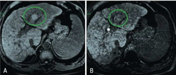
Figure 1. MRI with liver-specific contrast medium in a chronic liver disease patient showing a typical hepatocellular carcinoma on the left lobe. In the arterial phase (A) the lesion is predominantly hypervascular, while in the late hepatobiliary phase it presents a (B) predominant low signal intensity
einstein. 2015;13(2):326-9 328 Reis MA, Baroni RH
low signal intensity in the hepatobiliary phase can be
considered as such for therapy planning, because the
risk of malignant changes is very high.
hYpervascUlar hepaTocellUlar carcinoma
VERSUS
pseUDolesion wiTh arTerial enhancemenT
Arterioportal shunts can mimic hypervascular HCC in
conventional MRI and CT studies. These shunts
are
observed more often in cirrhotic livers as hypervascular
lesions ranging between 0.5 and 2.0cm in size, usually
without significant expression in any other sequence of
the exam.
(26,27)However, up to approximately 50% of hypervascular
foci in cirrhotic livers actually correspond to HCC
and their characterization without repeated exams
is a challenge. Today, this differentiation is possible
with liver-specific contrast medium, because the
shunts
correspond to areas of preserved parenchyma
(with isointensity to the remaining liver in the late
hepatobiliary phase), while most HCC do not show
functioning hepatocytes (with low signal intensity in late
hepatobiliary phase)
(27,28)(Figure 3).
of fibrosis and the prolonged enhancement peak and
washout period was demonstrated.
(29,30)Another use of Gd-EOB-dTPA still under investigation
is the quantitative assessment of liver function.
(31-37)Its
main advantages are the non-invasive assessment and
the regional quantification of liver function, potentially
useful to predict residual function in patients that
will undergo partial hepatectomy. Gd-EOB-dTPA
can also be used to diagnose early liver failure and
other parenchymal manifestations of post-transplant
complications.
(38)conclUsion
Gadoxetic acid as a liver-specific contrast medium has
been increasingly used in chronic liver disease patients,
mainly to assess hepatocellular carcinomas and to
differentiate it from other focal lesions.
Future perspectives include its use in quantification
of fibrosis and liver function.
reFerences
1 Chanyaputhipong J, Low SC, Chow PK. Gadoxetate Acid-Enhanced MR Imaging for HCC: A Review for Clinicians. Int J Hepatol. 2011;2011:489342. 2. Pauleit D, Textor J, Bachmann R, Conrad R, Flacke S, Layer G, et al. Hepatocellular
carcinoma: detection with gadolinium- and ferumoxides-enhanced MR imaging of the liver. Radiology. 2002;222(1):73-80.
3. Weinmann HJ, Schuhmann-Giampieri G, Schmitt-Willich H, Vogler H, Frenzel T, Gries H. A new lipophilic gadolinium chelate as a tissue-specific contrast medium for MRI. Magn Reson Med. 1991;22(2):233-7; discussion 242. 4. Hamm B, Staks T, Mühler A, Bollow M, Taupitz M, Frenzel T, et al. Phase
I clinical evaluation of Gd-EOB-DTPA as a hepatobiliary MR contrast agent: safety, pharmacokinetics, and MR imaging. Radiology. 1995;195(3):785-92. 5. Reimer P, Rummeny EJ, Shamsi K, Balzer T, Daldrup HE, Tombach B, et al.
Phase II clinical evaluation of Gd-EOB-DTPA: dose, safety aspects, and pulse sequence. Radiology. 1996;199(1):177-83.
6. van Montfoort JE, Stieger B, Meijer DK, Weinmann HJ, Meier PJ, Fattinger KE. Hepatic uptake of the magnetic resonance imaging contrast agent gadoxetate by the organic anion transporting polypeptide Oatp1. J Pharmacol Exp Ther. 1999;290(1):153-7.
7. Libra A, Fernetti C, Lorusso V, Visigalli M, Anelli PL, Staud F, et al. Molecular determinants in the transport of a bile acid-derived diagnostic agent in tumoral and nontumoral cell lines of human liver. J Pharmacol Exp Ther. 2006; 319(2):809-17.
8. Bartolozzi C, Battaglia V, Bozzi E. HCC diagnosis with liver-specific MRI--close to histopathology. Dig Dis. 2009;27(2):125-30. Review.
9. Ba-Ssalamah A, Uffmann M, Saini S, Bastati N, Herold C, Schima W. Clinical value of MRI liver-specific contrast agents: a tailored examination for a confident non-invasive diagnosis of focal liver lesions. Eur Radiol. 2009;19(2):342-57. Review.
10. Kim SH, Kim SH, Lee J, Kim MJ, Jeon YH, Park Y, et al. Gadoxetic acid-enhanced MRI versus triple-phase MDCT for the preoperative detection of hepatocellular carcinoma. AJR Am J Roentgenol. 2009;192(6):1675-81.
11. Frericks BB, Loddenkemper C, Huppertz A, Valdeig S, Stroux A, Seja M, et al. Qualitative and quantitative evaluation of hepatocellular carcinoma and cirrhotic liver enhancement using Gd-EOB-DTPA. AJR Am J Roentgenol. 2009; 193(4):1053-60.
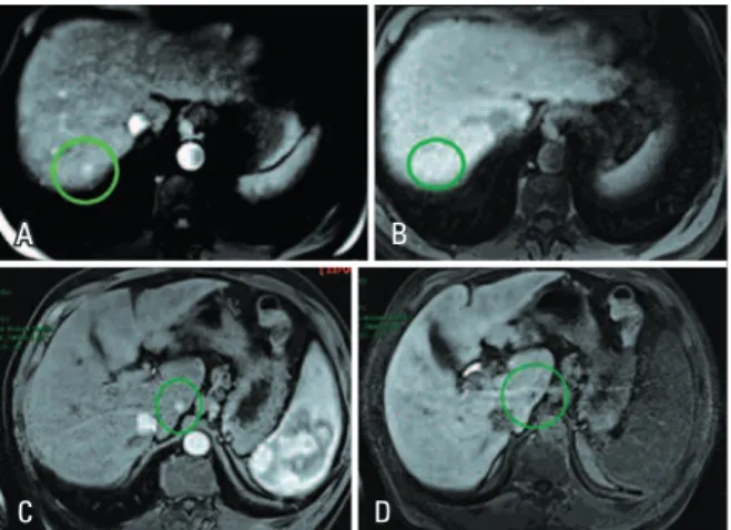
Figure 3. MRIs with liver-specific contrast medium of two chronic liver disease patients. The first patient shows nodular hypervascular focus on segment VII (A), with isointensity to the rest of the parenchyma in late hepatobiliary phase (B), indicating arterioportal shunt. The second patient shows a hypervascular nodular focus in the caudate lobe (C), with low signal intensity in late hepatobiliary phase (D), indicating a small hepatocellular carcinoma
A
C
B
D
perspecTive
329 Liver-specific magnetic resonance contrast medium in the evaluation of chronic liver disease
einstein. 2015;13(2):326-9
12. Ahn SS, Kim MJ, Lim JS, Hong HS, Chung YE, Choi JY. Added value of gadoxetic acid-enhanced hepatobiliary phase MR imaging in the diagnosis of hepatocellular carcinoma. Radiology. 2010;255(2):459-66.
13. Kudo M. Early hepatocellular carcinoma: definition and diagnosis. Liver Cancer. 2013;2(2):69-72.
14. The general rules for the clinical and pathological study of primary liver cancer. Liver Cancer Study Group of Japan. 3rd ed. Tokyo: Kanehara; 2010.
15. Theise ND, Park YN, Curado MP, Sakamoto M, Franceschi S, Torbenson M, et al. Hepatocellular carcinoma. In: Bosman FT, Carneiro F, Hruban RH, Theise ND, editors. WHO Classification of Tumours of the Digestive System. 4th ed. Lyon: International Agency for Research on Cancer; 2010. p 214-16.
16. Kojiro M. Diagnostic discrepancy of early hepatocellular carcinoma between Japan and West. Hepatol Res. 2007;37 Suppl 2:S121-4.
17. Chuma M, Sakamoto M, Yamazaki K, Ohta T, Ohki M, Asaka M, et al. Expression profiling in multistage hepatocarcinogenesis: identification of HSP70 as a molecular marker of early hepatocellular carcinoma. Hepatology. 2003;37(1):198-207.
18. Libbrecht L, Severi T, Cassiman D, Vander Borght S, Pirenne J, Nevens F, et al. Glypican-3 expression distinguishes small hepatocellular carcinomas from cirrhosis, dysplastic nodules, and focal nodular hyperplasia-like nodules. Am J Surg Pathol. 2006;30(11):1405-11.
19. Di Tommaso L, Franchi G, Park YN, Fiamengo B, Destro A, Morenghi E, et al. Diagnostic value of HSP70, glypican 3, and glutamine synthetase in hepatocellular nodules in cirrhosis. Hepatology. 2007;45(3):725-34.
20. Sano K, Ichikawa T, Motosugi U, Sou H, Muhi AM, Matsuda M, et al. Imaging study of early hepatocellular carcinoma: usefulness of gadoxetic acid-enhanced MR imaging. Radiology. 2011;261(3):834-44.
21. Kudo M. The 2008 Okuda lecture: Management of hepatocellular carcinoma: from surveillance to molecular targeted therapy. J Gastroenterol Hepatol. 2010;25(3):439-52.
22. Kitao A, Matsui O, Yoneda N, Kozaka K, Shinmura R, Koda W, et al. The uptake transporter OATP8 expression decreases during multistep hepatocarcinogenesis: correlation with gadoxetic acid enhanced MR imaging. Eur Radiol. 2011;21(10):2056-66.
23. Motosugi U, Ichikawa T, Sano K, Sou H, Onohara K, Muhi A, et al. Outcome of hypovascular hepatic nodules revealing no gadoxetic acid uptake in patients with chronic liver disease. J Magn Reson Imaging. 2011;34(1):88-94. 24. Kumada T, Toyoda H, Tada T, Sone Y, Fujimori M, Ogawa S, et al. Evolution of
hypointense hepatocellular nodules observed only in the hepatobiliary phase of gadoxetate disodium-enhanced MRI. AJR Am J Roentgenol. 2011;197(1): 58-63.
25. Kobayashi S, Matsui O, Gabata T, Koda W, Minami T, Ryu Y, et al. Gadolinium ethoxybenzyl diethylenetriamine pentaacetic Acid-enhanced magnetic resonance imaging findings of borderline lesions at high risk for progression to hypervascular classic hepatocellular carcinoma. J Comput Assist Tomogr. 2011;35(2):181-6.
26. Ahn JH, Yu JS, Hwang SH, Chung JJ, Kim JH, Kim KW. Nontumorous arterioportal shunts in the liver: CT and MRI findings considering mechanisms and fate. Eur Radiol. 2010;20(2):385-94.
27. Motosugi U, Ichikawa T, Sou H, Sano K, Tominaga L, Muhi A, et al. Distinguishing hypervascular pseudolesions of the liver from hypervascular hepatocellular carcinomas with gadoxetic acid-enhanced MR imaging. Radiology. 2010;256(1):151-8.
28. Sun HY, Lee JM, Shin CI, Lee DH, Moon SK, Kim KW, et al. Gadoxetic acid-enhanced magnetic resonance imaging for differentiating small hepatocellular carcinomas (< or = 2 cm in diameter) from arterial enhancing pseudolesions: special emphasis on hepatobiliary phase imaging. Invest Radiol. 2010;45(2): 96-103.
29. Tsuda N, Okada M, Murakami T. Potential of gadolinium-ethoxybenzyl-diethylenetriamine pentaacetic acid (Gd-EOB-DTPA) for differential diagnosis of nonalcoholic steatohepatitis and fatty liver in rats using magnetic resonance imaging. Invest Radiol. 2007;42(4):242-7.
30. Tsuda N, Okada M, Murakami T. New proposal for the staging of nonalcoholic steatohepatitis: evaluation of liver fibrosis on Gd-EOB-DTPA-enhanced MRI. Eur J Radiol. 2010;73(1):137-42.
31. Yamada A, Hara T, Li F, Fujinaga Y, Ueda K, Kadoya M, et al. Quantitative evaluation of liver function with use of gadoxetate disodium-enhanced MR imaging. Radiology. 2011;260(3):727-33.
32. Motosugi U, Ichikawa T, Sou H, Sano K, Tominaga L, Kitamura T, et al. Liver parenchymal enhancement of hepatocyte-phase images in Gd-EOB-DTPA-enhanced MR imaging: which biological markers of the liver function affect the enhancement? J Magn Reson Imaging. 2009;30(5):1042-6.
33. Motosugi U, Ichikawa T, Oguri M, Sano K, Sou H, Muhi A, et al. Staging liver fibrosis by using liver-enhancement ratio of gadoxetic acid-enhanced MR imaging: comparison with aspartate aminotransferase-to-platelet ratio index. Magn Reson Imaging. 2011;29(8):1047-52.
34. Cho SH, Kang UR, Kim JD, Han YS, Choi DL. The value of gadoxetate disodium-enhanced MR imaging for predicting posthepatectomy liver failure after major hepatic resection: a preliminary study. Eur J Radiol. 2011;80(2):e195-200. 35. Watanabe H, Kanematsu M, Goshima S, Kondo H, Onozuka M, Moriyama N,
et al. Staging hepatic fibrosis: comparison of gadoxetate disodium-enhanced and diffusion-weighted MR imaging--preliminary observations. Radiology. 2011;259(1):142-50.
36. Katsube T, Okada M, Kumano S, Hori M, Imaoka I, Ishii K, et al. Estimation of liver function using T1 mapping on Gd-EOB-DTPA-enhanced magnetic resonance imaging. Invest Radiol. 2011;46(4):277-83. Erratum in: Invest Radiol. 2013; 48(2):112.
37. Kim T, Murakami T, Hasuike Y, Gotoh M, Kato N, Takahashi M, et al. Experimental hepatic dysfunction: evaluation by MRI with Gd-EOB-DTPA. J Magn Reson Imaging. 1997;7(4):683-8.