2255-4823/$ – see front matter © 2013 Elsevier Editora Ltda. All rights reserved.
7PMVNFt/ÞNFSPt/PWFNCSP%F[FNCSPt*44/t*44/ 0OMJOF
www.ramb.org.br ARTIGOS
ARTIGOS ORIGINAIS _____________Qualidade da informação da internet disponível para pacientes em páginas em português ___________________________________________________________645 Acesso a informações de saúde na internet: uma questão de saúde pública? ______650 Maus-tratos contra a criança e o adolescente no Estado de São Paulo, 2009_______659 Obesidade e hipertensão arterial em escolares de Santa Cruz do Sul – RS, Brasil ___666 Bone mineral density in postmenopausal women with and without breast cancer ___673 Prevalence and factors associated with thoracic alterations in infants born prematurely __________________________________________________679 Análise espacial de óbitos por acidentes de trânsito, antes e após a Lei Seca, nas microrregiões do estado de São Paulo ___________________________________685 Sobrevida e complicações em idosos com doenças neurológicas em nutrição enteral ______________________________________________________691 Infliximab reduces cardiac output in rheumatoid arthritis patients without heart failure ______________________________________________________698 Análise dos resultados maternos e fetais dos procedimentos invasivos genéticos fetais: um estudo exploratório em Hospital Universitário _______________703 Frequência dos tipos de cefaleia no centro de atendimento terciário do Hospital das Clínicas da Universidade Federal de Minas Gerais __________________709
ARTIGO DE REVISÃO ______________Influência das variáveis nutricionais e da obesidade sobre a saúde e o metabolismo __714 EDITORIAL
Conclusão: como exibir a cereja do bolo 633 PONTO DE VISTA
Os paradoxos da medicina contemporânea 634 IMAGEM EM MEDICINAObstrução duodenal maligna: tratamento
endoscópico paliativo utilizando prótese metálica autoexpansível 636 Gossypiboma 638
DIRETRIZES EM FOCO Hérnia de disco cervical no adulto: tratamento cirúrgico 639 ACREDITAÇÃOAtualização em perda auditiva: diagnóstico radiológico 644 SEÇÕES ____________________________
www.ramb.org.br
Revista da
ASSOCIAÇÃO MÉDICA BRASILEIRA
Review article
Hepatocellular carcinoma: epidemiology, biology,
diagnosis, and therapies
q
Marcos António Gomes
a, Denise Gonçalves Priolli
b,
José Guilherme Tralhão
c,d, Maria Filomena Botelho
a,d,*
a Service of Biophysics, Instituto Biomédico de Investigação da Luz e Imagem, Coimbra, Portugal
b School of Medicine, Universidade de São Francisco, Bragança Paulista, SP, Brazil
c Faculty of Medicine, Department of Surgery, Surgery 3, Hospitais Universitários de Coimbra, Coimbra, Portugal
d Centro de Investigação em Meio Ambiente, Genética e Oncobiologia, Coimbra, Portugal
A RT I C L E I N F O
Article history:
Received 18 June 2012 Accepted 23 March 2013
Keywords:
Hepatocellular carcinoma Therapy
Diagnosis Hepatitis B Hepatitis C Liver neoplasms
q Study conducted at the Instituto Biomédico de Investigação da Luz e Imagem and at the Centro de Investigação em Meio Ambiente,
Genética e Oncobiologia, Coimbra, Portugal.
* Corresponding author.
E-mail: [email protected] (M.F. Botelho).
A B S T R A C T
Hepatocellular carcinoma is the fifth most common cancer in men and the seventh in women, as is diagnosed in more than half a million individuals worldwide every year. In Portugal, its incidence and mortality rates are low compared to other types of cancers. In Brazil, in the city of São Paulo, according to data released by the Brazilian Unified Health System (Sistema Único de Saúde – SUS), the incidence of primary liver cancer was 2.07/100,000 inhabitants. Although the vast majority of cases (85%) mainly affect developing countries, especially where infection by hepatitis B virus (HBV) is endemic, the incidence in developed countries is increasing. This pathology is associated with several risk factors, not only environmental but also genetic, generating an increasing interest in attaining a better understanding of this disease, which is still associated with very late diagnosis and poor prognosis. Of the available treatments, few patients benefit from their scanty advantages, increasingly stimulating research of new forms of treatment against this disease. This review aimed to briefly but fully identify risk factors, molecular and biochemical pathways, pathophysiology, diagnosis, and possible clinical approaches of hepatocellular carcinoma.
© 2012 Elsevier Editora Ltda. All rights reserved.
Carcinoma hepatocelular: epidemiologia, biologia, diagnóstico e terapias
R E S U M O
O carcinoma hepatocelular é o quinto tipo de câncer mais comum em homens e o sétimo em mulheres, diagnosticado todos os anos em mais de meio milhão de pessoas por todo o mundo. Em Portugal, sua incidência e mortalidade são baixas, comparativamente a outros tipos de cânceres. No Brasil, no município de São Paulo, segundo dados divulgados
Palavras-chave:
Carcinoma hepatocelular Terapia
pelo Sistema Único de Saúde (SUS), a incidência do câncer primário de fígado foi de 2,07/100.000 habitantes. Apesar de a grande maioria dos casos (85%) afetar principalmente países em desenvolvimento, sobretudo onde a infeção pelo vírus de hepatite B (HVB) é endêmica, a incidência em países desenvolvidos é cada vez maior. Esta patologia está associada a inúmeros fatores de risco não só ambientais, mas também genéticos, os quais, cada vez mais, despertam interesse na procura pelo melhor conhecimento da patologia, muito associada ainda a diagnósticos tardios e maus prognósticos. Dos tratamentos disponíveis, poucos doentes são aqueles que usufruem das suas escassas vantagens, estimulando cada vez mais a pesquisa de novas formas de terapêutica. Esta revisão pretende, de forma breve mas completa, identificar fatores de risco, vias moleculares e bioquímicas, fisiopatologia, diagnóstico e possíveis abordagens clínicas do carcinoma hepatocelular.
© 2012 Elsevier Editora Ltda. Todos os direitos reservados.
Hepatocellular carcinoma
Hepatocellular carcinoma (HCC) is the primary liver cancer, that is to say, derived from primary liver cells. As in all cancers, it appears when mutations in the genes of a cell make uncontrolled growth possible. This mutation can be caused by external agents, such as hepatitis virus, or by excessive multiplication of cells, as in chronic regeneration in chronic hepatitis, which increases the risk of replication errors in the genes. HCC is typically aggressive, with a high rate of death after symptom onset (most commonly jaundice and/or ascites). If detected only in the symptomatic phase, the patient has an untreated mean life expectancy of less than one month; even at this stage, the available treatments are limited and ineffective.
HCC represents 70-85% of primary liver cancers, and is the most frequently observed primary malignant liver tumors worldwide,; cholangiocarcinoma, which originates from cholangiocytes, i.e., the epithelial cells lining the bile ducts, constitute 10-15% of primary hepatic malignancies. The remaining 5% are uncommon tumors such as primary liver angiosarcoma, hepatic epithelioid hemangioendothelioma, hemangiopericytoma, or primary hepatic lymphoma.
Annually, HCC is diagnosed in more than half a million people worldwide. The latest figures showed an estimated 748,300 new cases of HCC and 695,900 deaths caused by this disease. In Europe, 60,200 new cases were diagnosed in 2008, making this type of cancer the fifth most common in men and
the seventh in women1,2 (Fig. 1).
In Portugal, the incidence rate of this disease is low compared with other types of cancer, according to data from GLOBOCAN 2008, representing 1.1% of all cancer types. Regarding mortality associated with this type of pathology,
it is responsible for 2% of all cancer-related deaths3; in the
year 2011, according to the National Institute of Statistics of Portugal, 979 deaths due to malignant liver and intrahepatic bile duct neoplasms were recorded, 84 more cases than in the
previous year.4
The World Health Organization (WHO) indicates HCC as the second leading cause of cancer-related death in humans due to its high incidence in the East, in areas of Africa, and
in the Western Pacific. In Brazil, the incidence of HCC is low, while higher in some states such as Espírito Santo and Bahia. In the state of São Paulo, HCC is the fifth in frequency among digestive tract tumors, according to the Brazilian Association of Liver Transplants and Liver Disease Carriers.
Approximately 85% of the cases occur in developing countries, and the highest incidence rates are described in regions where hepatitis B virus (HBV) infection is endemic: Southeast Asia and sub-Saharan Africa. HCC rarely occurs before 40 years of age and reaches its peak at approximately 70 years of age. The prevalence rate of liver cancer among men is two to four times higher than among females.
HCC related to hepatitis C virus (HCV) infection has become the fastest-growing cause of the disease in the United States, contributing to the rising incidence of HCC in that country, which has tripled; the five-year rate survival remained below 12%.
Epidemiological data regarding the HCC in some countries, such as Brazil and Portugal, are still scarce and dispersed, making the organization and planning of health-promoting activities with an impact on prevention and early diagnosis of this pathology difficult. According to data released by the Brazilian Unified Health System (Sistema Único de Saúde – SUS), the incidence of primary liver cancer in the city of São Paulo, Brazil, was 2.07/100,000 inhabitants. The mean age of patients was 54.7 years, with a male/female ratio of 3.4:1. HBV surface antigen (HBsAg) positivity was 41.6%, anti-HCV positivity was 26.9%, presence of chronic alcoholism was 37%,
and cirrhosis was 71.2%.5
Estimates made by GLOBOCAN for 2008 suggest that the incidence rate is 3.5/100,000 for males and 1.2/100,000 for females, resulting in 477 new cases per year. Mortality rates are, according to the same source, 3.4/100,000 and 1.1/100,000 for males and females, respectively.
Risk factors
The main risk factors for HCC include infection by HBV and HCV, liver diseases caused by alcohol consumption, aflatoxin exposure, and mainly non-alcoholic fatty liver
disease6 (NAFLD). Less common causes include hereditary
Hepatite B Hepatite C
hemochromatosis (HH), alpha-1-antitrypsin deficiency, autoimmune hepatitis, certain porphyria types, and Wilson’s disease. The distribution of these risk factors among patients with HCC is highly variable and depends on the geographic
region, race, and ethnic group.7
Most of these risk factors lead to the onset and progression of cirrhosis, which is present in 80-90% of patients with HCC. The cumulative risk at five years for patients with cirrhosis
to develop HCC varies between 5% and 30%, depending on the cause, with the greatest risk among those infected with HCV, those with specific risk due to geographic region or ethnic group (17% in USA and 30% in Japan, for instance), and the stage of cirrhosis; patients with decompensated disease
present the greatest risk.8
Worldwide, approximately 50% of all adult patients with HCC have chronic HBV infection, whereas in children
Fig. 1 – Regional variation in incidence rates of hepatocellular carcinoma (standardized age). The incidence rates shown (number of cases per 100,000 inhabitants) are for both genders and all age ranges.106,107
Cases/100,000 individuals
≤ 2.5
≤ 4.0
≤ 6.0
≤ 9.3
≤ 9.4
Fig. 2 – Molecular events in the development of hepatocellular carcinoma stages.39,46-48,50,53-56,58-61,63,65-67,70-74 HCC,
hepatocellular carcinoma; HCV, hepatitis C virus; TGF, transforming growth factor; VEGF, vascular endothelial growth factor.
Diabetes Obesity
Metabolic syndrome
Aflatoxins Alcohol Tabacco Vinyl chloride
Genetic predisposition
Other carcinogens
DNA lesion TP53 mutations
Chronic hepatitis Cirrhosis
HCV nuclear protein activation
p53 binding
p53 binding
Wnt/b-catenin activation Inhibits TGF-b
pathway Inhibits p21WAF
expression E-cadherin
decreased expression Wnt/b-catenin activation Activation of IL-8; TNF; TGF-b-1; EGFR VEGF overexpression
Displasia
Early HCC
Advanced/ metastatic
HCC TNF-a and INF-g
overexpression
Inflammatory reaction p53 mutation
Rb dysfunction
Rb dysfunction
b-catenin
activation Aberrant DNA
p53 mutation methylation
Liver regeneration
p16 inactivation
p21
WAF1/ClP1
decreased expression
E-caderin inactivation
NO increase
HBx transactivation Virus VHB/VHC
this association occurs in virtually all cases. In endemic areas of Asia and Africa, where HBV infection is vertical, approximately 90% of those infected undergo chronic evolution of the disease, with frequent virus integration into the host
DNA.9,10 The association between HBV and cancer varies
greatly depending on the country and the types of laboratory
tests used to diagnose the disease11; this scenario is changing
with the emergence of new diagnostic techniques, especially polymerase chain reaction (PCR).
Although HBV can cause HCC in the absence of cirrhosis, between 70% and 80% of the patients present liver cirrhosis as a consequence of HBV infection. The risk of HCC in individuals with HBV infection is higher in males, the elderly, those with long-term exposure to the virus, family history of HCC, exposure to aflatoxin-type mycotoxins, alcohol or tobacco consumption, HCV or hepatitis delta virus (HDV) co-infection, high levels of HBV viral replication, and/or infection by HBV
genotype C.10,12-15 The estimated risk of HCC is 15-20 times
higher among individuals infected with HCV compared with the non-infected; most of the additional risk is limited to those
with advanced liver fibrosis or cirrhosis.16
An evolutionary molecular analysis study demonstrated that HCV infection occurred in a large number of young adults in Japan in the 1920s, in southern Europe in the 1940s, and in North America in the 1960s and 1970s. These cases in North America occurred as a result of the sharing of contaminated
needles among drug addicts and due to blood transfusions17;
the first case was described in 1989.
HCV infection markers are found in 80-90% of patients with HCC in Japan, 44-66% in Italy, and 30-50% in the United
States.7 Risk factors for HCC among individuals infected with
HCV include older age at the time of infection, male gender, co-infection with the human immunodeficiency virus (HIV)
or HBV, and probably diabetes or obesity.18-21 The prolonged
and abusive consumption of alcohol, defined as daily ingestion of 40 to 60 g of beverages containing 13.7 g of alcohol is a well-established risk factor for HCC, whether independently, with a 1.5-2.0 times increased risk , or combined with other risk factors, such as infection by HCV, or, to a lesser extent,
infection with HBV.16
The development of effective screening methods in blood donors regarding the presence of HBV in the 1980s and of HCV in the 1990s drastically reduced the incidence of transfusion-related viral hepatitis, reducing its rates
from 33% to 0.3% in the United States.22 The development
of effective prophylactic pre-exposure vaccines against HBV and the combination with hepatitis B immunoglobulins for post-exposure prophylaxis dramatically reduced the incidence
of HBV infection.23,24 In Taiwan, HCC incidence in children
after universal vaccination against HBV reduced the number
of cases from 0.70 to 0.35 per 100,000 children.25
The incidence of HCV also decreased after screening of blood donors in the 1990s, but due to the 20-year latency time from the acute infection to onset of cirrhosis and HCC, the impact of the reduction in HCV will not be observed before the year 2015. It is expected that the incidence of cases of HCV-related HCC will continue to decrease. An effective vaccine against HCV infection has not yet been developed due to the high mutation rate of the virus. For patients with chronic
HBV or HCV infection, treatment with alpha interferon has
been associated with lower incidence of HCC.26-28
Several studies performed in Western countries have demonstrated that 30% to 40% of HCC patients had no chronic HBV or HCV infection, suggesting the presence of other causes. Some of these patients had clinical or biochemical alterations compatible with NAFLD, such as obesity or metabolic syndrome. In population-based cohort studies in the United States, Scandinavia, Taiwan, and Japan, HCC was 1.5 to 2.0 times more frequent in obese than in non-obese
patients.29-32 Several case-control studies and some cohort
studies have demonstrated that, on average, HCC is twice as
likely to develop in individuals with diabetes mellitus type 2 than
in non-diabetic patients.33,34 NAFLD, which is present in almost
90% of obese individuals and in almost 70% of individuals with
type 2 diabetes mellitus, appears to be a possible risk factor for
HCC.35 Due to the limited data establishing a direct association
between the progression of hepatic steatosis and HCC, the currently available estimates of risk are not clear. However, due to the high incidence of metabolic syndrome in Western countries, any increase, however small, in the risk of obesity or diabetes can result in a higher number of HCC cases.
The pathogenic mechanisms involved include lipid peroxidation and oxidative stress, production of free radicals as a precursor to tumor onset and progression; chronic activation
of the inhibitor of kappa-beta kinase (IKK-b), which leads to
inflammation; and hepatic fibrosis due to hyperinsulinemia
and hyperglycemia.36
Aflatoxins, particularly aflatoxin B1 (AFB-1), are potent liver
carcinogens produced by a fungus (Aspergillus flavus and A.
parasiticus), common contaminants of stored grain.37 Gene
mutation caused by AFB-1 at codon 249, exon 7 of the tumor suppressor gene TP53 (GC to TA transversion) was identified and positively correlated with exposure to aflatoxin in a
meta-analysis comprising 49 studies.38,39 In addition, aflatoxins
are synergistic with chronic HBV infection for the development
of HCC.11
Hepatocellular carcinoma molecular pathways
The association between hepatocarcinogenesis molecular pathways and the risk factors associated with this condition is the object of investigation, aiming at adequate, personalized, and effective chemoprevention and treatment of HCC (Fig. 2).
Chronic HBV infection involves three distinct mediation mechanisms in hepatocarcinogenesis. The first mechanism involves integration of viral DNA into the host genome,
inducing chromosome instability.40,41 The second proposed
mechanism involves multiple genetic mutations by insertion, resulting in the integration of HBV genome at specific sites that can activate endogenous genes, for instance, RAR (retinoic
acid receptor) b, cyclin A, and TRAP1.42-44 The third mechanism
involves modulation of cell proliferation by viral protein expression, particularly HBV X protein (HBx) with 154 amino acids (16.5-kDa), which can transactivate or overexpress a
variety of viral and cellular genes.44,45
shown that HBx can coactivate the transcription process of some important cell and viral genes, thus coordinating the
balance between cell proliferation and apoptosis.46,47 Cell
promoters of genes associated with proliferation, such as interleukin 8 (IL-8), tumor necrosis factor (TNF), transforming
growth factor (TGF-b1), and epidermal growth factor receptor
(EGFR), as well as transcription factors, are activated with HBx
transactivation.48,49
HBx also appears to be involved in the activation of signaling cascades involving the Ras/Raf/MAPK pathway,
contributing to disregulation of cell cycle checkpoints,50 as well
as to the activation of several oncogenes such as c-myc, c-jun
and c-fos in the cytoplasm.51,52 Studies have also demonstrated
two pathways by which HBx can activate the Wnt/b-catenin
pathway, in collaboration with the proto-oncogene protein
Wnt-1, through the stabilization of cytoplasmic b-catenin in
HCC cells,53 or by activation of the extracellular signal-regulated
kinase (Erk), which leads to phosphorylation, and in turn, to
the inactivation of kinase-3b of glycogen synthesis (GSK-3b),
stabilizing the b-catenin.54
Another alternative mechanism of Wnt activation in HCC is by the repression of E-cadherin by HBx in transcription,
through the hypermethylation of E-cadherin promoter.55
These mechanisms demonstrate the association of HBx
with decreased expression of E-cadherin and b-catenin
accumulation in the cytoplasm and/or nucleus, leading to
increased activation of b-catenin.56 In the normal state,
b-catenin is targeted for degradation by casein kinase Ia and
GSK-3b.57 20% to 90% of all HCC have evidence of b-catenin
activation, which has increased interest in the study of this
pathway as a therapeutic target.58-60
Similarly to what occurs with several proteins codified by virus DNA, HBx can bind to p53, forming protein-protein complexes, and thus inactivating p53-dependent critical
activities such as p53-mediated apoptosis,61 or even
repressing TP53 transcription.62 These modulatory
effects of HBx on p53 provide the basis for cell malignant transformation. In addition to this role in apoptosis, HBx can also contribute to tumorigenesis in HCC through the overexpression of the potent angiogenic factor of vascular endothelial growth factor (VEGF), demonstrated in studies
under hypoxic conditions.63,64
Clinical and epidemiological studies attribute a higher degree of aggressiveness to HCV than to HBV, as there is a higher frequency of cases of HCC in patients with HCV-induced cirrhosis. In contrast to HBV, HCV is an RNA virus that is not integrated in the host genome; nevertheless, several virus-host interactions occur, which are believed to be responsible for the indirect hepatocarcinogenesis of this virus. The core protein of HCV is highly conserved and has been widely studied, as it is believed that it plays an important role in hepatocarcinogenesis through modulation of cell proliferation, apoptosis, and immune response.
This core protein of HCV induces ROS formation through interaction with shock protein Hsp60, in addition to binding
to p53, p73, and Rb protein.47,65 This interaction with tumor
suppressor proteins appears to explain the fact that the protein
is associated with p21WAF1 inhibition, leading to apoptosis
inhibition and promoting cell cycle.47
The Raf1/MAPK and Wnt/b-catenin pathways are activated
by the HCV core protein, while it also inhibits TGF-b pathway
and activates TNF-a receptor and NF-kb pathway.58,66,67 Studies
have also demonstrated that the frequency of the b-catenin
gene mutation in HCC of patients with HCV is approximately
twice as high when compared to other causes.68
It is known that chronic inflammation and infection are often associated with increased risk of cancer. Infections caused by HBV and HCV cause inflammation with production of free radicals, cytokines, and chemokines, resulting in DNA damage, cell proliferation, fibrosis, and angiogenesis. An example of a response to inflammatory stress is the
p53 pathway itself.69 Free radicals, such as reactive oxygen
or nitrogen species, can directly damage DNA and proteins, and/or indirectly damage these macromolecules through lipid peroxidation.
Increasing evidence indicates that nitric oxide (NO), an important signaling and bioregulation molecule catalyzed by the NOS enzyme family, plays an important role in
carcinogenesis.70,71 In chronic hepatitis, the overexpression of
proinflammatory cytokines such as TNF-a, INF-g, and IL-1, lead
to increased concentrations of NO in human hepatocytes.72
NO can cause DNA damage and induce a stress response through p53 anti-carcinogenic pathway or cause mutations in
cancer-related genes, such as TP53.70
The mutational inactivation of TP53 has been widely described as one of the molecular mechanisms involved in HCC pathogenesis, especially in geographic areas where exposure to
aflatoxin B1 (AFB1) is prominent.39,73,74 However, and although
the G»T mutation in codon 249 of the P53 gene is closely related to the AFB1 exposure, most HCC cases show absence of TP53 mutations, so that this inactivation is associated with
other mechanisms, such as interactions with viral proteins.75
HCC has a high degree of genetic heterogeneity, suggesting that multiple molecular pathways may be involved in the genesis of hepatocellular cancer subsets. Understanding the molecular and cellular bases of neoplastic transformations that occur in the liver allow for the development of better strategies for the prevention of and/or more effective treatments for HCC.
Symptoms
HCC symptoms are not specific, generally indicate advanced HCC, and are more directly related to liver function impair-ment. There may be abdominal pain (between 40% and 60%), which may indicate spontaneous bacterial peritonitis; palpable mass in the abdomen on the right (23%); abdominal distension (45%) loss of appetite (45%); jaundice (16%); ascites (26%); weight loss (29%); malaise (60%); andsigns of hepatic encephalopathy, from drowsiness to coma, and
gastrointestinal bleeding76 (7%).
Pathology and diagnosis
HCC may display four degrees of differentiation (according to the classification of Edmondson and Steiner, 1954) and five different histological types:
• sclerosing HCC;
• fibrolamellar carcinoma (which is the most straightforward to undergo surgical resection, thus yielding a better prog -nosis);
• cholangiocellular carcinoma;
• hepatocholangiocarcinoma;
• hepatoblastoma (more common in children).
Malignant transformation of hepatocytes in HCC is a gradual process associated with genetic mutations, allelic loss, epigenetic alterations, and disruption of cellular and molecular pathways. The phenotypic expression of these alterations may manifest as precursor lesions that accompany HCC, called
dysplastic nodules.77,78 These nodules are distinct from benign
regenerative nodules associated with cirrhosis due to their high proliferative index and clonality.
The International Working Party of Terminology of the World Congress of Gastroenterology classified the dysplastic nodules, which are distinct nodular lesions larger than 5 mm in diameter, as low-grade dysplastic nodules (LGDN)
and high-grade dysplastic nodules79 (HGDN). LGDN nodules
show only mild dysplasia and do not express any relation to malignancies. HGDN nodules, in turn, are characterized by higher cell density (small-cell lesions) than the surrounding tissues, exhibiting characteristic formations such as “nodule inside nodule”, resembling well-differentiated HCC both at
radiological and pathological assessment.80,81
The distinction between HGDN nodules and early-stage HCC is a challenge yet to be resolved. A panel of three immunohistochemical markers of malignant transformation, such as heat shock protein (HSP) 70, glutamine synthetase, and
glypican-3 has been used to differentiate them.82,83
Additio-nally, molecular data based on quantitative gene expression profiles with the LYVE-1 gene, E-cadherin, and survivin allow
for a reliable diagnosis of early-stage HCC.84
The diagnosis of HCC can be attained through imaging tests, tumor markers (blood test), and anatomopathological examination (biopsy). As there is not always an increase in tumor markers and biopsy may not be possible (due to clotting deficiency caused by liver failure, to ascites, or to access difficulty due to tumor location) or recommended (there is a theoretical risk, from anecdotal reports, of tumor dissemination along the needle trajectory), the diagnosis of HCC can be achieved by imaging assessment presenting typical lesion and tumor marker increase, or by the presence of typical image in two different types of imaging assessment.
The guidelines for the diagnosis of HCC76 suggest that
patients with cirrhosis and radiologically-detected mass do not need biopsy confirmation of HCC diagnosis. Criteria for this diagnosis are: lesion of at least 2 cm in diameter with typical vascular patterns in dynamic images obtained from computed tomography (CT), ultrasound with contrast, or magnetic resonance imaging (MRI) showing enhancement in the arterial phase with rapid clearance of contrast in the portal phase.
For lesions 1-2 cm in diameter, at least two dynamic imaging studies are needed to highlight the typical features of
HCC. Lesions < 1 cm in diameter should be followed by contrast
ultrasound every 3-6 months. These guidelines are supported by the high incidence of regenerative nodules in cirrhotic livers that remain stable for many years, but can undergo malignant transformation to HCC due to changes in vascular supply, with shunting of blood from the portal vein to the hepatic artery and consequent increase in size. A percutaneous biopsy of liver injury in cirrhotic patients increases the risk of bleeding, dissemination of tumor cells, and false negative results due to inaccurate radiological location, particularly with small lesions. Levels of serum alpha-fetoprotein (AFP), a glycoprotein normally produced by the fetal liver and yolk sac, can be elevated with the development of HCC. AFP levels are
particularly indicative if > 500 mg/L85,86 (normal values 10 to
20 mg/L), although 20% of HCCs are not associated with high
serum AFP levels, and moderate increases in AFP can be found in other conditions such as active hepatitis of any etiology (virus-related, drug-related, autoimmune disease), pregnant women carrying fetuses with abnormalities such as spina bifida, and patients with germ-cell or gastric cancer. Serum AFP levels have also been used to enhance the sensitivity of non-contrast ultrasound when screening individuals at high
risk for HCC.87,88
Staging and treatment
Due to the close association between HCC and cirrhosis, prognosis of individual cases depends on cirrhosis severity and the degree of malignant involvement of HCC. Most staging criteria for HCC prognosis incorporate elements on tumor extent and cirrhosis severity; however, the tumor-node-metastasis (TNM) staging system includes size, multi focality, vascular involvement, and distant metastases, but does not
include the characteristics of cirrhosis.89
For the prognosis of patients with cirrhosis, the parameters of chronic liver disease evaluation, such as those used in the Child-Pugh classification, have been incorporated to most HCC classification criteria, including the Cancer of the Liver Italian Program score (CLIP), the Japan Integrated Staging Score (JIS), the Chinese University Prognostic Index (CUPI) and the French
Prognostic Classification.90-93
Okuda classification
chemoembolization techniques and percutaneous treatments, and with the aid of new instruments such as argon scalpels and Yag-laser, in addition to the improvement in hepatectomy techniques. The classification remains important regarding the prognosis of untreated patients as a basis for comparison with
the currently available treatments.94,95
The CLIP classification system encompasses the Child-Pugh classification, which assesses liver function, affected liver fraction (as in the Okuda classification), serum levels of alpha-fetoprotein, and hepatic vessel infiltration by cancer, especially the portal vein. With the obtained score, it is possible to compare the expected life expectancy in similar
conditions.96
The Barcelona Clinic Liver Cancer (BCLC) is an internatio-nally standardized system recommended by the American Association for the Study of Liver Diseases (AASLD) and the European Association for the Study of the Liver (EASL),
and is based solely on clinical parameters.76,97 Recently
(2012) the EASL, together with the European Organization for Research and Treatment of Cancer (EORTC), published a set of clinical guidelines for surveillance, diagnosis, and therapeutic strategies for HCC, thus updating the panel of
recommendations proposed in 200198 and used to date.99
The BCLC system classifies HCC as very early, early, intermediate, advanced, and terminal disease, based on the Child-Pugh criteria, functional status, tumor size, multifocality, and presence of portal vein invasion. Patients with very early and early stages are amenable to curative treatment modalities, including surgical resection, liver transplantation, percutaneous ethanol injection, or radiofrequency ablation.
Patients with intermediate-stage HCC are amenable to treatment by chemoembolization. For patients with advanced-stage HCC, which represent a large number of diagnosed cases, the recommended treatments include several tyrosine kinase inhibitors or participation in phase III and IV clinical trials, considering that the aforementioned treatments
are rarely effective. Critically-ill patients are not usually considered for specific treatment and palliative treatment is indicated for symptom control.
Early-stage HCC presents as a small nodule with a diameter
< 2 cm and can be classified into two types: small, nodular-type
HCC, which is well defined and sometimes encapsulated; or indistinct (vague) nodular-type that is poorly defined and has
no clear borders.100,101 Dysplastic nodules and early HCC are
usually asymptomatic and are usually incidental findings on radiographic studies or detected as a result of screening procedures. Early HCC can be indolent or slow-growing (up to one year), until it undergoes rapid tumor progression. In these early stages, HCC gradually progresses with increase in tumor volume, followed by invasion of the portal vein branches, extending to the main portal vein. The portal vein invasion is generally accompanied by a rapid increase in serum concentrations of AFP, even with no alterations in size of the primary liver tumor. The tumor can also directly invade the hepatic vein branches and the intrahepatic inferior vena cava, spreading to the superior vena cava or the right atrium as tumor thrombus. Hepatic artery invasion is much less common.
HCC dissemination occurs primarily through the blood to the lungs, bones, and brain at later stages. These metastatic lesions are typically hypervascular, as is the primary tumor, and predispose to bleeding (intracranial hemorrhage or hemoptysis). Bone metastases can be single or multiple and can produce isolated symptoms such as cranial nerve or peripheral nerve compression.
The standardized assessments for the detection of HCC metastasis are CT with contrast of the skull, chest, abdomen, and pelvis, as well as bone scintigraphy . Dynamic acquisitions with contrast are generally not necessary, but if performed, they can demonstrate arterial hypervascularity of the metastatic lesions similar to that of the primary tumor. Positron emission tomography (PET) has not shown sufficient
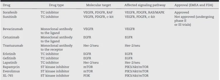
Drug Drug type Molecular target Affected signaling pathway Approval (EMEA and FDA) Sorafenib TC inhibitor VEGFR, PDGFR, RAF VEGFR, PDGFR, RAS/MAPK Approved
Sunitinib TC inhibitor VEGFR, PDGFR, c-kit VEGFR, PDGFR, c-kit Not approved (undergoing phase II
or III trials) Bevacizumab Monoclonal antibody
to the ligand VEGFR VEGFR Cetuximab Monoclonal antibody
to the ligand
EGFR EGFR Trastuzumab Monoclonal antibody
to the receptor Her-2/neu Her-2/neu Erlotinib TC inhibitor EGFR EGFR Gefitinib TC inhibitor EGFR EGFR Lapatinib TC inhibitor Her-2/neu Her-2/neu Rapamycin ST kinase inhibitor mTOR PIK3/Akt/mTOR Everolimus ST kinase inhibitor mTOR PIK3/Akt/mTOR XL-765 ST kinase inhibitor PI3K PIK3/Akt/mTOR
Akt, protein kinase B; EMEA, European Medicines Agency; FDA, Food and Drug Administration; MAPK, mitogen-activated protein kinase; mTOR, mammalian target of rapamycin; PDGFR, platelet-derived growth factor receptor; PIK3, phosphatidylinositol 3-kinase; ST, serine/ threonine; TC, tyrosine kinase; VEGFR, vascular endothelial growth factor receptor.
reliability to justify its inclusion in routine examinations in
HCC staging.102
Multiple tumors can arise in cirrhotic liver, particularly with chronic HCV infection, or may represent liver metastases consequent to thrombosis of the portal vein, from the primary tumor and hematogenous spread to the liver.
The clinical and genetic heterogeneity of this disease dictates the fact that there is little effective therapeutic response in HCC. Therapy directed at molecular targets, either genes or their receptors, aims to inactivate activated oncogenes, recover tumor suppressor genes, or any molecule or gene involved in the development of HCC, thereby repairing errors or abnormal functions or biological behavior. Recently, many genome-based molecules have been candidates for targeted therapy. They have been discovered by microarray studies, at the analysis of epigenetic aberrations of the total genome, from high-performance sequencing systems. Some genes or target molecules currently in study are VEGFR, EGFR, DDEFL, VANGL1, WDRPUH, Ephrin-A1, GPC3, and PFTK1,
among others.103,104 Many of these molecules for targeted
therapy, such as monoclonal antibodies, small molecules, and antisense molecules are now in phase II and III clinical trials, with promising data at this time. At the moment only sorafenib, a multikinase VEGFR and RAS kinase inhibitor, has been approved by the Food and Drug Administration and by
the European Medicines Agency104,105 (EMEA) (Table 1).
Conflicts of interest
All authors declare to have no conflicts of interest.
R E F E R E N C E S
1. Jemal A, Bray F, Center MM, Ferlay J, Ward E, Forman D. Global cancer statistics. CA Cancer J Clin. 2011;61:69-90.
2. Ferlay J, Parkin DM, Steliarova-Foucher E. Estimates of cancer incidence and mortality in Europe in 2008. Eur J Cancer. 2010; 46:765-81.
3. Ferlay J, Shin HR, Bray F, Forman D, Mathers C, Parkin DM. Estimates of worldwide burden of cancer in 2008: GLOBOCAN 2008. Int J Cancer. 2010; 127:2893-917.
4. INE. Óbitos pela causa tumor maligno do fígado e das vias biliares intra-hepáticas do CID 10: resultados 2002-2011. Lisboa; 2012.
5. Gonçalves CS, Pereira FEL, Gayotto LCC. Hepatocellular carcinoma in Brazil: report of a national survey (Florianópolis, SC, 1995). Rev Inst Med Trop São Paulo. 1997;39:165-70. 6. Baffy G, Brunt EM, Caldwell SH. Hepatocellular carcinoma
in non-alcoholic fatty liver disease: an emerging menace. J Hepatol. 2012;56:1384-91.
7. El-Serag HB, Rudolph KL. Hepatocellular carcinoma: epidemio-logy and molecular carcinogenesis. Gastroenteroepidemio-logy. 2007; 132:2557-76.
8. Fattovich G, Stroffolini T, Zagni I, Donato F. Hepatocellular carcinoma in cirrhosis: incidence and risk factors. Gastro-enterology. 2004;127(5Suppl1):S35-S50.
9. El-Serag HB. Epidemiology of viral hepatitis and hepatocellular carcinoma. Gastroenterology. 2012;142:1264-73.
10. Ott JJ, Stevens GA, Groeger J, Wiersma ST. Global epidemio logy of hepatitis B virus infection: new estimates of age-specific HBsAg seroprevalence and endemicity. Vaccine. 2012;30: 2212-9.
11. Lutwick, L. Relation between aflatoxin, hepatitis-B virus, and hepatocellular carcinoma. Lancet. 1979;313:755-7.
12. Chen CJ, Yang HI, Iloeje UH. Hepatitis B virus DNA levels and outcomes in chronic hepatitis B. Hepatology. 2009;49 (5 Suppl):S72-S84.
13. Yang HI, Yeh SH, Chen PJ, Iloeje UH, Jen CL, Su J, et al. Associations between hepatitis B virus genotype and mutants and the risk of hepatocellular carcinoma. J Natl Cancer Inst. 2008;100:1134-43.
14. Pollicino T, Saitta C, Raimondo G. Hepatocellular carcinoma: the point of view of the hepatitis B virus. Carcinogenesis. 2011;32:1122-32.
15. Fallot G, Neuveut C, Buendia M-A. Diverse roles of hepatitis B virus in liver cancer. Curr Opin Virol. 2012;2:467-73.
16. Donato F, Tagger A, Gelatti U, Parrinello G, Boffetta P, Albertini A, et al. Alcohol and hepatocellular carcinoma: the effect of lifetime intake and hepatitis virus infections in men and women. Am J Epidemiol. 2002;155:323.
17. Tanaka Y, Kurbanov F, Mano S, Orito E, Vargas V, Esteban JI, et al. Molecular tracing of the global hepatitis C virus epidemic predicts regional patterns of hepatocellular carcinoma mortality. Gastroenterology. 2006;130:703-14.
18. Davis GL, Alter MJ, El-Serag H, Poynard T, Jennings LW. Aging of hepatitis C virus (HCV)-infected persons in the United States: a multiple cohort model of HCV prevalence and disease progression. Gastroenterology. 2010;138:513-21.
19. Calle EE, Rodriguez C, Walker-Thurmond K, Thun MJ. Overweight, obesity, and mortality from cancer in a prospec-tively studied cohort of US adults. N Engl J Med. 2003;348: 1625-38.
20. Moller H, Mellemgaard A, Lindvig K, Olsen J. Obesity and cancer risk: a Danish record-linkage study. Eur J Cancer. 1994; 30A:344-50.
21. EASL. Clinical practice guidelines: management of hepatitis C virus infection. J Hepatol. 2011;55:245-64.
22. Alter HJ, Houghton M. Hepatitis C virus and eliminating post-transfusion hepatitis. Nature Med. 2000;6:1082-6. 23. Ni Y, Chang MH, Huang LM, Chen HL, Hsu HY, Chiu TY, et
al. Hepatitis B virus infection in children and adolescents in a hyperendemic area: 15 years after mass hepatitis B vaccination. Ann Intern Med. 2001;135:796-800.
24. Harpaz R, McMahon BJ, Margolis HS, Shapiro CN, Havron D, Carpenter G, et al. Elimination of new chronic hepatitis B virus infections: results of the Alaska immunization program. J Infect Dis. 2000;181:413-8.
25. Chang MH, Chen CJ, Lai MS, Hsu HM, Wu TC, Kong MS, et al. Universal hepatitis B vaccination in Taiwan and the incidence of hepatocellular carcinoma in children. N Engl J Med. 1997; 336:1855-9.
26. Lin SM, Sheen IS, Chien RN, Chu CM, Liaw YF. Long-term beneficial effect of interferon therapy in patients with chronic hepatitis B virus infection. Hepatology. 1999;29:971-5. 27. Brunetto M, Oliveri F, Koehler M, Zahm F. Effect of
interferon-alpha on progression of cirrhosis to hepatocellular carcinoma: a retrospective cohort study. Lancet. 1998;351: 1535-9. 28. Lin S, Yu M, Lee C, Chien, R, Sheen I. Interferon therapy in HBeAg
positive chronic hepatitis reduces progression to cirrhosis and hepatocellular carcinoma. J Hepatol. 2007;46:45-52.
30. Moller H, Mellemgaard A, Lindvig K. Obesity and cancer risk: a Danish record-linkage study. Eur J Cancer. 1994;30A: 344-50.
31. Wolk A, Gridley G, Svensson M. A prospective study of obesity and cancer risk (Sweden). Cancer Causes Control. 2001; 12:13-21.
32. Calle E, Kaaks R. Overweight, obesity and cancer: epidemio-logical evidence and proposed mechanisms. Nat Rev Cancer. 2004;4:579-91.
33. El-serag HB, Tran T, Everhart JE. Diabetes increases the risk of chronic liver disease and hepatocellular carcinoma. Gastroenterology. 2004;126:460-8.
34. El–Serag HB, Hampel H, Javadi F. The association between diabetes and hepatocellular carcinoma: a systematic review of epidemiologic evidence. Clin Gastroenterol Hepatol. 2006;4: 369-80.
35. Neuschwander-Tetri BA, Caldwell SH. Nonalcoholic steato-hepatitis: summary of an AASLD Single Topic Conference. Hepatology. 2003;37:1202-19.
36. Edmison J, McCullough AJ. Pathogenesis of non-alcoholic steatohepatitis: human data. Clin Liver Dis. 2007;11:75-104. 37. Peers F, Bosch X, Kaldor J, Linsell A, Pluijmen M. Aflatoxin
exposure, hepatitis B virus infection and liver cancer in Swaziland. Int J Cancer. 1987;39:545-53.
38. Hsu I, Metcalf RA, Sun T, Welsh JA, Wang NJ, Harris CC. Mutational hot spot in the p53 gene in human hepatocellular carcinomas. Nature. 1991;350:427-8.
39. Bressac B, Kew M, Wands J, Ozturk M. Selective G to T mutations of p53 gene in hepatocellular carcinoma from southern Africa. Nature. 1991;350:429-31.
40. Minami M, Daimon Y, Mori K, Takashima H, Nakajima T, Itoh Y, et al. Hepatitis B virus-related insertional mutagenesis in chronic hepatitis B patients as an early drastic genetic change leading to hepatocarcinogenesis. Oncogene. 2005;24:4340-8. 41. Shafritz DA, Shouval D, Sherman HI, Hadziyannis SJ, Kew MC.
Integration of hepatitis B virus DNA into the genome of liver cells in chronic liver disease and hepatocellular carcinoma. Studies in percutaneous liver biopsies and post-mortem tissue specimens. N Engl J Med. 1981;305:1067-73.
42. Paterlini-Bréchot P, Murakami Y, Saigo K, Chami M, Mugnier C, Lagorce D, et al. Hepatitis B virus-related insertional mutagenesis occurs frequently in human liver cancers and recurrently targets human telomerase gene. Oncogene. 2003; 22:3911-6.
43. Wang J, Chenivesse X, Henglein B, Bréchot C. Hepatitis B virus integration in a cyclin A gene in a hepatocellular carcinoma. Nature. 1990;343:555-7.
44.Gozuacik D, Murakami Y, Saigo K, Chami M, Mugnier C, Lagorce D, et al. Identification of human cancer-related genes by naturally occurring hepatitis B virus DNA tagging. Oncogene. 2001;20:6233-40.
45. Murakami S. Hepatitis B virus X protein: a multifunctional viral regulator. J Gastroenterol. 2001;36:651-60.
46. De Mitri MS, Cassini R, Bernardi M. Hepatitis B virus-related hepatocarcinogenesis: molecular oncogenic potential of clear or occult infections. Eur J Cancer. 2010;46:2178-86.
47. Tsai W-L, Chung RT. Viral hepatocarcinogenesis. Oncogene. 2010;29:2309-24.
48. Andrisani OM, Barnabas S. The transcriptional function of the hepatitis B virus X protein and its role in hepatocarcinogenesis (review). Int J Oncol. 1999;15:373-9.
49. Morris SM, Baek JY, Koszarek A, Kanngurn S, Knoblaugh SE, Grady WM. Transforming growth factor-beta signaling promotes hepatocarcinogenesis induced by p53 loss. Hepato-logy. 2012;55:121-31.
50. Tarn C, Lee S, Hu Y, Ashendel C, Andrisani OM. Hepatitis B virus X protein differentially activates RAS-RAF-MAPK and JNK pathways in X-transforming versus non-transforming AML12 hepatocytes. J Biol Chem. 2001;276:34671-80.
51. Natoli G, Avantaggiati ML, Chirillo P, Costanzo A, Artini M, Balsano C, et al. Induction of the DNA-binding activity of c-jun/c-fos heterodimers by the hepatitis B virus transactiva-tor pX. Mol Cell Biol. 1994;14:989-98.
52. Benn J, Schneider RJ. Hepatitis B virus HBx protein activates Ras-GTP complex formation and establishes a Ras, Raf, MAP kinase signaling cascade. Proc Natl Acad Sci USA. 1994;91: 10350-4.
53. Cha M-Y, Kim C-M, Park Y-M, Ryu W-S. Hepatitis B virus X protein is essential for the activation of Wnt/beta-catenin signaling in hepatoma cells. Hepatology. 2004;39:1683-93. 54. Ding Q, Xia W, Liu JC, Yang JY, Lee DF, Xia J, et al. Erk associates
with and primes GSK-3beta for its inactivation resulting in upregulation of beta-catenin. Mol Cell. 2005;19:159-70. 55. Lee J-O, Kwun HJ, Jung JK, Choi KH, Min DS, Jang KL. Hepatitis B
virus X protein represses E-cadherin expression via activation of DNA methyltransferase 1. Oncogene. 2005;24:6617-25. 56. Lian Z, Liu J, Li L, Li X, Clayton M, Wu MC, et al. Enhanced cell
survival of Hep3B cells by the hepatitis B x antigen effector, URG11, is associated with upregulation of beta-catenin. Hepatology. 2006;43:415-24.
57. Nelson WJ, Nusse R. Convergence of Wnt, beta-catenin, and cadherin pathways. Science. 2004;303:1483-7.
58. Thompson MD, Monga SPS. WNT/beta-catenin signaling in liver health and disease. Hepatology. 2007;45:1298-305. 59. Luu HH, Zhang R, Haydon RC, Rayburn E, Kang Q, Si W, et al.
Wnt/beta-catenin signaling pathway as a novel cancer drug target. Curr Cancer Drug Targets. 2004;4:653-71.
60. Kundu JK, Choi K-Y, Surh Y-J. beta-Catenin-mediated signaling: a novel molecular target for chemoprevention with anti-inflammatory substances. Bioch Biophys Acta. 2006; 1765:14-24.
61. Staib F, Hussain SP, Hofseth LJ, Wang XW, Harris CC. TP53 and liver carcinogenesis. Hum Mutat. 2003;21:201-16.
62. Lee SG, Rho HM. Transcriptional repression of the human p53 gene by hepatitis B viral X protein. Oncogene. 2000;19: 468-71.
63. Moon E-J. Hepatitis B virus X protein induces angiogenesis by stabilizing hypoxia-inducible factor-1alpha. FASEB J. 2004; 18:382-4.
64. Lee SW, Lee YM, Bae SK, Murakami S, Yun Y, Kim KW. Human hepatitis B virus X protein is a possible mediator of hypoxia-induced angiogenesis in hepatocarcinogenesis. Biochem Biophys Res Commun. 2000;268:456-61.
65. Bartosch B, Thimme R, Blum HE, Zoulim F. Hepatitis C virus-induced hepatocarcinogenesis. J Hepatol. 2009;51:810-20. 66. Block TM, Mehta AS, Fimmel CJ, Jordan R. Molecular viral
oncology of hepatocellular carcinoma. Oncogene. 2003;22: 5093-107.
67. Chen CM, You LR, Hwang LH, Lee YH. Direct interaction of hepatitis C virus core protein with the cellular lymphotoxin-beta receptor modulates the signal pathway of the lympho-toxin-beta receptor. J Virol. 1997;71:9417-26.
68. Huang H, Fujii H, Sankila A, Mahler-Araújo BM, Matsuda M, Cathomas G, et al. Beta-catenin mutations are frequent in human hepatocellular carcinomas associated with hepatitis C virus infection. Am J Pathol. 1999;155:1795-801.
70. Fukumura D, Kashiwagi S, Jain RK. The role of nitric oxide in tumour progression. Nat Rev Cancer. 2006; 6:521-34.
71. Lala PK, Chakraborty C. Role of nitric oxide in carcinogenesis and tumour progression. Lancet Oncol. 2001;2:149-56. 72. De Vera ME, Shapiro RA, Nussler AK, Mudgett JS, Simmons
RL, Morris SM, et al. Transcriptional regulation of human inducible nitric oxide synthase (NOS2) gene by cytokines: initial analysis of the human NOS2 promoter. Proc Natl Acad Sci USA. 1996;93:1054-9.
73. Blonski W, Kotlyar DS, Forde KA. Non-viral causes of hepatocellular carcinoma. World J Gastroenterol. 2010;16: 3603-15.
74. Ming L, Franchi G, Park YN, Fiamengo B, Destro A, Morenghi E, et al. Dominant role of hepatitis B virus and cofactor role of aflatoxin in hepatocarcinogenesis in Qidong, China. Hepatology. 2002;36:1214-20.
75. Aguilar F, Harris CC, Sun T, Hollstein M, Cerutti P. Geographic variation of p53 mutational profile in nonmalignant human liver. Science. 1994;264:1317-9.
76. Bruix J, Sherman M. Management of hepatocellular carcinoma. Hepatology. 2005;42:1208-36.
77. Theise ND. Macroregenerative (dysplastic) nodules and hepatocarcinogenesis: theoretical and clinical considerations. Sem Liver Dis. 1995;15:360-71.
78. Theise ND, Schwartz M, Miller C, Thung SN. Macroregenerative nodules and hepatocellular carcinoma in forty-four sequen-tial adult liver explants with cirrhosis. Hepatology. 1992;16: 949-55.
79. Wanless IR. Terminology of nodular hepatocellular lesions. Hepatology. 22:983-93.
80. Kojiro M, Roskams T. Early hepatocellular carcinoma and dysplastic nodules. Semin Liver Dis. 2005;25:133-42.
81. Roncalli M, Park Y, Di Tommaso L. Histopathological classification of hepatocellular carcinoma. Dig Liver Dis. 2010; 42(Suppl 1):S228-S34.
82. Suriawinata A, Thung SN. Molecular signature of early hepatocellular carcinoma. Oncology. 2010;78(Suppl 1):36-9. 83. Di Tommaso L, Franchi G, Park YN, Fiamengo B, Destro A,
Morenghi E, et al. Diagnostic value of HSP70, glypican 3, and glutamine synthetase in hepatocellular nodules in cirrhosis. Hepatology. 2007;45:725-34.
84. Llovet JM, Chen Y, Wurmbach E, Roayaie S, Fiel MI, Schwartz M, et al. A molecular signature to discriminate dysplastic nodules from early hepatocellular carcinoma in HCV cirrhosis. Gastroenterology. 2006;131:1758-67.
85. Wu JT. Serum alpha-fetoprotein and its lectin reactivity in liver diseases: a review. Ann Clin Lab Sci. 1990;20:98-105. 86. Chan SL, Chan ATC, Yeo W. Role of alpha-fetoprotein in
hepato cellular carcinoma: prognostication, treatment monito ring or both? Future Oncol. 2009;5:889-99.
87. Singal A, Wolk ML, Waljee A, Salgia R, Higgins P, Rogers MA, et al. Meta-analysis: surveillance with ultrasound for early-stage hepatocellular carcinoma in patients with cirrhosis. Aliment Pharmacol Ther. 2009;30:37-47.
88. Sherman M, Peltekian KM, Lee C. Screening for hepatocellular carcinoma in chronic carriers of hepatitis B virus: incidence and prevalence of hepatocellular carcinoma in a North American urban population. Hepatology. 1995.22:432-8. 89. Vauthey J-N, Lauwers GY, Esnaola NF, Do KA, Belghiti J, Mirza
N, et al. Simplified staging for hepatocellular carcinoma. J Clin Oncol. 2002;20:1527-36.
90. Daniele B, Annunziata M, Barletta E, Tinessa V, Di Maio M. Cancer of the Liver Italian Program (CLIP) score for staging hepatocellular carcinoma. Hepatol Res. 2007;37(Suppl 2): S206-9.
91. CLIP Prospective validation of the CLIP score: a new prognostic system for patients with cirrhosis and hepatocellular carcinoma. Hepatology. 2000;31:840-5.
92. Kudo M, Chung H, Osaki Y. Prognostic staging system for hepatocellular carcinoma (CLIP score): its value and limita-tions, and a proposal for a new staging system, the Japan Integrated Staging Score (JIS score). J Gastroenterol. 2003;38: 207-15.
93. Leung TWT, Tang AM, Zee B, Lau WY, Lai PB, Leung KL, et al. Construction of the Chinese University Prognostic Index for hepatocellular carcinoma and comparison with the TNM staging system, the Okuda staging system, and the Cancer of the Liver Italian Program staging system: a study based on 926 patients. Cancer. 2002;:92:1760-9.
94. Okuda K, Ohtsuki T, Obata H, Tomimatsu M, Okazaki N, Hasegawa H, et al. Natural history of hepatocellular carcino ma and prognosis in relation to treatment study of 850 patients. Cancer. 1985;56:918-28.
95. Okuda K. Natural history of hepatocellular carcinoma including fibrolamellar and hepato-cholangiocarcinoma variants. J Gastroenterol Hepatol. 2002;17:401-5.
96. A new prognostic system for hepatocellular carcinoma: a retrospective study of 435 patients: the Cancer of the Liver Italian Program (CLIP) investigators. Hepatology. 1998;28: 751-5.
97. Llovet JM, Brú C, Bruix J. Prognosis of hepatocellular carcinoma: the BCLC staging classification. Semin Liver Dis. 1999;19:329-38.
98. Bruix J, Sherman M, Llovet JM, Beaugrand M, Lencioni R, Burroughs AK, et al. Clinical management of hepatocellular carcinoma. Conclusions of the Barcelona-2000 EASL conference. European Association for the Study of the Liver. J Hepatol. 2011;35:421-30.
99. European Association for the Study of the Liver & European Organisation for Research and Treatment of Cancer EASL-EORTC Clinical practice guidelines: management of hepato-cellular carcinoma. J Hepatol. 2012;56:908-4.
100. International Concensus Group for Hepatocellular Neoplasia. Pathologic diagnosis of early hepatocellular carcinoma: a report of the international consensus group for hepatocellular neoplasia. Hepatology. 2009;49:658-64.
101. Kojiro M. Diagnostic discrepancy of early hepatocellular carcinoma between Japan and West. Hepatol Res. 2007;37 (Suppl 2):S121-4.
102. Wolfort RM, Papillion PW, Turnage RH, Lillien DL, Ramaswamy MR, Zibari GB. Role of FDG-PET in the evaluation and staging of hepatocellular carcinoma with comparison of tumor size, AFP level, and histologic grade. Int Surg. 2010;95:67-75. 103. Llovet JM, Bruix J. Molecular targeted therapies in
hepato-cellular carcinoma. Hepatology. 2008;48:1312-27.
104. Midorikawa Y, Sugiyama Y, Aburatani H. Molecular targets for liver cancer therapy: from screening of target genes to clinical trials. Hepatol Res. 2010;40:49-60.
105. Thomas M. Molecular targeted therapy for hepatocellular carcinoma. J Gastroenterol. 2009;44(Suppl 1):136-41.
106. Ferlay J, Shin HR, Bray F, Forman D, Mathers C, Parkin DM. Cancer incidence and mortality worldwide: IARC CancerBase No. 10. GLOBOCAN 2008 v1.2, Lyon, France: International Agency for Research on Cancer; 2010. Lancet Oncol. 2012;13(6): 607-15.
107. Bray F, Ren J, Masuyer E, Ferlay J. Estimates of global cancer prevalence in 2008 for 27 sites in the adult population. Int J Cancer. 2013;132:1133-45.
109. Thomas MB, Chadha R, Glover K, Wang X, Morris J, Brown T, et al. Phase 2 study of erlotinib in patients with unresectable hepatocellular carcinoma. Cancer. 2007;110:1059-67.
110. Zhu AX, Stuart K, Blaszkowsky LS, Muzikansky A, Reitberg DP, Clark JW, et al. Phase 2 study of cetuximab in patients with advanced hepatocellular carcinoma. Cancer. 2007;110:581-9. 111. Liu L, Cao Y, Chen C, Zhang X, McNabola A, Wilkie D, et
al. Sorafenib blocks the RAF/MEK/ERK pathway, inhibits tumor angiogenesis, and induces tumor cell apoptosis in
hepatocellular carcinoma model PLC/PRF/5. Cancer Res. 2006; 66:11851-8.
112. Faivre S, Raymond E, Boucher E, Douillard J, Lim HY, Kim JS, et al. Safety and efficacy of sunitinib in patients with advanced hepatocellular carcinoma: an open-label, multicentre, phase II study. Lancet Oncol. 2009;10:794-800.