Helicobacter Pylory infection in patients with esophageal
squamous cell carcinoma
Omer Bilgehan Poyrazoglu,IAhmet Cumhur Dulger,II,* Bilge Sumbul GultepeIII
ILokman Hekim Hospital, General Surgery, Van, Turkey.IIYuzuncu Yil University, School of Medicine, Department of Gastroenterology, Van, Turkey. IIIBezmialem Vakif University, School of Medicine, Microbiology, Istanbul, Turkey.
OBJECTIVE: Esophageal squamous cell carcinoma is one of the most common esophageal diseases in the
developing world, but the relationship between esophageal squamous cell carcinoma and Helicobacter pylori infection remains a neglected topic. The primary objective of this study was to determine the association between Helicobacter pylori infection and esophageal squamous cell carcinoma. A second purpose was to determine the incidence and factors associated with Helicobacter pylori infection following esophagectomy. METHOD: The microorganism was identified by testing the gastric biopsy materials from 95 esophageal squamous cell carcinoma patients (66 females; 39 were esophagectomized) for urease activity in a medium containing urea and a power of hydrogen detection reagent and comparing the results with those from a healthy population. Differences in patient characteristics were assessed with chi-square tests and t-tests for categorical and continuous factors, respectively.
RESULTS: The patients with esophageal squamous cell carcinoma had a significantly lower prevalence of Helicobacter pylori compared with the healthy population (po0.001). The naive and esophagectomized
patients, in contrast, showed no significant differences in Helicobacter pylori infection (p40.005). Patients with
esophageal squamous cell carcinoma showed a significant association between leukocytosis and hypoglobu-linemia and the presence of Helicobacter pylori infection (p=0.023 andp=0.045, respectively).
CONCLUSION: These results suggest that Helicobacter pylori is not an etiological factor in patients with esophageal squamous cell carcinoma. We found a statistically significant negative correlation between esoph-ageal squamous cell cancer and Helicobacter pylori infection. These findings may guide new strategies for esophageal squamous cell carcinoma therapy.
KEYWORDS: Helicobacter pylori; Esophageal Squamous Cell Carcinoma; Turkey.
Poyrazoglu OB, Dulger AC, Gultepe BS.Helicobacter Pylory infection in patients with esophageal squamous cell carcinoma. Clinics. 2017; 72(3):150-153
Received for publication onJuly 12, 2016;First review completed onOctober 10, 2016;Accepted for publication onDecember 19, 2016
*Corresponding author. E-mail: [email protected]
’ INTRODUCTION
Esophageal squamous cell carcinoma (ESCC), one of the most aggressive digestive system tumors, is associated with numer-ous factors, including advanced age, achalasia, Plummer-Vinson syndrome, low socioeconomic status, high-starch diets lacking in fruits and vegetables, alcohol abuse, tobacco use, previous head and neck squamous cell carcinoma, and radiation ther-apy (1). It is also an important cause of mortality in the Asian esophageal cancer belt and in the eastern part of Turkey (2).
Helicobacter pylori (HP), a gram-negative bacterium found on the gastric mucosa, was first isolated 30 years ago (3). Infection with HP is one cause of duodenal or gastric ulcers (reported to develop in 1 to 10% of infected patients), gastric
cancer (in 0.1 to 3%), and gastric mucosa-associated lym-phoid-tissue (MALT) lymphoma (in o0.01%) (4). Recent
studies have demonstrated a high frequency of HP infection in gastric cancer patients, suggesting that presence of HP may function as a driver of the events contributing to oncogen-esis in gastric adenocarcinomas (5,6). Furthermore, an inverse association has been established between Cag A-positive HP infection and the risk of esophageal adenocarcinoma (7). HP infection plays crucial roles in gastric carcinogenesis; however, the impact of HP on ESCC is not well understood.
The available data, derived from studies using serologic tests, are conflicting with respect to any association between HP and ESCC (8-10). The aim of the present study was to examine the potential correlation between ESCC and HP infection and to compare the presence of HP in naive ESCC patients and their esophagectomized counterparts.
’ PATIENTS AND METHODS
This retrospective trial was conducted at a university medical center in a large metropolitan area near the Iranian border of Turkey, where both ESCC and HP infection are DOI:10.6061/clinics/2017(03)04
Copyright&2017CLINICS–This is an Open Access article distributed under the terms of the Creative Commons License (http://creativecommons.org/licenses/by/ 4.0/) which permits unrestricted use, distribution, and reproduction in any medium or format, provided the original work is properly cited.
No potential conflict of interest was reported.
150
endemic. In total, 95 ESCC patients (65 women, aged 32-92 years) were evaluated in our clinic from July 2012 to July 2015. All the patients were diagnosed with ESCC based on established endoscopic and histopathological criteria. Of these, 39 had undergone a subtotal esophagectomy. In this esophagectomy group, esophageal reconstruction was per-formed through a subcutaneous route in 10 patients, through a retrosternal route in 25, and through a posterior mediast-inal route in 4. The reconstructed esophagus was a wide gastric tube, as described by Holscher (11).
Postoperatively, the patients typically underwent upper gastrointestinal endoscopy 6 weeks after surgery to deter-mine the presence or absence of HP infection.
Gastric biopsy samples were tested for urease activity using a commercial Hp-fast test kit (GI Supplys
, Camp Hill, PA, USA) consisting of a urea-containing medium and a power of hydrogen (pH) detection reagent.
The control group comprised 151 dyspeptic subjects (100 women and 51 men, aged 30–85 years). Controls were also
required to be medical-treatment free for at least 6 months from the time of study entry. Antrum biopsies with normal endoscopic evaluations were examined for HP using the same method. The prognostic values of the presence of HP and other clinicopathologic factors were also evaluated.
Statistical analysis
The two groups were compared using the Mann-Whitney U-test. The differences were considered statistically signifi-cant atpo0.005.
’ RESULTS
The mean age was 52.96±11.81 years in the control group
(100 women), 59.53±13.93 years in the naive ESCC group
(36 females), and 55.95±11.53 years in esophagectomized
group (28 females). Nearly two-thirds of the ESCC patients were female. Descriptive statistics and comparison results according to the presence of HP are shown in Table 1.
HP infection was observed in 39 (68.4%) of the 57 naive ESCC patients, 27 (69.2%) of the 39 esophagectomized patients, and 128 (85%) of the 151 dyspeptic control patients (Figure 2). We found a significantly lower rate of HP in patients with ESCC compared with the dyspeptic subjects (po0.0001), whereas no statistically significant difference was
detected between the naive ESCC patients and their esopha-gectomized counterparts (p40.005). We also found no gender
differences between the groups (p40.0005).
Significantly higher levels of leukocytes and serum glob-ulin were also found in the ESCC patients diagnosed with HP compared with those without HP infection (p=0.023 for
leukocytes andp=0.045 for serum globulin).
’ DISCUSSION
Our study revealed a strong association between HP infec-tion and a reduced risk of ESCC. In the Turkish adult popula-tion, the prevalence of HP infection is higher than reported in Western countries. A previous investigation of 4622 Turkish subjects indicated an HP infection prevalence of 82.5% (12). The prevalence of HP infection in the present study was
Table 1-Descriptive statistics and comparison results according to Helicobacter pylori status.
HP1 n Mean Std. Dev Min. Max.
p
Age (years) + 65 57.02 11.514 32 92 0.239
- 30 60.43 15.939 23 87
Total 95 58.09 13.084 23 92
Hemoglobin (gr/dL) + 63 12.50 2.025 8 17 0.371
- 30 12.11 1.876 9 17
Total 93 12.38 1.976 8 17
Hematocrit + 63 37.20 5.616 24 49 0.427
- 30 36.21 5.502 28 49
Total 93 36.88 5.569 24 49
Leukocytes (/mm3)
+ 63 7.598 3.8817 1.8 25.0 0.023
- 30 5.803 2.4689 2.0 11.0
Total 93 7.019 3.5760 1.8 25.0
Platelets (/mm3) + 63 270.000 123.839 67.000 702.000 0.150
- 30 616.687 324.542 79.000 178.000
Total 93 443.343 184.309 67.000 178.000
ALT2(U/L)
+ 63 18.94 17.766 6 142 0.284
- 30 15.27 8.026 6 39
Total 93 17.75 15.362 6 142
AST3(U/L)
+ 62 33.02 48.837 2 341 0.089
- 30 17.57 6.976 9 38
Total 92 27.98 40.833 2 341
Albumin (g/dL) + 58 3.71 0.734 2 5 0.882
- 25 3.69 0.667 2 5
Total 83 3.71 0.711 2 5
Globulin (g/dL) + 47 3.07 0.533 2 4 0.045
- 19 2.73 0.772 1 4
Total 66 2.97 0.625 1 4
Calcium (mg/dL) + 57 8.92 1.362 1 13 0.370
- 27 9.17 0.639 8 11
Total 84 9.00 1.181 1 13
Std. Dev: Standard deviation 1Helicobacter pylori, 2Alanine transaminase, 3Aspartate transaminase
151
similar (85%). Squamous cell carcinomas are usually detected in the proximal two-thirds of the esophagus. They predomi-nantly affect elderly people and usually present as dysphagia, odynophagia, and unintentional weight loss (13). Worldwide, esophageal cancer ranks fifth in mortality among all malig-nancies, and ESCC remains the most common type.
In Asia, upper gastrointestinal cancers constitute a major group of malignancies with high rates of morbidity and mortality. The esophageal cancer belt, originating in the Far East and extending through middle Asia and the Near East, encompasses many countries, including northern China, northern Iran and the eastern part of Turkey (14). The pre-dominant histopathological type of esophageal cancer is the squamous cell type in the endemic Asian regions, with incidence rates that may vary 200-fold among different populations within the same defined region due to cultural practices. More than 80% of ESCC patients in the rural areas of Asia also present at advanced stages that are not amenable to curative therapies; hence the need for novel preventive strategies is urgent (15).
The Van region of eastern Turkey is located in the western end of the esophageal cancer belt. Esophageal and gastric
cancers are the most prevalent malignancies in both females and males in eastern part of Turkey (16). The probable culprit factors for ESCC in this region are low educational and socioeconomic status; the consumption of smoked, salted, hot, fatty foods; overconsumption of hot tea; cigarette smok-ing; poor intake of fresh fruits and vegetables, and poor hygienic conditions (17). Previous studies have shown that the eastern part of Turkey has one of the highest rates of both ESCC and HP infection. An HP infection rate of 36% was reported for gastric biopsy specimens in patients with gastric carcinoma from the Van region (18).
Various environmental factors, including cigarette smok-ing and excessive alcohol intake, can be associated with an increased risk of developing ESCC (19). HP is considered one of the most important human carcinogens for the upper gastrointestinal tract and the stomach (20). At present, a few reports have indicated a possible relationship between HP and ESCC, but most of these were performed using non-endoscopic (serologic) techniques (7). A recent Chinese study reported an HP seropositivity of 35.3% in ESCC patients, which was lower than that of the control groups (40% and 59%) (21). A recent meta-analysis also found an association between CagA-negative HP strains and a marginally signif-icant increased risk of ESCC (7). In contrast, a prospective and serological study from China showed no association between HP and ESCC (22). Another recent meta-analysis from China showed an association between HP infection and a decreased risk of ESCC in Eastern populations and a decreased risk of esophageal adenocarcinoma (EAC) in the overall population (23).
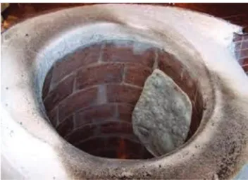
The current study revealed no association between HP infection and ESCC among people living in the eastern part of Turkey. The low prevalence of HP in these ESCC patients was similar to that reported in the Chinese meta-analyses. We also found a predominance of ESCC in female patients, in agreement with a previous Turkish study (17). This phe-nomenon has been linked to the use of the traditional large ovens (tandirs) that use smoke to cook meals (Figure 1). The mechanisms by which these ovens induce esophageal carci-nogenesis remain undefined, but previous work suggests that smoke may play role similar to that of cigarette smoking (17). We conclude that an effective follow-up strategy for HP-negative female Asian adults will be necessary if ESCC screening is to yield public health benefits. The higher levels of leucocytes and serum globulin among ESCC patients with HP may also reflect an emerging phenomenon that requires additional investigation to determine the underlying causa-tive factors.
The H. pylori-fast test (Hp-fast test) is based on the detection of HP urease activity and has a high sensitivity (85%) and specificity (495%) for detecting HP infection.
As this test is considered cost-effective and suitable for endoscopy units (24), we used the Hp-fast test to establish HP infection.
The current data are limited regarding any changes in the prevalence of HP in patients who have undergone esopha-gectomy. We therefore performed gastroduodenoscopy with pathological examination of the biopsy specimens obtained from the gastric conduit. We observed that the rate of HP infection was lower in esophagectomized ESCC patients than in the control subjects (19% and 78%, respectively;
po0.001). The rate of HP infection was similar, however,
between the naive ESCC patients and the esophagectomized patients (p40.005). A Japanese study reported that the HP Figure 1 -Traditional large oven (Tandir).
Figure 2 -Rate of HP according to groups.
152 HP in esophageal squamous cell carcinoma
infection status changed from preoperatively positive to postoperatively negative and that this changing pattern was linked to the eradication of HP via the perioperative administration of antibiotics (25). Conversely, a report from China showed a low incidence of HP infection in the gastric conduit in patients who underwent esophagectomy, pylor-oplasty, and reconstruction. The authors of that study con-cluded that this phenomenon was mostly due to the chronic reflux of bile after pyloroplasty (26).
Our findings were in concordance with previous reports on HP status and gastric tube cancer patients. In our Turkish population, HP infection was not associated with ESCC and had a similar pattern to that reported for Asian populations. HP infection may not contribute to the development of ESCC in patients who reside in rural areas of Asia, especially not in females. Furthermore, esophageal damage could be dimin-ished by the prior presence of HP in areas with a high risk of ESCC. HP treatment has been associated with impro-ved thrombocyte levels. An increase in platelet counts was observed in only 6.7% of treated patients. In the current study, we found no relationship between HP status and thrombocyte levels (27,28).
Finally, further studies are needed to address the impact of HP infection on ESCC and its natural history.
’ AUTHOR CONTRIBUTIONS
Poyrazoglu OB was involved in developing the concept and design of the study and writing the manuscript. Dulger AC was involved in imple-menting the study and collecting data. Dulger AC and Gultepe BS were involved in collecting and processing the data. All the authors read and
approved thefinal manuscript.
’ REFERENCES
1. Layke JC, Lopez PP. Esophageal cancer: a review and update. Am Fam Physician. 2006;73(12):2187-94.
2. Onuk MD, Oztopuz A, Memik F. Risk factors for esophageal cancer in eastern Anatolia. Hepatogastroenterology. 2002;49(47):1290-2.
3. Unidentified curved bacilli on gastric epithelium in active chronic gas-tritis. Lancet. 1983;1(8336):1273-5.
4. Vakil N, Malfertheiner P, Chey WD. Helicobacter pylori infection. N Engl J Med. 2010;363(6):595, http://dx.doi.org/10.1056/NEJMc1006158. 5. Suerbaum S, Michetti P. Helicobacter pylori infection. N Engl J Med.
2002;347(15):1175-86, http://dx.doi.org/10.1056/NEJMra020542. 6. Peek RM Jr, Blazer MJ. Helicobacter pylori and gastrointestinal tract
adenocarcinomas. Nat Rev Cancer. 2002;2(1):28-37, http://dx.doi.org/ 10.1038/nrc703.
7. Islami F, Kamangar F. Helicobacter pylori and esophageal cancer risk: a meta-analysis. Cancer Prev Res. 2008;1(5):329-38, http://dx.doi.org/ 10.1158/1940-6207.CAPR-08-0109.
8. Ye W, Held M, Lagergren J, Engstrand L, Blot WJ, McLaughlin JK, et al. Helicobacter pylori infection and gastric atrophy: risk of adenocarcinoma and squamous-cell carcinoma of the esophagus and adenocarcinoma of the gastric cardia. J Natl Cancer Inst. 2004;96(5):388-96, http://dx.doi. org/10.1093/jnci/djh057.
9. Wu DC, Wu IC, Lee JM, Hsu HK, Kao EL, Chou SH, et al. Helicobacter pylori infection: a protective factor for esophageal squamous cell
carcinoma in a Taiwanese population. Am J Gastroenterol. 2005;100 (3):588-93, http://dx.doi.org/10.1111/j.1572-0241.2005.40623.x. 10. Henrik Simán J, Forsgren A, Berglund G, Floren CH. Helicobacter pylori
infection is associated with a decreased risk of developing oesophageal neoplasms. Helicobacter. 2001;6(4):310-6, http://dx.doi.org/10.1046/j.1523-5378.2001.00041.x.
11. Holscher AH, Voit H, Buttermann G, Siewert JR. Function of the intra-thoracic stomach as esophageal replacement. World J Surg. 1998;12(6): 835-44, http://dx.doi.org/10.1007/BF01655491.
12. Ozaydin N, Turkyilmaz SA, Cali S. Prevalence and risk factors of Heli-cobacter pylori in Turkey: a nationally-representative, cross-sectional, screening with the13C-Urea breath test. BMC Public Health. 2013;13:1215,
http://dx.doi.org/10.1186/1471-2458-13-1215.
13. Enzinger PC, Mayer RJ. Esophageal cancer. N Engl J Med. 2003;349(23): 2241-52, http://dx.doi.org/10.1056/NEJMra035010.
14. Lambert R, Hainaut P. The multidisciplinary management of gastro-intestinal cancer. Epidemiology of oesophagogastric cancer. Best Pract Res Clin Gastroenterol. 2007;21(6):921-45, http://dx.doi.org/10.1016/ j.bpg.2007.10.001.
15. Ke L. Mortality and incidence trends from esophagus cancer in selected geographic areas of China circa 1970-90. Int J Cancer. 2002;102(3):271-4, http://dx.doi.org/10.1002/ijc.10706.
16. Koca T, Arslan D, Basaran H, Cerkesli AK, Tastekin D, Sezen D, et al. Dietary and demographical risk factors for oesophageal squamous cell carcinoma in the Eastern Anatolian region of Turkey where upper gas-trointestinal cancers are endemic. Asian Pac J Cancer Prev. 2015;16(5): 1913-7, http://dx.doi.org/10.7314/APJCP.2015.16.5.1913.
17. Turkdogan MK, Akman N, Tuncer I, Uygan I, Kösem M, Ozel S, et al. Epidemiological aspects of endemic upper gastrointestinal cancers in eastern Turkey. Hepatogastroenterology. 2005;52(62):496-500.
18. Türkdog˘an K, Alici S, I˙lhan M, Dilek H, Akman E, Ayakta H, et al.
Helicobacter pylori infection in gastric carcinoma in the Van region of Turkey. Turk J Gastroenterol. 1999;10(1):36-9.
19. Lambert R, Hainaut P. Esophageal cancer: cases and causes (part I). Endoscopy. 2007;39(6):550-5, http://dx.doi.org/10.1055/s-2007-966530. 20. Goodwin CS, Mendall MM, Northfield TC. Helicobacter pylori infection.
Lancet. 1997;349(9047):265-9, http://dx.doi.org/10.1016/S0140-6736(96) 07023-7.
21. Wu IC, Wu DC, Yu FJ, Wang JY, Kuo CH, Yang SF, et al. Association between Helicobacter pylori seropositivity and digestive tract cancers. World J Gastroenterol. 2009;15(43):5465-71, http://dx.doi.org/10.3748/ wjg.15.5465.
22. Kamangar F, Qiao YL, Blaser MJ, Sun XD, Katki H, Fan JH, et al. Heli-cobacter pylori and oesophageal and gastric cancers in a prospective study in China. Br J Cancer. 2007;96(1):172-6, http://dx.doi.org/10.1038/ sj.bjc.6603517.
23. Xie FJ, Zhang YP, Zheng QQ, Jin HC, Wang FL, Chen M, et al. Helico-bacter pylori infection and esophageal cancer risk: an updated meta-analysis. World J Gastroenterol. 2013;19(36):6098-107, http://dx.doi.org/ 10.3748/wjg.v19.i36.6098.
24. Onders RP. Detection methods of Helicobacter pylori: accuracy and costs. Am Surg. 1997;63(8):665-8.
25. Mori N, Fujita H, Sueyoshi S, Aoyama Y, Yanagawa T, Shirouzu K. Helicobacter pylori infection influences the acidity in the gastric tube as an esophageal substitute after esophagectomy. Dis Esophagus. 2007;20(4): 333-40, http://dx.doi.org/10.1111/j.1442-2050.2007.00718.x.
26. Kise Y, Kijima H, Shimada H, Tanaka H, Kenmochi T, Chino O, et al. Gastric tube cancer after esophagectomy for esophageal squamous cell cancer and its relevance to Helicobacter pylori. Hepatogastroenterology. 2003;50(50):408-11.
27. Ahn ER, Tiede MP, Jy W, Bidot CJ, Fontana V, Ahn YS. Platelet activation in Helicobacter pylori-associated idiopathic thrombocytopenic purpura: eradication reduces platelet activation but seldom improves platelet counts. Acta Haematol. 2006;116(1):19-24, http://dx.doi.org/10.1159/ 000092343.
28. Sultan S, Irfan SM, Kaker J, Hasan M. Efficacy of helicobacter pylori eradication as an upfront treatment of secondary immune thrombocyto-penia: an experience from Pakistan. Med J Malaysia. 2016;71(2):53-6.
153