CLINICAL SCIENCE
Helicobacter pylori as a potential target for the
treat-ment of central serous chorioretinopathy
Antonio Marcelo Barbante Casella,IRodrigo Fabri Berbel,IGla´ucio Luciano Bressanim,IIMarcus Rudolph Malaguido,IJose´ Augusto CardilloIII
IUniversidade Estadual de Londrina, Department of Ophthalmology, Londrina, Brazil.IIInstituto da Visa˜o de Cascavel, Department of Ophthalmology, Cascavel, Brazil.IIIHospital de Olhos de Araraquara, Department of Ophthalmology, Araraquara, Brazil.
OBJECTIVES: The objective of this study was to evaluate the relationship between the treatment ofHelicobacter pylorigastric infection and changes in best-corrected visual acuity and macular detachment in patients with chronic central serous chorioretinopathy.
METHODS:Seventeen patients diagnosed with central serous chorioretinopathy were examined for gastric infection withHelicobacter pyloriusing the urease test and gastric biopsy.Helicobacter pylori-positive patients were treated with the appropriate medication. The response to therapy was monitored by evaluating the best-corrected visual acuity and optical coherence tomography. The data were analyzed using Student’s t-test before and after treatment.
RESULTS:Fourteen patients (15 eyes) aged 30-56 years (mean 43.4¡8.3 years) were positive forHelicobacter pylori. Most of the positive patients had gastric symptoms (78.5%); one had bilateral central serous chorioretinopathy. The mean baseline best-corrected visual acuity was 20/98 (logMAR = 0.53¡0.28). Three months after starting treatment with antibiotics, the serous detachment had resolved in 14 of 15 eyes, but two cases required laser treatment. The follow-up period ranged from 6 to 27 months. The mean final best-corrected visual acuity differed significantly from baseline.
CONCLUSION:Our findings suggest thatHelicobacter pyloriinfection may be present in many chronic central serous chorioretinopathy patients and that treatment for the infection may have a favorable effect on the outcome of chronic central serous chorioretinopathy. Due to the possibility of the spontaneous regression of chronic central serous chorioretinopathy and the high prevalence of the infection in the general population, prospective and masked clinical trials are necessary to confirm that treatment forHelicobacter pyloriinfection may benefit patients with chronic central serous chorioretinopathy.
KEYWORDS: Risk Factors; Central Serous Chorioretinopathy; Helicobacter Pylori; Treatment; Macula.
Casella AM, Berbel RF, Bressanim GL, Malaguido MR, Cardillo JA. Helicobacter pylori as a potential target for the treatment of central serous chorioretinopathy. Clinics. 2012;67(9):1047-1052.
Received for publication onMay 1, 2012;First review completed onMay 17, 2012;Accepted for publication onMay 17, 2012
E-mail: [email protected]
Tel.: 55 43 3324-1177
INTRODUCTION
Central serous chorioretinopathy (CSC) causes a serous neurosensory detachment of the macula associated with one or more idiopathic leaks at the level of the retinal pigment epithelium (RPE). It predominantly affects male adults aged 40-60 years (1,2). Although the condition spontaneously improves in at least 90% of those affected, recurrence affects approximately one third of patients (3). Among patients with CSC, the chronic form of central serous chorioretino-pathy (CCSC) is defined as the repeating variant of the
disease or diffuse retinal pigment epitheliopathy for more than six months (4,5). These patients are difficult to treat, with the most successful modality appearing to be photo-dynamic therapy (PDT) guided by optical coherence tomography (OCT). However, patients may lose vision due to atrophic changes in the RPE/photoreceptor layer (5). The potential risk factors for CSC include hypertension, pregnancy, allergic respiratory disease, antibiotic or alcohol use, corticosteroid therapy and sympathomimetic drugs (1,2,6-8). A correlation between CSC and infection with
Helicobacter pylori (HP) was recently suggested (9-15). In a prospective pilot study of 16 patients with CSC and diffuse retinal pigment epitheliopathy, the prevalence ofHelicobacter pyloriinfection was significantly higher in patients with CSC than in the general population (10). The prevalence was also significantly higher in CSC patients than in a control population from the same country (11). Other studies also demonstrated this correlation (14,15). One recent article
Copyrightß2012CLINICS– This is an Open Access article distributed under
the terms of the Creative Commons Attribution Non-Commercial License (http:// creativecommons.org/licenses/by-nc/3.0/) which permits unrestricted non-commercial use, distribution, and reproduction in any medium, provided the original work is properly cited.
found good outcomes in patients with CSC and receiving treatment for HP eradication therapy (16). Thus, the objective of the present study was to assess the best-corrected visual acuity (BCVA) and macular detachment using OCT in patients with CCSC who were receiving conventional drug therapy to treat HP infection.
MATERIALS AND METHODS
This was an interventional, non-blinded case series of patients with proven chronic CSC. The study was approved by the research ethics committee of the State University of Londrina, and all patients provided informed written consent. CSC was defined as an idiopathic leakage at the level of the retinal pigment epithelium leading to the accumulation of subretinal fluid. Subjects diagnosed with long-lasting (.6 months) or recurring (2 or more times over the preceding 12 months) CSC and serous macular detach-ment involving the fovea were followed by the authors from September 2003 to November 2008. Twenty patients were invited to participate in the study, but only 17 accepted. None of the subjects used corticosteroids or sympathomi-metic drugs or had a systemic disease associated with CSC. The patients had a complete ophthalmological examina-tion, including best-corrected Snellen visual acuity (BCVA), slit-lamp biomicroscopy, tonometry, indirect binocular ophthalmoscopy, optical coherence tomography (time domain OCT: Stratus OCT-3000; Carl Zeiss Meditec, Dublin, CA, USA) and, in the absence of confirmed or suspected allergy and clinical contraindication, fluorescein angiography. Patients with clinically and image-confirmed serous detachment were asked about gastric symptoms and referred for HP screening with urease tests and gastric biopsy by endoscopy when the urease test was negative (17).
HP-positive patients were treated with a combination of clarithromycin (500 mg twice a day), amoxicillin (1 g twice a day) and lansoprazole (30 mg twice a day) for seven days. The treatment was prescribed and followed by a gastro-enterologist who performed the endoscopic examination of the upper digestive tract. Following drug therapy, patients underwent complete ophthalmological examinations at 30-day intervals to assess visual acuity and ocular status. OCT images were used to assess the resolution of disease (absence of subretinal fluid). The follow-up period ranged from 6 to 27 months. A second endoscopic examination and gastric biopsy was performed three months after drug therapy to confirm the eradication of the bacteria. Follow-up was continued through November 30, 2008, or ended when the patient decided to undergo another treatment. The variables are presented in a descriptive form. Student’s paired t test was used to compare the baseline and end-point BCVA values, andp-values,0.05 were considered to be statistically significant.
RESULTS
The study population consisted of three women and 14 men. Eighteen eyes of 17 patients, aged 30-56 years (mean: 43.4¡8.7 years), were studied. The follow-up period varied from six to 27 months (mean 15¡8.1 months). Fifteen eyes tested positive for HP. Table 1 shows the patients according to age, gender, the affected eye, disease duration, the presence of gastric symptoms, HP examination and visual acuity at baseline and after treatment. One case (Case 2) presented with bilateral CCSC. In four patients, serous detachment recurred (Cases 1, 8, 11 and 14).
Case 1 had CCSC in one eye that occurred in September 2003. After HP detection, antibiotic treatment was started, and the serous detachment resolved completely after two
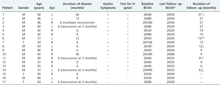
Table 1 -The results of the follow-up of 17 patients (18 eyes) diagnosed with central serous chorioretinopathy (CSC) who were screened and treated for gastric infection withHelicobacter pylori(HP).
Patient Gender Age (years) Eye
Duration of disease (months)
Gastric Symptoms
Test for H. pylori
Baseline BCVA
Last follow- up BCVA*
Duration of follow- up (months)
1 M 56 L 36 + + 20/40 20/30 27{
2 M 46 L 13 + + 20/80 20/50 27
2 M 46 R 6 (multiple recurrences) + + 20/200 20/30 27
3 M 56 L 6 (recurrence at 3 months) + + 20/80 20/20 27
4 M 42 R 6 + + 20/30 20/20 19
5 M 42 R 6 + + 20/80 20/20 13
6 M 31 L 12 - + 20/50 20/50 13**
7 F 50 R 6 + + 20/100 20/30 13
8 M 43 L 6 + + 20/30 20/20 12{
9 M 36 R 6 + + 20/50 20/20 9
10 M 45 R 36 + + 20/200 20/80 9
11 M 30 R 6 (recurrence at 3 months) - + 20/40 20/25 9{{
12 M 37 R 6 - + 20/60 20/25 9
13 M 55 R 6 + + 20/40 20/25 6
14 M 37 L 6 (recurrence at 3 months) + + 20/400 20/25 6{{
15 F 45 R 6 - - 20/30 20/30
---16 M 40 L 6 - - 20/50 20/50
---17 F 43 L 6 (recurrence at 3 months) - _ 20/80 20/20
---*The baseline visual acuity was 20/98 (logMAR = 0.53 standard deviation [¡] 0.28); the final visual acuity was 20/31 (log MAR 0.16¡0.17) {(p= 0.001) paired Student’s t test (for patients 1-14)
{
Recurrence with serous detachment. Second biopsy positive. Retreated with improvement of serous detachment. **Persistent serous detachment treated with laser photocoagulation
{{
Recurrence after anti-HP drug therapy and a negative gastric biopsy for HP. Patient treated with laser photocoagulation.
{{
months (Figure 1). This patient experienced a recurrence after 20 months and again tested positive for HP. After retreatment, the serous detachment resolved. Case 3 was positive for HP after the first treatment and underwent retreatment. The patient experienced a complete resolution of the serous detachment one month after the treatment ended. All of the other patients experienced resolution of their serous detachment after the first month of treatment except Case 6, who required laser photocoagulation. Case 2 had bilateral serous detachment but no recurrence during the 27 months of follow-up (Figure 2). Case 4 (Figure 3), Case 5 (Figure 4), Case 13 and Case 14 had leaks near the fovea, precluding regular laser treatment. Case 6, despite testing positive for HP, did not improve after antibiotic treatment and was referred for laser photocoagulation of the leakage site. Case 11 experienced complete resolution of the serous detachment after treatment with antibiotics but experienced a CSC recurrence two months later. The gastric biopsy was negative for HP, and the patient was treated with laser coagulation. Case 9 experienced a serous detachment recurrence seven months after successful treatment and was subsequently found to have HP infection. The serous detachment resolved after HP retreatment, and the patient’s
visual acuity improved from 20/40 to 20/20. In Case 14, the serous detachment resolved but recurred after five months. The second biopsy was HP positive, but the patient requested treatment with both anti-HP medication and photodynamic therapy, which was successful. The mean baseline visual acuity was 20/98 (logMAR = 0.57¡0.28). The mean final visual acuity was 20/31 (log MAR 0.16¡0.17) and differed
significantly from baseline (p,0.001).
DISCUSSION
To our knowledge, this is the first study in the literature that treated HP in chronic cases of CSC and reported improvement following an evaluation of BCVA and an OCT examination. A similar study was performed in 2002 (10); however, in that study, HP was not eradicated, the sample size (16 eyes) was smaller than the current study (18 eyes), and improvements in BCVA and OCT were not evaluated. A French study found HP to be significantly more prevalent in patients diagnosed with CSC than in the normal population (11). In our study, a higher incidence of
Helicobacter pyloriinfection (82.35% - 15 patients and 18 eyes) was found than the previously reported prevalence in southern Brazil (63%) (18,19). Interestingly, 13 of 14 patients showed improvement of CCSC following treatment for HP. However, one of these cases recurred, and laser photo-coagulation was required after a negative gastric biopsy. In three patients, CCSC recurrence was associated with the recurrence of HP infection, and serous detachment resolved upon anti-HP retreatment in two of the cases; in the third case, the patient asked for both photodynamic therapy and anti-HP medication. In most of the responding cases, serous detachment resolved within one month after antibiotic treatment. One previously reported case of CCSC improved clinically when the gastric infection was eradicated, and recurrences of CCSC in this patient were associated with the return of the bacterial infection (9). The recurrence rate of 27% (5 of 18 eyes) indicates that it is a frequent problem associated with CSC, and this has been previously reported (3,5).
Recently, a decrease in the subretinal fluid reabsorption time was demonstrated in a group that received anti-HP treatment compared to a control group (16).
HP chronically infects the gastric mucosa of more than half of the human population (20). However, only 10-20% of HP-infected patients develop known gastrointestinal com-plications, such as peptic ulcer, gastric cancer and MALT lymphoma, during their lifetime (21). The clinical outcome of HP infection is determined by a complex interaction between the bacterium and the host (22). HP has also been associated with extra-gastric disorders, including a strong association with idiopathic thrombocytopenic purpura and, to a lesser degree, iron deficiency anemia, cardiovascular disease and atherosclerosis (23). The explanations proposed for these observed associations involve one or more mechanisms with direct pathogenic effects, including molecular mimicry between host and Helicobacter pylori
antigens, the chronic release of inflammatory mediators, malabsorption and the abnormal production of vasoactive substances (20). Indeed, Helicobacter pylori infection may alter vascular function by increasing endothelin 1, inducible nitric oxide synthase and nitric oxide, which may affect a condition such as CSC (24,25). Although all of these data suggest that HP infection is a systemic disease, we found
only one case with bilateral CCSC among HP-positive patients. However, if we were to evaluate angiogram abnormalities in the contralateral eye, they would be present in a significant percentage of patients (3). Another hypoth-esis is that the medications used to treat HP infection interfere with cortisol metabolism; for example, a decrease in systemic cortisol levels was found after the use of clarithromycin (26). One interesting finding of this research is that 78.5% of the HP-positive patients and 64.7% of all patients had gastric symptoms. This was previously noted by other authors but was not observed in other studies (1,2,6-13,27).
We evaluated the initial BCVA, final BCVA and complete resolution of fluid by OCT and fluorescein angiography. The OCT examination is particularly important in CCSC cases because RPE and photoreceptors may be damaged, and BCVA may not be the best parameter to evaluate the possible benefits of HP treatment, as stated by other authors (28).
This study was limited by its design. The retrospective analysis of a multifactorial condition with a considerable potential for spontaneous resolution, such as CCSC, is problematic. Additionally, the high prevalence of HP infection in the general population is another potential confounding factor to be considered in a retrospective study with a lack of controls. The observed changes in the macular pathology may simply be due to chance or placebo effects or
even indirectly to changes in the emotions of the patients with HP infection. However, the natural history of CCSC observed in practice is different from that reported in this study. Additionally, our findings suggest the possibility that HP infection plays a role in the outcome of patients with CCSC: serous detachment generally improved shortly after initiating HP eradication treatment; the recurrence of CCSC often coincided with the recurrence of HP infection; and the retreatment of recurrent infection led to the resolution of recurrent serous detachment.
Our findings suggest that HP infection may be present in many CCSC patients and that eradication therapy may have a favorable effect on the outcome of the disease. Due to the multifactorial causes of CCSC, the possibility of spontaneous regression and the high prevalence of the infection in the general population, prospective and masked clinical trials are necessary to confirm that treatment for HP infection may benefit patients with CCSC. Such a suggestion was pre-viously made by Abreu et al. in 2008 in their reply (29).
AUTHOR CONTRIBUTIONS
REFERENCES
1. Tittl MK, Spaide RF, Wong D, Pilotto E, Yannuzzi LA, Fisher YL, et al. Systemic and ocular findings in central serous chorioretinopathy. Am J Ophthalmol. 1999;128(1):63-8, http://dx.doi.org/10.1016/S0002-9394(99)00075-6.
2. Haimovici R, Koh S, Gagnon DR, Lehrfeld T, Wellik S. Risk factors for central serous chorioretinopathy: a case-control study. Ophthalmology. 2004;111(20):244-9, http://dx.doi.org/10.1016/j.ophtha.2003.09.024. 3. Guyer DR, Gagroudas ES. Central Serous Chorioretinopathy. In: Albert
DM, Jakobiec FA. Principles and Practice of Ophthalmology. Philadelphia: Saunders, 1994;818-25.
4. Spaide RF, Campeas L, Haas A, Yannuzzi LA, Fisher YL, Guyer DR, et al. Central serous chorioretinopathy in younger and older adults. Ophthalmology. 1996;103(12):2070-80.
5. Yannuzzi LA, Slakter JS, Gross NE, Spaide RF, Costa DL, Huang SJ, et al. Indocyanine green angiography-guided photodynamic therapy for treatment of chronic central serous chorioretinopathy: a pilot Study. Retina. 2003;23(3):288-98, http://dx.doi.org/10.1097/00006982-200306000-00002.
6. Carvalho-Recchia CA, Yannuzzi LA, Negra˜o S, Spaide RF, Freund KB, Rodriguez-Coleman H, et al. Corticosteroids and central serous chorioretinopathy. Ophthalmology. 2002;109(1):1834-7, http:// dx.doi.org/10.1016/S0161-6420(02)01117-X.
7. Koyama M, Mizota A, Igarashi Y, Adachi-Usami E. Seventeen cases of central serous chorioretinopathy associated with systemic corticosteroid therapy. Ophthalmologica. 2004;218(2):107-10, http://dx.doi.org/ 10.1159/000076145.
8. Michael JC, Pak J, Pulido J, de Venecia G. Central serous chorioretino-pathy associated with administration of sympathomimetic agents. Am J Ophthalmol. 2003;136(1):182-5, http://dx.doi.org/10.1016/S0002-9394(03)00076-X.
9. Giusti C. Central serous chorioretinopathy: a new extragastric manifes-tation of Helicobacter pylori? Analysis of a clinical case. Clin Ter. 2001;152(6):393-7.
10. Mauget-Fay¨sse M, Kodjikian L, Quaranta M, Ben Ezra D, Trepsat C, Mion F, et al. Roˆle de l’Helicobacter pylori dans la choriore´tinopathie se´reuse centrale et l’e´pithe´liopathie re´tinienne diffuse. Re´sultats de la premie`re e´tude prospective pilote. J Fr Ophtalmol. 2002;25(10):1021-5. 11. Ahnoux-Zabsonre A, Quaranta M, Mauget-Fay¨sse M. Pre´valence de
l’Helicobacter pylori dans la choriore´tinopathie se´reuse centrale et l’e´pithe´liopathie re´tinienne diffuse. J Fr Ophtalmol. 2004;27(10):1129-33, http://dx.doi.org/10.1016/S0181-5512(04)96281-X.
12. Asensio-Sa´nchez VM, Rodrı´guez-Delgado B, Garcı´a-Herrero E, Cabo-Vaquera V, Garcı´a-Loygorri C. Coriorretinopatı´a serosa central como manifestacio´n extradigestiva de infeccio´n ga´strica por Helicobacter pylori. Arch Soc Esp Oftalmol. 2008;83(3):177-82.
13. Giusti C. Association of Helicobacter pylori with central serous chorior-etinopathy: hypotheses regarding pathogenesis. Med Hypotheses. 2004;63(3):524-7, http://dx.doi.org/10.1016/j.mehy.2004.02.020. 14. Cotticelli L, Borrelli M, D’Alessio AC, Menzione M, Villani A, Piccolo G,
et al. Central serous chorioretinopathy and Helicobacter pylori. European journal of ophthalmology. 2006;16(2):274-8.
15. Misiuk-Hojło M, Michałowska M, Turno-Krecicka A. Helicobacter pylori--a risk factor for the development of the central serous chorioretinopathy. Klin Oczna. 2009;111(1-3):30-2.
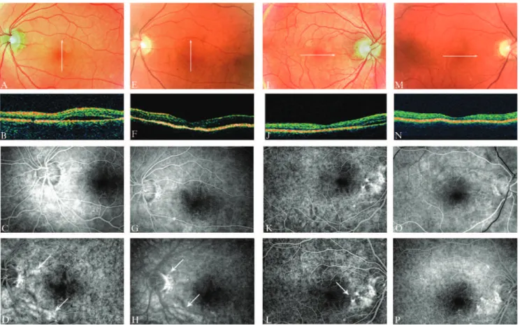
16. Rahbani-Nobar MB, Javadzadeh A, Ghojazadeh L, Rafeey M, Ghorbanihaghjo A. The effect of Helicobacter pylori treatment on remission of idiopathic central serous chorioretinopathy. Mol Vis. 2011;17:99-103. Figure 3 -A patient with a six-month history of CSC and macular
serous detachment (A-B). Fluorescein angiography showed RPE changes and a juxtafoveal leak (arrow), precluding regular laser treatment (C-D). The serous detachment improved after anti-HP treatment (E-F), and fluorescein angiography showed prominent resolution of the leakage with persistence of the RPE changes (G-H).
17. Ogata SK, Kawakami E, Patrı´cio FRS, Pedroso MZ, Santos AM. Evolution of invasive and non-invasive methods for the diagnosis of Helicobacter pylori in symptomatic children and adolescents. Sa˜o Paulo Med J. 2001;119(2):67-71, http://dx.doi.org/10.1590/S1516-31802001000200006. 18. Santos IS, Boccio J, Santos AS, Valle NC, Halal CS, Bachilli MC, et al.
Prevalence of Helicobacter pylori infection and associated factors among adults in Southern Brazil: a population-based cross-sectional study. BMC Public Health. 2005;5:118, http://dx.doi.org/10.1186/1471-2458-5-118. 19. Zaterka S, Eisig JN, Chinzon D, Rothstein W. Factors related to
Helicobacter pylori prevalence in an adult population in Brazil. Helicobacter. 2007;12(1):82-8.
20. Mitchell HM. The epidemiology of Helicobacter pylori. Curr Top Microbiol Immunol. 1999;241:11-30, http://dx.doi.org/10.1007/978-3-642-60013-5_2. 21. D’Elios MM, Andersen LP. Helicobacter pylori: Inflammation, Immunity, and Vaccines. Helicobacter. 2007;12(Suppl.1):15-9, http:// dx.doi.org/10.1111/j.1523-5378.2007.00530.x.
22. Maeda S, Mentis AF. Pathogenesis of Helicobacter pylori Infection. Helicobacter. 2007;12 (Suppl.1):10-4.
23. Bohr URM, Annibale B, Franceschi F, Roccarina D, Gasbarrini A. Extragastric Manifestations of Helicobacter pylori Infection – Other Helicobacters. Helicobacter. 2007;12(Suppl. 1):45-53.
24. Whittle BJR, Morschl E, Pozsa´r J, Moran AP, La´szlo´ F. Helicobacter pylori lipopolysaccharide provokes iNOS-mediated acute systemic microvascular inflammatory responses in rat cardiac, hepatic, renal and pulmonary tissues. J Physiol Paris. 2001;95(1-6):257-9, http:// dx.doi.org/10.1016/S0928-4257(01)00035-3.
25. Giusti C, Mauget-Fay¨sse M. Helicobacter pylori and idiopathic central serous chorioretinopathy. Swiss Med Wkly. 2004;134(27-28):395-8. 26. Ushiama H, Echizen H, Nachi S, Ohnishi A. Dose-dependent inhibition
of CYP3A activity by clarithromycin during Helicobacter pylori eradication therapy assessed by changes in plasma lansoprazole levels and partial cortisol clearance to 6beta-hydroxycortisol. Clin Pharmacol Ther. 2002;72(1):33-43, http://dx.doi.org/10.1067/mcp.2002.125559. 27. Mansuetta CC, Mason JO 3rd, Swanner J, Feist RM, White MF Jr,
Thomley ML, et al. An association between central serous chorioretino-pathy and gastroesophageal reflux disease. Am J Ophthalmol. 2004;137(6):1096-100, http://dx.doi.org/10.1016/j.ajo.2004.01.054. 28. Aggio FB, Roisman L, Melo GB, Lavinsky D, Cardillo JA, Farah ME.
Clinical factors reralted to the visual outcome in central serous choriorretinopathy. Retina. 2010;30(7):128-34.