.
Prognostic value of the neutrophil to lymphocyte
ratio in lung cancer: A meta-analysis
Yongmei Yin,IJun Wang,II,*Xuedong Wang,ILan Gu,IHao Pei,II Shougang Kuai,II Yingying Zhang,II Zhongbo ShangII
IJiangnan University, The Fifth People
’s Hospital of Wuxi, Radiology Department, Wuxi, Jiangsu, China.IIJiangnan University, The Fifth People
’s Hospital of Wuxi, Center of Clinical Laboratory, Wuxi, Jiangsu, China.
Recently, a series of studies explored the correlation between the neutrophil to lymphocyte ratio and the prognosis of lung cancer. However, the current opinion regarding the prognostic role of the neutrophil to lymphocyte ratio in lung cancer is inconsistent.
We performed a meta-analysis of published articles to investigate the prognostic value of the neutrophil to lymphocyte ratio in lung cancer. The hazard ratio (HR) and its 95% confidence interval (CI) were calculated. An elevated neutrophil to lymphocyte ratio predicted worse overall survival, with a pooled HR of 1.243 (95%CI:
1.106-1.397; Pheterogeneity=0.001) from multivariate studies and 1.867 (95%CI: 1.487-2.344; Pheterogeneity=0.047) from univariate studies. Subgroup analysis showed that a high neutrophil to lymphocyte ratio yielded worse overall survival in non-small cell lung cancer (NSCLC) (HR=1.192, 95%CI: 1.061-1.399; Pheterogeneity=0.003) as well as small cell lung cancer (SCLC) (HR=1.550, 95% CI: 1.156-2.077; Pheterogeneity=0.625) in multivariate studies. The synthesized evidence from this meta-analysis of published articles demonstrated that an elevated neutrophil to lymphocyte ratio was a predictor of poor overall survival in patients with lung cancer.
KEYWORDS: NLR; Lung Cancer; Overall Survival; Meta-Analysis.
Yin Y, Wang J, Wang X, Gu L, Pei H, Kuai S, et al. Prognostic value of the neutrophil to lymphocyte ratio in lung cancer: A meta-analysis. Clinics. 2015;70(7):524-530
Received for publication onJanuary 27, 2015;First review completed onMarch 18, 2015;Accepted for publication onApril 30, 2015
E-mail: [email protected]
*Corresponding author
’ INTRODUCTION
Lung cancer is one of the leading causes of cancer-related deaths (1,2). Non-small cell lung cancer (NSCLC) accounts for approximately 85% of lung cancer cases, and small-cell lung cancer (SCLC) accounts for nearly 13% of overall lung cancer cases. Despite continue efforts and progress in diagnosis and treatment, the overall survival (OS) for lung cancer patients remains poor (1,2). Prognostic factors influencing survival have been previously identified, includ-ing tumor stage, performance status, weight loss, age, sex, histopathology, and plasma lactate dehydrogenase (LDH) and carcinoembryonic antigen (CEA) levels (3-7). Although novel immunological and histological biomarkers such as intercellular adhesion molecule-1 (IDM-1) and epidermal growth factor receptor (EGFR) have been identified (8,9), these marks are expensive and often time-consuming to
measure. Thus, there remains no promising prognostic factor that can be easily detected and closely linked to clinical outcomes for lung cancer patients (10).
The tumor immune environment plays an important role in tumor progression by promoting tumor angiogenesis, tumor metastasis, and cancer cell proliferation and by interfering with the response to systemic treatment (11,12). Neutrophils and T and B lymphocytes have been suggested to play vital roles in tumor inflammation (13,14), and the imbalance between neutrophils and lymphocytes is thought to be secondary to tumor hypoxia or necrosis and associated with anti-apoptotic effects (15). The neutrophil to lympho-cyte ratio (NLR), representing a combination of circulating neutrophil and lymphocyte counts, can reflect the imbalance between neutrophils and lymphocytes in patients with tumors and serves as a representative index of systemic inflammation.
Recently, an elevated preoperative or pretreatment NLR, calculated from peripheral blood tests, was identified as an independent and readily available prognostic biomarker related to poor survival in numerous cancers, including colorectal cancer, breast cancer, gastric cancer and esopha-geal cancer (16-19). Additionally, a series of studies have explored the correlation between the NLR and the prognosis of lung cancer. However, according to their results, the
DOI:10.6061/clinics/2015(07)10
Copyright&2015CLINICS–This is an Open Access article distributed under the terms of the Creative Commons Attribution Non-Commercial License (http:// creativecommons.org/licenses/by-nc/3.0/) which permits unrestricted non-commercial use, distribution, and reproduction in any medium, provided the original work is properly cited.
current opinion on the prognostic role of the NLR in lung cancer is inconsistent and inconclusive. Thus, we performed this meta-analysis, which is the first systematic study on the subject, to investigate the prognostic value of the NLR in lung cancer.
’ MATERIALS AND METHODS
Search strategy and study selection
To identify eligible studies regarding the NLR for predicting the prognosis of lung cancer, a systematic review was conducted. Relevant studies were identified by searching the PubMed and Web of Science databases using the following search terms: NLR, neutrophil-to-lymphocyte ratio, neutrophil lymphocyte ratio or neutrophil-lymphocyte ratio with lung cancer, carcinoma of the lung, pulmonary carcinoma and prognosis, prognostic, survival or outcome. The last search was updated on October 31, 2014. Both Medical subheadings (MeSH) and free text terms were used as keywords. The reference lists of papers of interest and published review articles were also explored to potentially retrieve additional studies. The inclusion criteria for the studies were as follows: (a) provided clear information on lung cancer confirmation and the included patients; (b) investigated the association of the pre-treatment NLR with OS; and (c) full text articles in English. The exclusion criteria were as follows: (a) letters, reviews, expert opinions, case reports or laboratory studies; (b) studies with overlapping or duplicate data; and (c) a lack of key information for evaluating the hazard ratio (HR) for further analysis.
Data extraction
All searches were conducted independently by two investigators. The same two authors independently extracted data on the name of the first author, the year of publication, the country of origin, ethnicity, the total number of cases, cancer types, stages, cut-off values, follow ups and HRs of the NLR for OS with 95% confidence intervals (CIs). Any discrepancy was resolved by consensus and, if needed, by consultation with the third author.
Statistical analysis
OS results were evaluated as HRs for each included study. HRs and 95% CIs were obtained directly from each publication. If not available, the necessary data were extracted to calculate the HR using the method reported by Tierney et al. (20). The heterogeneity of pooled results was estimated using Cochran’s Q test and Higgins’I-squared statistic. A p-valueo0.10 for the
Q-test indicated significant heterogeneity, and a random-effects model (DerSimonian-Laird method) was used to calculate the pooled HRs (21). Otherwise, a fixed-effects model (Mantel-Haenszel method) was applied (22). Egger’s linear regression and Begg’s funnel plot test were applied to evaluate publication bias in the literature, and a p-value o0.05 was considered
significant. A trim and fill method was performed to estimate asymmetry in the funnel plot. Meta-regression was performed to explore the potential source of heterogeneity using variables such as the year of publication, ethnicity, cancer type, cutoff value and sample size. To validate the credibility of outcomes in this meta-analysis, sensitivity analysis was performed by sequential omission of each individual study using the
‘‘metainf’’ STATA command. All statistical analyses were
conducted with STATA software version 12.0 (STATA Corpora-tion, College StaCorpora-tion, TX, USA), and all p-values were two-sided.
’ RESULTS
Study characteristics
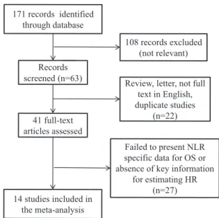
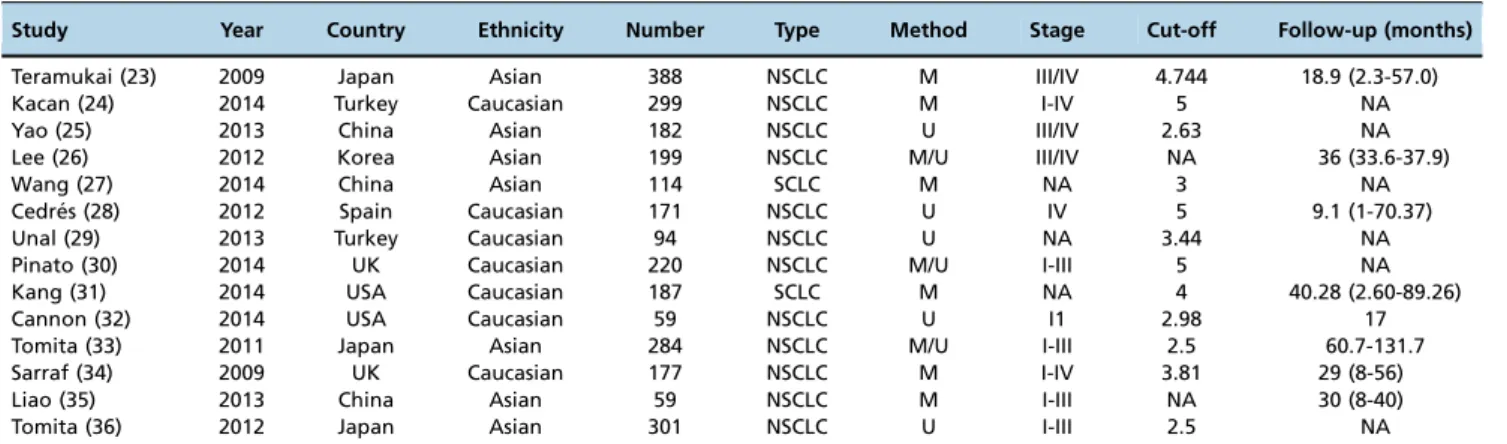
We identified fourteen studies according to the inclusion and exclusion criteria (23-36). The detailed screening process is shown in Figure 1. All of these articles were published in English. The characteristics of the included studies are shown in Table 1. Three studies were performed in China and Japan, whereas two each were performed in the USA, UK and Turkey and one each in Spain and Korea. Among the included studies, participants were Asian in seven studies and Caucasian in the other seven studies. A total of twelve studies explored the NLR in the prognosis of NSCLC, and two studies explored the NLR in SCLC. The cut-off value used in each study was not consistent, ranging from 2.5 to 5.0. The number of patients in each study ranged from 59 to 388. Six studies calculated HRs by multivariate analysis, five studies calculated HRs by univariate analysis, and the other three studies calculated HRs by both multivariate and univariate analyses. In total, nine studies contained HRs calculated from multivariate analysis, and eight studies contained HRs calculated from univariate analysis.
Outcome from eligible studies
As shown in Table 2, fourteen studies evaluating OS were classified into two groups: nine multivariate studies with HRs and 95% CIs acquired from multivariate analysis, and eight univariate studies with data from univariate analysis. In both the multivariate and univariate analysis groups, an elevated NLR predicted a worse outcome of OS, with a pooled HR of 1.243 (95%CI: 1.106-1.397; Pheterogeneity=0.001) and 1.867 (95%
CI: 1.487-2.344; Pheterogeneity=0.047), respectively (Figure 2).
Subgroup analysis by cancer type in the multivariate studies showed that a high NLR yielded worse OS in NSCLC (HR=1.192, 95%CI: 1.061-1.399; Pheterogeneity=0.003) and
SCLC (HR=1.550, 95%CI: 1.156-2.077; Pheterogeneity=0.625).
The cancer type in the univariate studies was NSCLC only. In the subgroup analysis by ethnicity, regardless of whether the patients were Asian or Caucasian, an elevated NLR remained a poor predictor of OS in multivariate studies
171 records identified through database
Records screened (n=63)
108 records excluded (not relevant)
Review, letter, not full text in English, duplicate studies
(n=22)
Failed to present NLR specific data for OS or absence of key information
for estimating HR (n=27) 41 full-text
articles assessed
14 studies included in the meta-analysis
(Caucasian: HR=1.545, 95%CI: 1.052-2.269; Pheterogeneity=0.005;
Asian: HR=1.261, 95%CI: 1.092-1.547; Pheterogeneity=0.021)
and univariate studies (Caucasian: HR=1.722, 95%CI: 1.360-2.179; Pheterogeneity=0.133; Asian: HR=1.661, 95%CI:
1.419-1.945; Pheterogeneity=0.036).
Considering different cut-off values, these studies used two subsets of NLR cut-offs and revealed similar results. The NLR was found to be a negative prognostic marker for the outcome of OS in multivariate studies (NLRX4:
HR=1.646, 95%CI: 1.319-2.053; Pheterogeneity=0.247; NLRo4:
HR=1.221, 95%CI: 1.016-1.468; Pheterogeneity=0.082) and
univariate studies (NLRX4: HR=1.500, 95%CI: 1.111-2.025;
Pheterogeneity=0.262; NLRo4: HR=2.043, 95%CI: 1.497-2.789;
Pheterogeneity=0.017).
Further analysis of studies evaluating OS by sample size (studies with more than 200 cases were classified as‘‘large’’, and studies with less than 200 cases were classified as ‘‘small’’) also revealed that a high NLR remained a worse prognostic marker regardless of the sample size (large:
HR=1.608, 95%CI: 1.186-2.179; Pheterogeneity=0.082; small:
HR=1.090, 95%CI: 1.034-1.131; Pheterogeneity=0.103) in
multi-variate studies and (large: HR=2.018, 95%CI: 1.229-3.315;
Pheterogeneity=0.016; small: HR=1.736, 95%CI: 1.403-2.148;
Pheterogeneity=0.211) in univariate studies.
Heterogeneity
Meta-regression analysis was performed to explore the potential source of heterogeneity among multivariate and univariate studies for OS using variables such as the year of publication, ethnicity, cancer type, cut-off value and sample size. In multivariate studies, the results showed that year of publication (p=0.193), ethnicity (p=0.573), cancer type (p=0.407), cut-off value (0.116) and sample size (p=0.183) did not contribute to the source of heterogeneity. The same results were shown in the univariate studies; the year of publication (p=0.146), ethnicity (p=0.963), cut-off (0.457) and sample size (p=0.795) also did not contribute to the source of heterogeneity.
Table 1-Characteristics of all included studies.
Study Year Country Ethnicity Number Type Method Stage Cut-off Follow-up (months)
Teramukai (23) 2009 Japan Asian 388 NSCLC M III/IV 4.744 18.9 (2.3-57.0)
Kacan (24) 2014 Turkey Caucasian 299 NSCLC M I-IV 5 NA
Yao (25) 2013 China Asian 182 NSCLC U III/IV 2.63 NA
Lee (26) 2012 Korea Asian 199 NSCLC M/U III/IV NA 36 (33.6-37.9)
Wang (27) 2014 China Asian 114 SCLC M NA 3 NA
Cedre´s (28) 2012 Spain Caucasian 171 NSCLC U IV 5 9.1 (1-70.37)
Unal (29) 2013 Turkey Caucasian 94 NSCLC U NA 3.44 NA
Pinato (30) 2014 UK Caucasian 220 NSCLC M/U I-III 5 NA
Kang (31) 2014 USA Caucasian 187 SCLC M NA 4 40.28 (2.60-89.26)
Cannon (32) 2014 USA Caucasian 59 NSCLC U I1 2.98 17
Tomita (33) 2011 Japan Asian 284 NSCLC M/U I-III 2.5 60.7-131.7
Sarraf (34) 2009 UK Caucasian 177 NSCLC M I-IV 3.81 29 (8-56)
Liao (35) 2013 China Asian 59 NSCLC M I-III NA 30 (8-40)
Tomita (36) 2012 Japan Asian 301 NSCLC U I-III 2.5 NA
NSCLC: non-small cell lung cancer; SCLC: small-cell lung cancer; M: multivariate; U: univariate.
Table 2-Meta-analysis results.
Outcome Variable Number of studies Model HR(95%Cl) Pheterogeneity
OS All 14
MULTIVARIATE 9 Random 1.243 (1.106-1.397) 0.001
Cancer type
NSCLC 7 Random 1.192 (1.061-1.339) 0.003
SCLC 2 Fix 1.550 (1.156-2.077) 0.625
Ethnicity
Asian 5 Random 1.261 (1.029-1.547) 0.021
Caucasian 4 Random 1.545 (1.052-2.269) 0.005
Cut-off
X4 4 Fix 1.646 (1.319-2.053) 0.247
o4 3 Random 1.221 (1.016-1.468) 0.082
Sample size
Large 4 Random 1.608 (1.186-2.179) 0.082
Small 5 Fix 1.090 (1.034-1.131) 0.103
UNIVARIATE 8 Random 1.867 (1.487-2.344) 0.047
Ethnicity
Asian 4 Random 1.890 (1.301-2.744) 0.036
Caucasian 4 Fix 1.722 (1.360-2.179) 0.133
Cut-off
X4 2 Fix 1.500 (1.111-2.025) 0.262
o4 5 Random 2.043 (1.497-2.789) 0.017
Sample size
Large 3 Random 2.018 (1.229-3.315) 0.016
Small 5 Fix 1.736 (1.403-2.148) 0.211
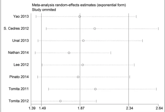
Publication bias and sensitivity analysis
We conducted leave-one-out sensitivity analysis by remov-ing one study per time to check if any individual study affected the results. The result patterns were not obviously affected by any single study in either the univariate or multivariate group (Figures 3, 4). Begg’s funnel plot and Egger’s linear regression tests were used to evaluate publication bias. In the univariate studies, the results did not show any evidence of publication bias (p=0.711 for Begg’s test, andp=0.141 for Egger’s test). However, publica-tion bias was found in the multivariate studies (p=0.002 for Egger’s test and p=0.251 for Begg’s test and). Therefore, a trim and fill method was used to evaluate the asymmetry in the funnel plot. The recalculated pooled HRs with presumed missing studies did not significantly change for multivariate studies (HR=1.118, 95%CI: 1.002–1.233; Pheterogeneity=0.026;
Figure 5), indicating the stability of the results.
’ DISCUSSION
Systemic inflammation appears to play a pivotal role in the progression of numerous cancers by promoting tumor angiogenesis, tumor metastasis and cancer cell proliferation and by affecting the tumor response to systemic treatment
(11). The NLR, a combination of circulating neutrophil and lymphocyte counts, serves as a representative index of systemic inflammation. Moreover, because it is calculated from peripheral blood test results, the NLR is a readily available biomarker of systemic inflammation that may predict the prognostic outcome of patients. Indeed, recent studies have evaluated the predictive value of the NLR in various types of cancers (16-19). Our current study aimed to evaluate the role of the NLR in lung cancer, and to the best of our knowledge, it is the first meta-analysis to investigate the prognostic role of the NLR in lung cancer.
This meta-analysis, including 14 studies with 2,734 lung cancer cases, showed that an elevated NLR indeed predicted worse OS, regardless of whether the HRs were calculated from multivariate or univariate analysis. Subgroup analysis showed that a high NLR yielded a worse OS in NSCLC and SCLC based on multivariate analysis. The cancer type of the studies using univariate analysis was NSCLC only. In the subgroup analyses by ethnicity, we found that regardless of whether patients were Asian or Caucasian, an elevated NLR was still a poor predictor of OS in both multivariate and univariate analyses. Considering different cut-off values, these studies using two subsets of NLR cut-offs revealed similar results, showing that the NLR was a negative
NOTE: Weights are from random effects analysis .
.
Overall (I-squared = 79.7%, p = 0.000) S. Cedres (2012)
Subtotal (I-squared = 69.0%, p = 0.001) Tomita (2011) Lee (2012) Unal (2013) Tomita (2011) Kang (2014) Sarraf (2009)
Subtotal (I-squared = 50.8%, p = 0.047)
Wang (2014) Nathan (2014) Study Tomita (2012) Yao (2013) Liao (2013) Pinato (2014) Kacan (2014) Satoshi (2009) Pinato (2014) ID Multivate Univariate Lee (2012)
1.51 (1.32, 1.72) 1.40 (1.10, 2.10)
1.24 (1.11, 1.40) 1.46 (1.21, 1.76) 1.09 (0.04, 1.14) 1.81 (1.16, 2.81)
1.29 (1.05, 1.57) 1.47 (1.01, 2.12)
1.10 (1.03, 1.17) 1.87 (1.49, 2.34)
1.70 (1.05, 2.75) 3.50 (1.66, 7.37)
2.80 (1.83, 4.28) 2.00 (1.29, 3.11)
1.00 (0.40, 2.49) 3.80 (1.60, 8.90) 1.70 (1.00, 2.70) 1.56 (1.09, 2.24) 2.30 (1.00, 5.00) HR (95% CI)
1.05 (1.00, 1.10)
100.00 7.25 61.14 10.30 0.59 5.24 9.86 6.39 12.56 38.86 4.74 2.50 % 5.49 5.29 1.78 1.98 4.54 6.56 2.21 Weight 12.73
1.51 (1.32, 1.72) 1.40 (1.10, 2.10)
1.24 (1.11, 1.40) 1.46 (1.21, 1.76) 1.09 (0.04, 1.14) 1.81 (1.16, 2.81)
1.29 (1.05, 1.57) 1.47 (1.01, 2.12)
1.10 (1.03, 1.17) 1.87 (1.49, 2.34)
1.70 (1.05, 2.75) 3.50 (1.66, 7.37)
2.80 (1.83, 4.28) 2.00 (1.29, 3.11)
1.00 (0.40, 2.49) 3.80 (1.60, 8.90) 1.70 (1.00, 2.70) 1.56 (1.09, 2.24) 2.30 (1.00, 5.00) HR (95% CI)
1.05 (1.00, 1.10)
100.00 7.25 61.14 10.30 0.59 5.24 9.86 6.39 12.56 38.86 4.74 2.50 % 5.49 5.29 1.78 1.98 4.54 6.56 2.21 Weight 12.73 1
.04 1 25
Figure 3 -Effect of univariate studies on the pooled HR for the NLR and OS of patients.
prognostic marker for the outcome of OS regardless of the analysis method. Further analysis by sample size also revealed the same results. Meta-regression analysis was performed using variables such as the year of publication, ethnicity, cancer type, cut-off value and sample size; however, none of these variables contributed to heterogeneity.
The NLR has been related to patient prognosis in numerous cancers, although the specific mechanism for this relationship remains incompletely understood. Myeloid cells are known to play a critical role in tumor pathogenesis by promoting cancer cell proliferation, tumor angiogenesis, cell invasion, and metastasis (37). In particular, tumor-derived inflammation can increase myelopoiesis with defective myeloid cell differentiation and proliferation by regulating the bone marrow and spleen, leading to the accumulation of immature myeloid cells in the peripheral circulation (38). In the context of cancer-mediated myelopoiesis, the neutrophil precursors myelocytes and promyelocytes proliferate and are released into the peripheral blood. Neutrophils are the most abundant granulocytes, which account for most peripheral white blood cells (37). Thus, the prognostic and predictive value of peripheral neutrophils as an independent index or as part of the NLR in cancers is apparent, and enhanced neutrophil responses and/or lymphocyte suppression, lead-ing to a high NLR, might promote tumor progression and inhibit the antitumor immune response.
Our study has several limitations that should be carefully considered. First, the studies included in the analysis were full texts in English and were identified by searching the PubMed and Web of Science databases. Thus, publication bias cannot be excluded, although it did not affect the results according to the trim and fill method. In addition, marked heterogeneity of the studies was found; this may have been
caused by the year of publication, ethnicity, cancer type, cut-off value and sample size. However, no variables listed above that were analyzed in the meta-regression analysis contributed to the observed heterogeneity. In fact, the existence of heterogeneity may have resulted from a variety of other factors. Due to the lack of detailed data, we could not use other clinical parameters in the meta-regression analysis. Additionally, the number of included studies was not large enough for part of the subgroup analysis; for example, only two studies investigated the NLR for OS in SCLC, and only two univariate studies with a small sample size (less than 100 cases was classified as‘‘small’’) yielded a trend of a poor prognostic role of the NLR for OS. In the future, more well-designed studies are needed to present more reliable results.
In conclusion, despite the limitations listed above, the synthesized evidence from published articles revealed that elevated NLR was a poor predictor of survival in patients with lung cancer. The NLR is an easily available blood test and may serve as a useful prognostic biomarker in lung cancer that does not require any additional resources for routine use. Nevertheless, the clinical utility of the NLR must still be confirmed in future analyses.
’ ACKNOWLEDGMENTS
The authors thank all of the patients and clinical investigators who were involved in the studies included in this meta-analysis.
’ AUTHOR CONTRIBUTIONS
Wang J conceived and designed the experiments. Wang J, Yin YM, Wang XD, Kuai SG and Shang ZB performed the experiments. JW, YMY,
Wang XD, Gu L, Pei H and Zhang YY analyzed the data. Thefirst three authors contributed equally to this article.
’ REFERENCES
1. Siegel R, Naishadham D, Jemal A. Cancer statistics. CA Cancer J Clin. 2013;63(1): 11–30, http://dx.doi.org/10.3322/caac.v63.1.
2. Jemal A, Bray F, Center MM, Ferlay J, Ward E, Forman D. Global cancer statistics. CA Cancer J Clin. 2011;61(2):69–90, http://dx.doi.org/10.3322/ caac.v61:2.
3. Gail MH, Eagan RT, Feld R, Ginsberg R, Goodell B, Hill L, et al. Prog-nostic factors in patients with resected stage I non-small cell lung cancer. A report from the Lung Cancer Study Group. Cancer. 1984; 54(9):1802–13, http://dx.doi.org/10.1002/(ISSN)1097-0142.
4. Hoang T, Xu R, Schiller JH, Bonomi P, Johnson DH. Clinical model to predictsurvival in chemonaive patients with advanced non-small-cell lung cancer treated with third-generation chemotherapyregimens based on eastern cooperative oncology group data. J Clin Oncol. 2005; 23(1): 175–83, http://dx.doi.org/10.1200/JCO.2005.04.177.
5. Simon GR, Sharma S, Cantor A, Smith P, Bepler G. ERCC1 expression is a predictor of survival in resected patients with non-small cell lung cancer. Chest. 2005;127(3):978–83, http://dx.doi.org/10.1378/chest.127.3.978. 6. Riquet M, Bagan P, Le Pimpec Barthes F, Banu E, Scotte F, Foucault C,
et al. Completely resected non-small cell lung cancer: reconsidering prognostic value and significance of N2 metastases. Ann Thorac Surg. 2007;84(6):1818–24, http://dx.doi.org/10.1016/j.athoracsur.2007.07.015. 7. Albain KS, Swann RS, Rusch VW, Turrisi AT 3rd, Shepherd FA, Smith C,
et al. Radiotherapy plus chemotherapy with or without surgical resection for stage III non-small-cell lung cancer: a phase III randomised controlled trial. Lancet. 2009;374(9687):379–86, http://dx.doi.org/10.1016/S0140-6736 (09)60737-6.
8. Gu X, Ma C, Yuan D, Song Y. Circulating soluble intercellular adhesion molecule-1 in lung cancer: a systematic review. Transl Lung Cancer Res. 2012;1(1):36–44.
9. Tanner NT, Sherman CA, Silvestri GA. Biomarkers in the selection of maintenance therapy in non-small cell lung cancer. Transl Lung Cancer Res. 2012;1(2):96–8.
10. Donnem T, Bremnes RM, Busund LT, Andersen S, Pezzella F. Gene expression assays as prognostic and predictive markers in early stage non-small cell lung cancer. J Thorac Dis. 2012;4(2):212–3.
11. Mantovani A, Allavena P, Sica A, Balkwill F. Cancer-related inflammation. Nature. 2008;454(7203):436–44, http://dx.doi.org/10.1038/nature07205. 12. Hanahan D, Weinberg RA. Hallmarks of cancer: the next generation. Cell.
2011;144(5):646–74, http://dx.doi.org/10.1016/j.cell.2011.02.013. 13. Lin X, Li W, Lai J, Okazaki M, Sugimoto S, Yamamoto S, et al. Five-year
update on the mouse model of orthotopic lung transplantation: scientific uses, tricks of the trade, and tips for success. J Thorac Dis. 2012;4(3): 247–58.
14. Schreiber RD, Old LJ, Smyth MJ. Cancer immunoediting: integrating immunity’s roles in cancer suppression and promotion. Science. 2011;331 (6024):1565–70, http://dx.doi.org/10.1126/science.1203486.
15. Roxburgh CS, McMillan DC. Role of systemic inflammatory response in predicting survival in patients with primary operable cancer. Future Oncol. 2010;6(1):149–63, http://dx.doi.org/10.2217/fon.09.136. 16. Walsh SR, Cook EJ, Goulder F, Justin TA, Keeling NJ.
Neutrophil-lymphocyte ratio as a prognostic factor in colorectal cancer. J Surg Oncol. 2005;91(3):181–4, http://dx.doi.org/10.1002/(ISSN)1096-9098.
17. Azab B, Bhatt VR, Phookan J, Murukutla S, Kohn N, et al. Usefulness of the neutrophil-to -lymphocyte ratio in predicting short- and long-term mortality in breast cancer patients. Ann Surg Oncol. 2012;19(1):217–24, http://dx.doi.org/10.1245/s10434-011-1814-0.
18. Gwak MS, Choi SJ, Kim JA, Ko JS, Kim TH, Lee SM, et al. Effects of gender on white blood cell populations and neutrophil-lymphocyte ratio following gastrectomy in patients with stomach cancer. J Korean Med Sci. 2007;22(Suppl):S104–8, http://dx.doi.org/10.3346/jkms.2007. 22.S.S104.
19. Sharaiha RZ, Halazun KJ, Mirza F, Port JL, Lee PC, Neugut AI et al. Elevated preoperative neutrophil: lymphocyte ratio as a predictor of postoperative disease recurrence in esophageal cancer. Ann Surg Oncol. 2011;18(12):3362–9, http://dx.doi.org/10.1245/s10434-011-1754-8.
20. Tierney JF, Stewart LA, Ghersi D, Burdett S, Sydes MR. Practical methods for incorporating summary time-to-event data into meta-analysis. Trials. 2007;7(8):16, http://dx.doi.org/10.1186/1745-6215-8-16.
21. DerSimonian R, Laird N. Meta–analysis in clinical trials. Control Clin Trials. 1986;7(3): 177–88, http://dx.doi.org/10.1016/0197-2456(86)90046-2. 22. Mantel N, Haenszel W. Statistical aspects of the analysis of data from
retrospective studies of disease. J Natl Cancer Inst. 1959;22(4): 719–48. 23. Teramukai S, Kitano T, Kishida Y, Kawahara M, Kubota K, Komuta K,
et al. Pretreatment neutrophil counts as an independent prognostic factor in advanced non-small cell lung cancer-an analysis of Japan multinational trial organization LC00-03. Eur J Cancer. 2009; 45(11):1950–8, http://dx. doi.org/10.1016/j.ejca.2009.01.023.
24. Kacan T, Babacan NA, Seker M, Yucel B, Bahceci A, Eren AA, et al. Could the neutrophil to lymphocyte ratio be a poor prognostic factor for non small cell lung cancers? Asian Pac J Cancer Prev. 2014; 15(5):2089–94, http://dx.doi.org/10.7314/APJCP.2014.15.5.2089.
25. Yao Y, Yuan D, Liu H, Gu X, Song Y. Pretreatment neutrophil to lym-phocyte ratios associated with response to therapy and prognosis of advanced non-small cell lung cancer patients treated with first-line pla-tinum-based chemotherapy. Cancer Immunol Immunother. 2013; 62(3): 471–9, http://dx.doi.org/10.1007/s00262-012-1347-9.
26. Lee Y, Kim SH, Han JY, Kim HT, Yun T, Soo LJ. Early neutrophil-to-lymphocyte ratio reduction as a surrogate marker of prognosis in never smokers with advanced lung adenocarcinoma receiving gefitinib or standard chemotherapyas first-line therapy. J Cancer Res Clin Oncol. 2012;138(12):2009–16, http://dx.doi.org/10.1007/s00432-012-1281-4. 27. Wang X, Jiang R, Li K. Prognostic significance of pretreatment laboratory
parameters in Combined Small-Cell Lung Cancer. Cell Biochem Biophys. 2014;69(3):633–40, http://dx.doi.org/10.1007/s12013-014-9845-3. 28. Cedrés S, Torrejon D, Martínez A, Martinez P, Navarro A, Zamora E, et al.
Neutrophil to lymphocyte ratio (NLR) as an indicator of poor prognosis in stage IV non-small cell lung cancer. Clin Transl Oncol. 2012;14(11):864–9, http://dx.doi.org/10.1007/s12094-012-0872-5.
29. Unal D, Eroglu C, Kurtul N, Oguz A, Tasdemir A. Are neutrophil/lym-phocyte and platelet/lymneutrophil/lym-phocyte rates in patients with non-small cell lung cancer associated with treatment response and prognosis? Asian Pac J Cancer Prev. 2013;14(9):5237–42, http://dx.doi.org/10.7314/APJCP.2013. 14.9.5237.
30. Pinato DJ, Shiner RJ, Seckl MJ, Stebbing J, Sharma R, Mauri FA. Prognostic performance of inflammation based prognostic indices in primary oper-able non-small cell lung cancer. Br J Cancer. 2014;110(8):1930–5, http://dx. doi.org/10.1038/bjc.2014.145.
31. Kang MH, Go SI, Song HN, Lee A, Kim SH, Kang JH, et al. The prognostic impact of the neutrophil-to lymphocyte ratio in patients with small-cell lung cancer. Br J Cancer. 2014;111(3):452–460, http://dx.doi.org/10.1038/ bjc.2014.317.
32. Cannon NA, Meyer J, Iyengar P, Ahn C, Westover KD, Choy H, et al. T Neutrophil-lymphocyte and platelet-lymphocyte ratios as prognostic factors following stereotactic radiation therapy for early-stage non-small cell lung cancer. J Thorac Oncol 2015; 10(2):280–285, http://dx.doi.org/ 10.1097/JTO.0000000000000399.
33. Tomita M, Shimizu T, Ayabe T, Yonei A, Onitsuka T. Preoperative neu-trophil to lymphocyte ratio as a prognostic predictor after curative resection for non-small cell lung cancer. Anticancer Res. 2011; 31(9):2995–8. 34. Sarraf KM, Belcher E, Raevsky E, Nicholson AG, Goldstraw P, Lim E.
Neutrophil/lymphocyte ratio and its association with survival after complete resection in non-small cell lung cancer. J Thorac Cardiovasc. Surg 2009;137(2):425–8.
35. Liao Y, Ni Y, He R, Liu W, Du J. Clinical implications of fibroblast acti-vation protein-a in non-small cell lung cancer after curative resection: a new predictor for prognosis. J Cancer Res Clin Oncol. 2013;139(9):1523–8, http://dx.doi.org/10.1007/s00432-013-1471-8.
36. Tomita M, Shimizu T, Ayabe T, Nakamura K, Onitsuka T. Elevated pre-operative inflammatory markers based on neutrophil-to-lymphocyte ratio and C-reactive protein predict poor survival in resected non-small cell lung cancer. Anticancer Res. 2012; 32(8):3535–8.
37. Diakos CI, Charles KA, McMillan DC, Clarke SJ. Cancer-related inflam-mation and treatment effectiveness. Lancet Oncol. 2014; 15(11):e493–503, http://dx.doi.org/10.1016/S1470-2045(14)70263-3.