Prognostic Significance of Combination of
Preoperative Platelet Count and
Neutrophil-Lymphocyte Ratio (COP-NLR) in Patients with
Non-Small Cell Lung Cancer: Based on a
Large Cohort Study
Hua Zhang1,2,3, Lianmin Zhang1,2,3, Kaikai Zhu4, Bowen Shi1,2,3, Yuesong Yin1,2,3, Jinfang Zhu1,2,3, Dongsheng Yue1,2,3, Bin Zhang1,2,3, Changli Wang1,2,3*
1Department of Lung Cancer, Tianjin Medical University Cancer Institute and Hospital, National Clinical Research Center for Cancer, Tianjin, China,2Key Laboratory of Cancer Prevention and Therapy, Tianjin, China,3Tianjin Lung Cancer Center, Tianjin, China,4Department of Pediatrics, Tianjin Medical University General Hospital, Tianjin, China
Abstract
Introduction
The aim of this study was to investigate the prognostic significance of the combination of the preoperative platelet count and neutrophil-lymphocyte ratio (COP-NLR) for predicting postoperative survival of patients undergoing complete resection for non-small cell lung cancer (NSCLC).
Methods
The preoperative COP-NLR was calculated on the basis of data obtained.Patients with both an increased platelet count (>30.0×104mm-3) and an elevated NLR (>2.3) were assigned a
score of 2, and patients with one or neither were assigned as a score of 1 or 0, respectively.
Results
A total of 1238 NSCLC patients were enrolled in this analysis. Multivariate analysis using the 15 clinicolaboratory variables selected by univariate analyses demonstrated that the preoperative COP-NLR was an independent prognostic factor for DFS (HR: 1.834, 95%CI: 1.536 to 2.200,P<0.001) and OS (HR: 1.810, 95%CI: 1.587 to 2.056,P<0.001). In sub-analyses by tumor stage (I, II, IIIA), a significant association was found between DFS and OS and level of COP-NLR in each subgroup (P<0.001,P=0.002,P<0.001 for DFS, respec-tively;P<0.001,P=0.001,P<0.001 for OS). When the subgroup of patients with high-risk COP-NLR (score of 2) was analyzed, no benefit of adjuvant chemotherapy could be found (P=0.237 for DFS andP=0.165 for OS).
a11111
OPEN ACCESS
Citation:Zhang H, Zhang L, Zhu K, Shi B, Yin Y, Zhu J, et al. (2015) Prognostic Significance of
Combination of Preoperative Platelet Count and Neutrophil-Lymphocyte Ratio (COP-NLR) in Patients with Non-Small Cell Lung Cancer: Based on a Large Cohort Study. PLoS ONE 10(5): e0126496. doi:10.1371/journal.pone.0126496
Academic Editor:Prasad S. Adusumilli, Memorial Sloan-Kettering Cancer Center, UNITED STATES
Received:January 13, 2015
Accepted:April 3, 2015
Published:May 7, 2015
Copyright:© 2015 Zhang et al. This is an open access article distributed under the terms of the
Creative Commons Attribution License, which permits
unrestricted use, distribution, and reproduction in any medium, provided the original author and source are credited.
Data Availability Statement:All relevant data are within the paper.
Conclusions
The preoperative COP-NLR is able to predict the prognosis of patients with NSCLC and di-vide these patients into three independent groups before surgery. Our results also demon-strate that high-risk patients based on the COP-NLR do not benefit from adjuvant
chemotherapy. Independent validation of our findings is warranted.
Introduction
Lung cancer is the most common cause of cancer-related death worldwide, with 5-year survival rates of less than 15% [1]. Non-small-cell lung cancer (NSCLC) accounts for approximately 80% of all lung cancers. Recently, a large number of prognostic predictors have been reported in the literature for patients with NSCLC, however, the majority of these factors cannot be ob-tained preoperatively. Moreover, some of these factors are available only as research tools.
Recently, there has been a growing interest in the host inflammatory response to tumors, and systemic inflammatory response has been shown to reflect the promotion of angiogenesis, DNA damage and tumor invasion through up-regulation of cytokines [2–4]. Based on this, a number of inflammation-based prognostic markers have been identified, such as the Glasgow Prognostic Score (GPS) and platelet to lymphocyte ratio (PLR) [5,6]. In addition, there is in-creasing evidence that the neutrophil to lymphocyte ratio (NLR) and thrombocytosis can be used for prognostication in lung cancer [7–10]. Recently, two studies investigated a novel in-flammation-based prognostic system named the combination of platelet count and NLR (COP-NLR). They revealed that COP-NLR was a useful predictor of postoperative survival in patients with colorectal cancer and gastric cancer [11,12]. However, to the best of our knowl-edge, no studies regarding COP-NLR in patients with NSCLC are available. Therefore, in this study, we evaluated the clinical utility of a novel inflammation-based prognostic system (COP-NLR) in patients with NSCLC undergoing complete resection.
Patients and Methods
Patients
A retrospective study of patients withNSCLC undergoing surgery was conducted at the Tianjin Medical University Cancer Institute and Hospital from January 2005 to December 2009. The study was approved by the Institutional Review Board of Tianjin Medical University Cancer Institute and Hospital. All patients provided written informed consent. The inclusion criteria were (1) complete pulmonary resection and systematic node dissection of the hilar and medias-tinal lymph nodes and (2) histologically confirmed NSCLC. Patients who met the following cri-teria were excluded from the study: (1) preoperative chemotherapy or radiotherapy;
(2) positive surgical margins; (3) advanced disease (e.g., malignant pleural effusion/
involvement or distant metastasis); (4) clinical evidence of infection or other bone marrow, he-matological or autoimmune disease; (5) a history of lung cancer or a second primary cancer di-agnosed within 5 years of the lung cancer index; and (6) patients who died within 1 month after surgery. Based on the inclusion and exclusion criteria, a total of 1238 NSCLC patients were analyzed in the present study. All of the patients were Chinese. The preoperative evalua-tion included a detailed clinical history and physical examinaevalua-tion with a series of biochemistry tests, complete blood cell counts and coagulation tests. Further investigations included radiog-raphy, flexible bronchoscopy, chest and upper abdominal computed tomography (CT),
radionuclide bone scan, and CT or magnetic resonance imaging (MRI) of the brain. Tumor stages were based on the 7th edition of the TNM Classification [13]. The main adjuvant treat-ment that patients underwent after operation was chemotherapy or radiotherapy, either alone or in combination. The chemotherapy was routine program for NSCLC, including vinorelbine, paclitaxel, or gemcitabine plus carboplatin or cisplatin.
Definition of COP-NLR and other variables
Venous blood sampling was taken in a week prior to surgery, and collected in ethylenediamine-tetraacetic acid (EDTA) containing tube. NLR was defined as follows: NLR = percentage of neutrophils for whole white blood cells (WBC)/percentage of lymphocytes for whole WBC or NLR = peripheral neutrophil count/peripheral lymphocyte count. The cut-off values of the pre-operative platelet count and NLR were decided using receiver operating characteristic (ROC) curve analyses. For the 1238 NSCLC patients in our study, an NLR of 2.3 corresponded to the maximum joint sensitivity and specificity on the ROC plot. The area under the curve (AUC) for NLR was 0.650. Thus, the recommended cut-off value for NLR was 2.3. Similarly, the prom-inent point on the ROC curve indicated a cut-off value of 29.0 ×104mm-3for the platelet count,and the area under the ROC curve was 0.562. Therefore, the recommended cut-off value of preoperative platelet count was defined as 30.0 ×104mm-3.
The COP-NLR was calculated on the basis of data obtained. Patients with both an increased platelet count (>30×104mm-3) and an elevated NLR (>2.3) were assigned a score of 2, and pa-tients with one or neither were assigned as a score of 1 or 0, respectively.
Statistical analysis
All statistical analyses were carried out using SPSS 13.0 software (SPSS Inc., Chicago, USA). Overall survival (OS) was defined as the interval between the date of surgery and the date of death or last follow-up. Disease-free survival (DFS) was defined as the duration of time be-tween the date of surgery and the date of first recurrence or last follow-up. Data are presented as the mean ± s.d. The differences between the three COP-NLR groups and the clinicolabora-tory variables were analyzed by chi-square test and Kruskal-Wallis test. Hazard ratios (HR) and 95% confidence intervals (95% CI) were calculated by univariate and multivariate analysis using the Cox proportional hazards model.
Twenty-three clinicolaboratory variables, including age, sex, smoking status, resection type, histological subtype, tumor location, lesion, TNM stage, adjuvant chemotherapy, adjuvant ra-diotherapy, WBC count, platelet count, neutrophil ratio, lymphocyte ratio, monocyte ratio, NLR, albumin, intraoperative blood loss, hemoglobin (Hb), lactate dehydrogenase (LDH), fi-brinogen, D-dimer, and COP-NLR were entered in the univariate analysis. In order to classify the patients into two groups, the cut-off values of clinicolaboratory variables were determined using ROC curve analyses. The value of the maximum joint sensitivity and specificity on the ROC plot was defined as the recommended cut-off value. Clinicolaboratory variables with a
P-value<0.05 were further analyzed in the multivariate analysis to determine the independent predictors of DFS and OS. The Kaplan-Meier method and the log-rank test were used to com-pare the survival curves of the three COP-NLR groups.
Results
24 to 82 years. The distribution of pathological stages was as follows: stage I, 536; stage II, 264; and stage IIIA, 438. The follow-up period ranged from 2 month to 96 months (median, 45.0 months; mean, 44.1 months). At the end of the last follow up, 686 patients had died. The 5-year OS rate was 47.3% for the whole study population.
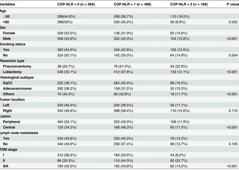
The distribution of the clinical background characteristics of the studied patients in the three groups is shown inTable 1. As shown inTable 1, significant differences between COP-NLR and age (P= 0.002), sex (P<0.001), smoking status (P= 0.004), resection type (P<0.001), histological subtype (P<0.001), lesion (P<0.001), and TNM stage (I/II/IIIA) (P<0.001) were demonstrated.
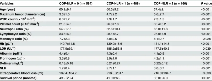
The relationships between COP-NLR and clinicolaboratory varibles are shown inTable 2. Significant differences between COP-NLR and age (P<0.001), maximum tumor diameter (P<0.001), WBC count (P<0.001), platelet count (P<0.001), neutrophil ratio (P<0.001), lym-phocyte ratio (P<0.001), monocyte ratio (P= 0.028), Hb (P<0.001), LDH (P= 0.039), albumin (P<0.001), fibrinogen (P<0.001), D-dimer (P= 0.001), NLR (P<0.001), intraoperative blood loss (P= 0.030), and survival period (P<0.001) were shown.
Table 1. Correlations between COP-NLR and clinical characteristics.
Variables COP-NLR = 0 (n = 584) COP-NLR = 1 (n = 488) COP-NLR = 2 (n = 166) Pvalue
Age
60 298(44.8%) 258 (38.7%) 110 (16.5%)
>60 286(50%) 230 (40.2%) 56 (8.8%) 0.002
Sex
Female 228 (53.5%) 136 (31.9%) 62 (14.6%)
Male 356 (43.9%) 352 (43.3%) 104 (12.8%) <0.001
Smoking status
Yes 360 (44.6%) 346 (42.8%) 102 (12.6%)
No 224 (52.1%) 142 (33.0%) 64 (14.9%) 0.004
Resection type
Pneumonectomy 38 (25.7%) 76 (51.4%) 34 (22.9%)
Lobectomy 546 (50.1%) 412 (37.8%) 132 (12.1%) <0.001
Histological subtype
SqCC 222 (38.1%) 264 (45.4%) 96 (16.5%)
Adenocarcinoma 292 (58.2%) 158 (31.5%) 52 (10.3%)
Others 70 (45.5%) 66 (42.8%) 18 (11.7%) <0.001
Tumor location
Left 250 (49.4%) 200 (39.5%) 56 (11.1%)
Right 334 (45.6%) 288 (39.4%) 110 (15.0%) 0.110
Lesion
Peripheral 464 (52.1%) 320 (40.0%) 106 (11.9%)
Central 120 (34.5%) 168 (48.3%) 60 (17.2%) <0.001
lymph node metastasis
Yes 240 (43.8%) 230 (42.0%) 78 (14.2%)
No 344 (49.9%) 258 (37.4%) 88 (12.7%) 0.105
TNM stage
I 312 (58.2%) 180 (33.6%) 44 (8.2%)
II 88 (33.3%) 116 (44.0%) 60 (22.7%)
IIIA 184 (42.0%) 192 (43.8%) 62 (14.2%) <0.001
Abbreviations: Squamous cell carcinoma = SqCC;COP-NLR = combination of preoperative platelet count and neutrophil-lymphocyte ratio.
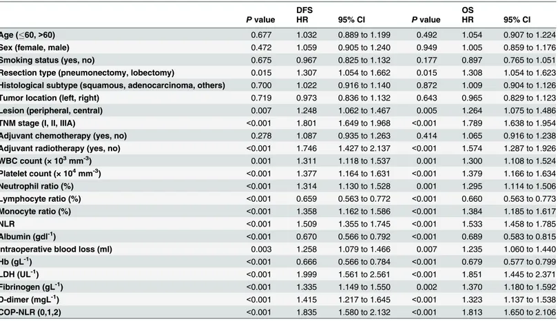
Based on the cut-off value, we separated the patients into different groups. Survival analyses in relation to NLR and platelet count and patient prognosis were performed. In the univariate analysis of OS, NLR and platelet countwere both found to be significant factors, with hazard ratio (HR) = 1.533 (95% confidence interval [CI]: 1.458 to 1.785) for NLR, and HR = 1.379 (95% CI: 1.166 to 1.634) for platelet count. To identify the optimal factor for patient prognosis, we evaluated the prognostic value of COP-NLR. The HR for COP-NLR was 1.813 (95% CI: 1.650 to 2.106), implying a more important prognostic value. For DFS, the HR for COP-NLR was 1.835 (95% CI: 1.580 to 2.132) (Table 3). Other identified prognostic factors for DFS and OS included resection type, lesion, TNM stage, adjuvant radiotherapy, WBC count, neutrophil ratio, lymphocyte ratio, monocyte ratio, albumin, intraoperative blood loss, Hb, LDH, fibrino-gen, and D-dimer.
Multivariate analysis using the 15 clinicopathological characteristics selected above (exclud-ing platelet count and NLR) demonstrated that preoperative COP-NLR was associated with DFS and OS (HR: 1.834, 95%CI: 1.536 to 2.200,P<0.001 and HR: 1.810, 95%CI: 1.587 to 2.056,
P<0.001, respectively) along with TNM stage, LDH level, and D-dimer (Table 4). At the same time, multivariate analysis also demonstrated that COP-NLR remained an independent prog-nostic marker in SqCC or adenocarcinoma (Tables5–8).
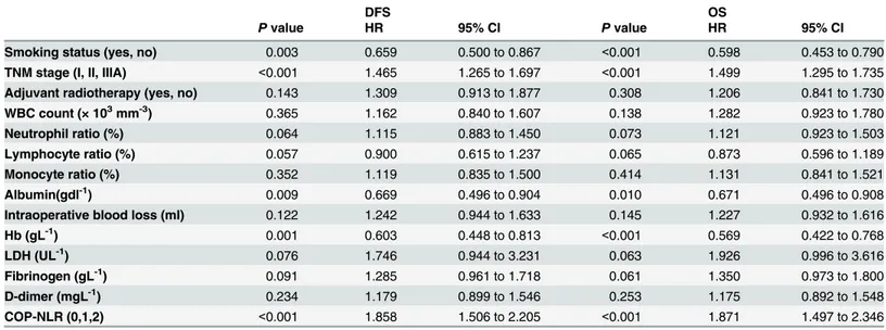
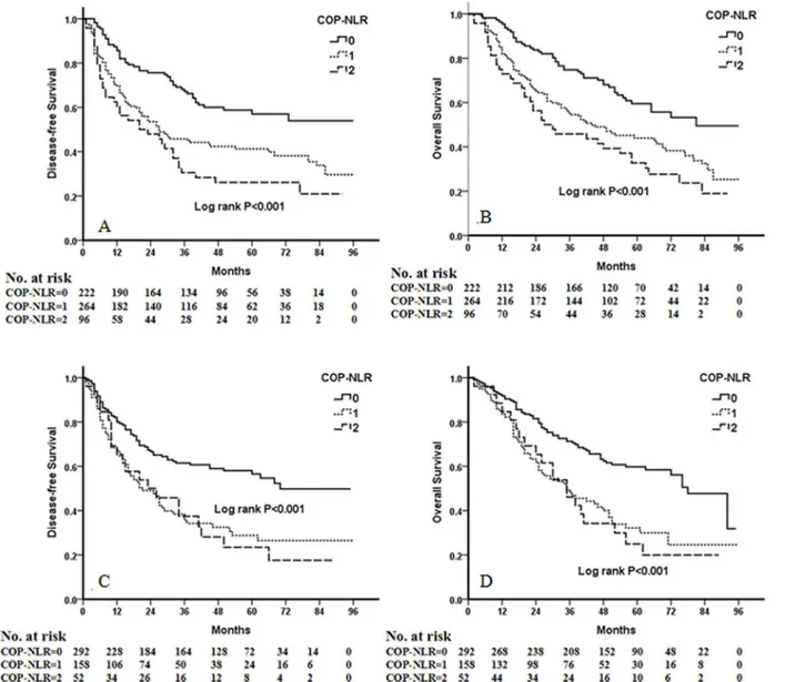
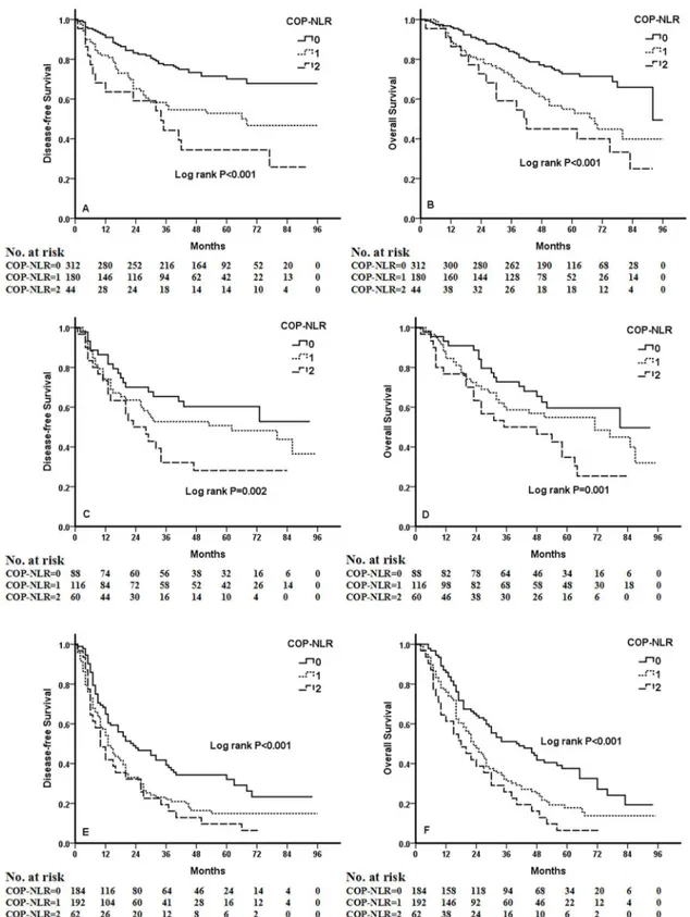
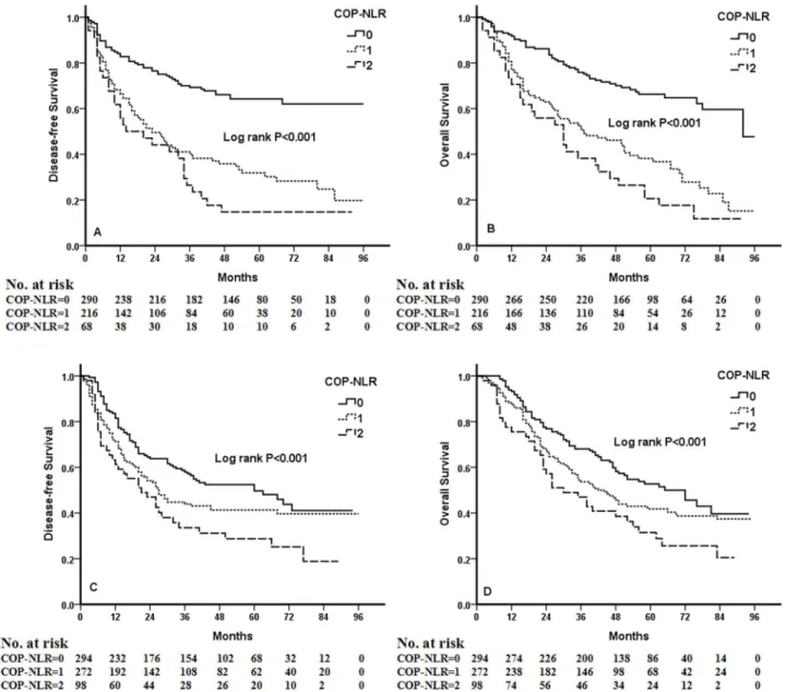
Kaplan-Meier analysis and log-rank test demonstrated that there were significant differ-ences in DFS and OS among the three COP-NLR groups (P<0.001 andP<0.001, respectively) (Fig 1). Patients with COP-NLR = 0 had better prognoses than those with COP-NLR = 1 or COP-NLR = 2. The 5-year survival rate for COP-NLR = 0, COP-NLR = 1, and COP-NLR = 2 was 59.5%, 40.2% and 25.3%, respectively. Thus, the preoperative COP-NLR was able to divide the patients into three independent groups. Similarly, COP-NLR could also separate the pa-tients into three independent groups in SqCC or adenocarcinoma (Fig 2). When the analysis was stratified by stage (I, II, IIIA), we found that DFS and OS were better in the lower COP-NLR group than in the higher COP-NLR group in the stage I, II, and IIIA subgroups (Fig 3). Furthermore, subgroup analyses also revealed that decreased preoperative COP-NLR
Table 2. Correlations between COP-NLR and clinicolaboratory characteristics.
Variables COP-NLR = 0 (n = 584) COP-NLR = 1 (n = 488) COP-NLR = 2 (n = 166) Pvalue
Age (year) 60.9±9.4 60.5±9.2 57.4±9.1 <0.001
Maximum tumor diameter (cm) 3.6±1.5 4.7±2.2 5.6±2.7 <0.001
WBC count (× 103mm-3) 6.3±1.7 7.3±1.7 7.3±1.5 <0.001
Platelet count (× 104mm-3) 21.8±4.5 26.0±7.8 33.4±6.2 <0.001
Neutrophil ratio (%) 54.9±7.5 62.6±10.4 66.0±11.8 <0.001
Lymphocyte ratio (%) 33.6±6.3 28.1±2.7 25.0±7.9 <0.001
Monocyte ratio (%) 7.7±2.3 8.0±2.5 8.1±2.7 0.028
Hb (gL-1) 140.7±14.8 139.9±16.6 131.1±14.5 <0.001
LDH (UL-1) 177.9±39.1 185.2±55.8 177.5±40.3 0.039
Albumin (gdl-1) 4.4±0.4 4.3±0.4 4.1±0.5 <0.001
Fibrinogen (gL-1) 3.3±0.8 3.9±1.0 4.2±1.1 <0.001
D-dimer (mgL-1) 0.18±0.18 0.21±0.27 0.22±0.12 0.001
NLR 1.7±0.4 2.7±1.1 3.0±0.7 <0.001
Intraoperative blood loss (ml) 182.4±104.2 216.5±231.1 216.0±184.7 0.030
Survival period (months) 49.2±23.4 41.0±26.2 36.0±26.0 <0.001
Abbreviations: COP-NLR = combination of preoperative platelet count and neutrophil-lymphocyte ratio; WBC = white blood cell; Hb = hemoglobin; LDH = lactate dehydrogenase; NLR = neutrophil/lymphocyte ratio.
Table 3. Univariate analysis of DFS and OS for all NSCLC patients.
DFS OS
Pvalue HR 95% CI Pvalue HR 95% CI
Age (60,>60) 0.677 1.032 0.889 to 1.199 0.492 1.054 0.907 to 1.224
Sex (female, male) 0.472 1.059 0.905 to 1.240 0.949 1.005 0.859 to 1.176
Smoking status (yes, no) 0.675 0.967 0.825 to 1.132 0.177 0.897 0.765 to 1.051
Resection type (pneumonectomy, lobectomy) 0.015 1.307 1.054 to 1.662 0.015 1.308 1.054 to 1.623
Histological subtype (squamous, adenocarcinoma, others) 0.700 1.022 0.916 to 1.140 0.872 1.009 0.904 to 1.126
Tumor location (left, right) 0.719 0.973 0.836 to 1.132 0.643 0.965 0.829 to 1.123
Lesion (peripheral, central) 0.007 1.248 1.062 to 1.467 0.005 1.264 1.075 to 1.486
TNM stage (I, II, IIIA) <0.001 1.801 1.649 to 1.968 <0.001 1.789 1.638 to 1.954
Adjuvant chemotherapy (yes, no) 0.278 1.087 0.935 to 1.263 0.414 1.065 0.916 to 1.238
Adjuvant radiotherapy (yes, no) <0.001 1.746 1.427 to 2.137 <0.001 1.574 1.287 to 1.926
WBC count (× 103mm-3) 0.001 1.311 1.118 to 1.537 0.001 1.300 1.108 to 1.524
Platelet count (× 104mm-3) <0.001 1.377 1.164 to 1.631 <0.001 1.379 1.166 to 1.634
Neutrophil ratio (%) <0.001 1.314 1.130 to 1.528 0.001 1.295 1.114 to 1.506
Lymphocyte ratio (%) <0.001 0.659 0.563 to 0.772 <0.001 0.660 0.563 to 0.773
Monocyte ratio (%) <0.001 1.358 1.162 to 1.586 <0.001 1.384 1.185 to 1.617
NLR <0.001 1.509 1.355 to 1.745 <0.001 1.533 1.458 to 1.785
Albumin (gdl-1) <0.001 0.670 0.566 to 0.792 <0.001 0.689 0.583 to 0.815
Intraoperative blood loss (ml) 0.003 1.258 1.079 to 1.466 0.007 1.235 1.060 to 1.440
Hb (gL-1) <0.001 0.666 0.566 to 0.784 <0.001 0.679 0.577 to 0.799
LDH (UL-1) <0.001 1.999 1.561 to 2.561 <0.001 1.851 1.445 to 2.371
Fibrinogen (gL-1) <0.001 1.335 1.149 to 1.550 0.002 1.370 1.180 to 1.592
D-dimer (mgL-1) <0.001 1.415 1.217 to 1.645 <0.001 1.323 1.137 to 1.538
COP-NLR (0,1,2) <0.001 1.835 1.580 to 2.132 <0.001 1.813 1.650 to 2.106
Abbreviations: DFS = disease-free survival; OS = overall survival; HR = hazard ratio; CI = confidence interval; WBC = white blood cell; Hb = hemoglobin; LDH = lactate dehydrogenase; NLR = neutrophil/lymphocyte ratio; COP-NLR = combination of preoperative platelet count and neutrophil-lymphocyte ratio.
doi:10.1371/journal.pone.0126496.t003
Table 4. Multivariate analysis of DFS and OS for all NSCLC patients.
DFS OS
Pvalue HR 95% CI Pvalue HR 95% CI
Resection type (pneumonectomy, lobectomy) 0.940 1.105 0.783 to 1.289 0.952 0.987 0.765 to 1.268
Lesion (peripheral, central) 0.568 1.055 0.868 to 1.285 0.851 1.025 0.832 to 1.240
TNM stage (I, II, IIIA) <0.001 1.630 1.473 to 1.812 <0.001 1.658 1.523 to 1.841
Adjuvant radiotherapy (yes, no) 0.057 1.341 0.976 to 1.651 0.065 1.236 0.978 to 1.491
WBC count (× 103mm-3) 0.175 1.136 0.945 to 1.370 0.350 1.085 0.912 to 1.310
Neutrophil ratio (%) 0.105 1.330 0.955 to 1.628 0.110 1.329 0.930 to 1.665
Lymphocyte ratio (%) 0.155 0.812 0.653 to 1.107 0.110 0.841 0.673 to 1.052
Monocyte ratio (%) 0.090 1.190 0.908 to 1.420 0.112 1.245 0.945 to 1.469
Albumin (gdl-1) 0.063 0.812 0.663 to 1.098 0.070 0.823 0.671 to 1.131
Intraoperative blood loss (ml) 0.283 1.100 0.932 to 1.295 0.376 1.082 0.934 to 1.256
Hb (gL-1) 0.101 0.864 0.721 to 1.035 0.095 0.857 0.713 to 1.045
LDH (UL-1) 0.001 1.441 1.333 to 1.780 0.002 1.536 1.361 to 1.834
Fibrinogen (gL-1) 0.135 0.871 0.730 to 1.053 0.410 0.935 0.778 to 1.119
D-dimer (mgL-1) 0.002 1.289 1.073 to 1.512 0.006 1.258 1.066 to 1.481
COP-NLR (0,1,2) <0.001 1.834 1.536 to 2.200 <0.001 1.810 1.587 to 2.056
Abbreviations: DFS = disease-free survival; OS = overall survival; HR = hazard ratio; CI = confidence interval; WBC = white blood cell; Hb = hemoglobin; LDH = lactate dehydrogenase; COP-NLR = combination of preoperative platelet count and neutrophil-lymphocyte ratio.
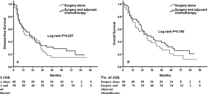
was significantly associated with better clinical outcomes for patients regardless of whether the patients received adjuvant chemotherapy (Fig 4). To evaluate whether high-risk patients based on COP-NLR = 2 would benefit from adjuvant chemotherapy compared with surgery alone, a Kaplan-Meier analysis and log-rank test were performed. The results of this high-risk subgroup demonstrated no significance difference in DFS and OS (P= 0.237 andP= 0.165, respectively) (Fig 5).
Discussion
Immune response within the tumor microenvironment is a critical component of tumor pro-gression and aggressiveness. Understanding these responses has allowed researchers to use im-mune factors to stratify the prognosis of patients with cancer. Previous studies found that several immune markers could predict the clinical outcome of patients with NSCLC. Using tis-sue microarray and immunohistochemistry, Suzuki and colleagues found that tumor interleu-kin-12 receptorβ2 (IL-12Rβ2), IL-7R, and stromal FoxP3/CD3 ratio are independent
predictors of recurrence in patients with stage I lung adenocarcinoma [14]. Furthermore, in 1290 patients with NSCLC, tumor-infiltrating lymphocytes (mostly CD8+) in the tumor corre-lated with a better OS [15]. Other immune markers in the tumor microenvironment and pe-ripheral blood of patients with NSCLC have also been reported [16]. In the present study, we investigated the prognostic value of a novel inflammation-based prognostic system, COP-NLR,
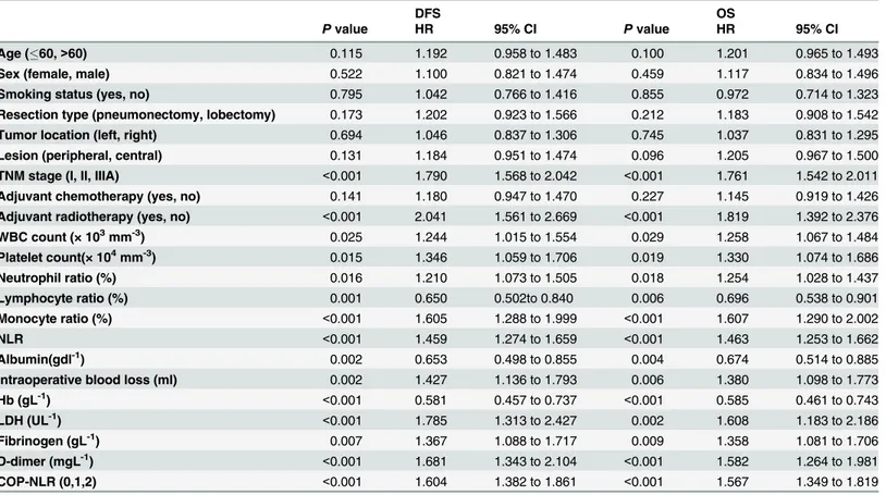
Table 5. Univariate analysis of DFS and OS for SqCC patients.
DFS OS
Pvalue HR 95% CI Pvalue HR 95% CI
Age (60,>60) 0.115 1.192 0.958 to 1.483 0.100 1.201 0.965 to 1.493
Sex (female, male) 0.522 1.100 0.821 to 1.474 0.459 1.117 0.834 to 1.496
Smoking status (yes, no) 0.795 1.042 0.766 to 1.416 0.855 0.972 0.714 to 1.323
Resection type (pneumonectomy, lobectomy) 0.173 1.202 0.923 to 1.566 0.212 1.183 0.908 to 1.542
Tumor location (left, right) 0.694 1.046 0.837 to 1.306 0.745 1.037 0.831 to 1.295
Lesion (peripheral, central) 0.131 1.184 0.951 to 1.474 0.096 1.205 0.967 to 1.500
TNM stage (I, II, IIIA) <0.001 1.790 1.568 to 2.042 <0.001 1.761 1.542 to 2.011
Adjuvant chemotherapy (yes, no) 0.141 1.180 0.947 to 1.470 0.227 1.145 0.919 to 1.426
Adjuvant radiotherapy (yes, no) <0.001 2.041 1.561 to 2.669 <0.001 1.819 1.392 to 2.376
WBC count (× 103mm-3) 0.025 1.244 1.015 to 1.554 0.029 1.258 1.067 to 1.484
Platelet count(× 104mm-3) 0.015 1.346 1.059 to 1.706 0.019 1.330 1.074 to 1.686
Neutrophil ratio (%) 0.016 1.210 1.073 to 1.505 0.018 1.254 1.028 to 1.437
Lymphocyte ratio (%) 0.001 0.650 0.502to 0.840 0.006 0.696 0.538 to 0.901
Monocyte ratio (%) <0.001 1.605 1.288 to 1.999 <0.001 1.607 1.290 to 2.002
NLR <0.001 1.459 1.274 to 1.659 <0.001 1.463 1.253 to 1.662
Albumin(gdl-1) 0.002 0.653 0.498 to 0.855 0.004 0.674 0.514 to 0.885
Intraoperative blood loss (ml) 0.002 1.427 1.136 to 1.793 0.006 1.380 1.098 to 1.773
Hb (gL-1) <0.001 0.581 0.457 to 0.737 <0.001 0.585 0.461 to 0.743
LDH (UL-1) <0.001 1.785 1.313 to 2.427 0.002 1.608 1.183 to 2.186
Fibrinogen (gL-1) 0.007 1.367 1.088 to 1.717 0.009 1.358 1.081 to 1.706
D-dimer (mgL-1) <0.001 1.681 1.343 to 2.104 <0.001 1.582 1.264 to 1.981
COP-NLR (0,1,2) <0.001 1.604 1.382 to 1.861 <0.001 1.567 1.349 to 1.819
Abbreviations: DFS = disease-free survival; OS = overall survival; HR = hazard ratio; CI = confidence interval; WBC = white blood cell; Hb = hemoglobin; LDH = lactate dehydrogenase; NLR = neutrophil/lymphocyte ratio; COP-NLR = combination of preoperative platelet count and neutrophil-lymphocyte ratio.
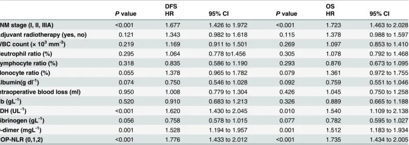
Table 6. Multivariate analysis of DFS and OS for SqCC patients.
DFS OS
Pvalue HR 95% CI Pvalue HR 95% CI
TNM stage (I, II, IIIA) <0.001 1.677 1.426 to 1.972 <0.001 1.723 1.463 to 2.028
Adjuvant radiotherapy (yes, no) 0.121 1.343 0.982 to 1.618 0.115 1.378 0.988 to 1.597
WBC count (× 103mm-3) 0.219 1.169 0.911 to 1.501 0.269 1.097 0.853 to 1.410
Neutrophil ratio (%) 0.295 1.064 0.778 to1.456 0.305 1.078 0.792 to 1.468
Lymphocyte ratio (%) 0.318 0.835 0.586 to 1.190 0.293 0.876 0.673 to 1.095
Monocyte ratio (%) 0.055 1.378 0.965 to 1.782 0.079 1.361 0.972 to 1.755
Albumin(g dl-1) 0.074 0.750 0.546 to 1.028 0.092 0.759 0.551 to 1.046
Intraoperative blood loss (ml) 0.950 1.008 0.779 to 1.304 0.426 1.045 0.750 to 1.258
Hb (gL-1) 0.520 0.910 0.683 to 1.213 0.326 0.889 0.665 to 1.188
LDH (UL-1) <0.001 1.620 1.430 to 2.045 0.010 1.540 1.109 to 2.138
Fibrinogen (gL-1) 0.056 0.758 0.578 to 1.015 0.077 0.782 0.595 to 1.027
D-dimer (mgL-1) 0.001 1.528 1.194 to 1.957 0.001 1.512 1.183 to 1.934
COP-NLR (0,1,2) <0.001 1.776 1.433 to 2.012 <0.001 1.735 1.434 to 2.005
Abbreviations: DFS = disease-free survival; OS = overall survival; HR = hazard ratio; CI = confidence interval; WBC = white blood cell; Hb = haemoglobin; LDH = lactate dehydrogenase; COP-NLR = combination of preoperative platelet count and neutrophil-lymphocyte ratio.
doi:10.1371/journal.pone.0126496.t006
Table 7. Univariate analysis of DFS and OS for adenocarcinoma patients.
DFS OS
Pvalue HR 95% CI Pvalue HR 95% CI
Age (60,>60) 0.785 0.967 0.761 to 1.229 0.928 1.011 0.796 to 1.284
Sex (female, male) 0.679 1.052 0.828 to 1.335 0.871 0.980 0.772 to 1.245
Smoking status (yes, no) 0.027 0.765 0.604 to 0.970 0.003 0.699 0.552 to 0.886
Resection type (pneumonectomy, lobectomy) 0.437 1.222 0.737 to 2.027 0.328 1.288 0.776 to 2.139
Tumor location (left, right) 0.728 1.044 0.819 to 1.330 0.764 1.038 0.815 to 1.322
Lesion (peripheral, central) 0.232 1.217 0.882 to 1.681 0.258 1.206 0.872 to 1.667
TNM stage (I, II, IIIA) <0.001 1.694 1.485 to 1.934 <0.001 1.683 1.475 to 1.920
Adjuvant chemotherapy (yes, no) 0.481 0.919 0.725 to 1.163 0.305 0.884 0.698 to 1.119
Adjuvant radiotherapy (yes, no) 0.019 1.475 1.041 to 2.090 0.012 1.362 0.962 to 1.930
WBC count (× 103mm-3) 0.003 1.517 1.153 to 1.995 <0.001 1.686 1.281 to 2.219
Platelet count(× 104mm-3) 0.017 1.406 1.064 to 1.862 0.012 1.430 1.082 to 1.894
Neutrophil ratio (%) 0.005 1.419 1.108 to 1.817 0.003 1.462 1.142 to 1.871
Lymphocyte ratio (%) <0.001 0.651 0.513 to 0.825 <0.001 0.624 0.492 to 0.791
Monocyte ratio (%) 0.037 1.264 1.005 to 1.513 0.024 1.311 1.112 to 1.612
NLR <0.001 1.731 1.519 to 2.103 <0.001 1.756 1.580 to 2.053
Albumin(gdl-1) <0.001 0.628 0.489 to 0.805 0.001 0.646 0.503 to 0.830
Intraoperative blood loss (ml) 0.043 1.247 1.057 to 1.455 0.037 1.196 1.015 to 1.440
Hb (gL-1) 0.002 0.675 0.525 to 0.867 0.004 0.694 0.540 to 0.891
LDH (UL-1) <0.001 2.603 1.611 to 4.205 <0.001 2.862 1.769 to 4.630
Fibrinogen (gL-1) <0.001 1.592 1.243 to 2.039 <0.001 1.673 1.305 to 2.145
D-dimer (mgL-1) 0.026 1.311 1.033 to 1.664 0.034 1.266 1.006 to 1.610
COP-NLR (0,1,2) <0.001 1.769 1.542 to 2.035 <0.001 1.807 1.582 to 2.093
Abbreviations: DFS = disease-free survival; OS = overall survival; HR = hazard ratio; CI = confidence interval; WBC = white blood cell; Hb = haemoglobin; LDH = lactate dehydrogenase; NLR = neutrophil/lymphocyte ratio; COP-NLR = combination of preoperative platelet count and neutrophil-lymphocyte ratio.
in 1238 NSCLC patients undergoing complete resection. To the best of our knowledge, this is the first study to show COP-NLR as an independent prognostic factor in patients with NSCLC.
In our study, the prominent point on the ROC curve indicated a cut-off value of 29.0 ×104 mm-3for the platelet count. Although the normal high value for platelet count was 30.0×104 mm-3, the cut-off value for reactive thrombocytosis is not clearly defined. Thus, based on the ROC curve, we set 30.0×104mm-3as the cut-off value of the platelet count. In physiology, platelets have been recognized as mediators of regulatory functions such as immunomodula-tion, wound healing and angiogenesis [17]. Serving as dynamic reservoirs of various factors,
Table 8. Multivariate analysis of DFS and OS for adenocarcinoma patients.
DFS OS
Pvalue HR 95% CI Pvalue HR 95% CI
Smoking status (yes, no) 0.003 0.659 0.500 to 0.867 <0.001 0.598 0.453 to 0.790
TNM stage (I, II, IIIA) <0.001 1.465 1.265 to 1.697 <0.001 1.499 1.295 to 1.735
Adjuvant radiotherapy (yes, no) 0.143 1.309 0.913 to 1.877 0.308 1.206 0.841 to 1.730
WBC count (× 103mm-3) 0.365 1.162 0.840 to 1.607 0.138 1.282 0.923 to 1.780
Neutrophil ratio (%) 0.064 1.115 0.883 to 1.450 0.073 1.121 0.923 to 1.503
Lymphocyte ratio (%) 0.057 0.900 0.615 to 1.237 0.065 0.873 0.596 to 1.189
Monocyte ratio (%) 0.352 1.119 0.835 to 1.500 0.414 1.131 0.841 to 1.521
Albumin(gdl-1) 0.009 0.669 0.496 to 0.904 0.010 0.671 0.496 to 0.908
Intraoperative blood loss (ml) 0.122 1.242 0.944 to 1.633 0.145 1.227 0.932 to 1.616
Hb (gL-1) 0.001 0.603 0.448 to 0.813 <0.001 0.569 0.422 to 0.768
LDH (UL-1) 0.076 1.746 0.944 to 3.231 0.063 1.926 0.996 to 3.616
Fibrinogen (gL-1) 0.091 1.285 0.961 to 1.718 0.061 1.350 0.973 to 1.800
D-dimer (mgL-1) 0.234 1.179 0.899 to 1.546 0.253 1.175 0.892 to 1.548
COP-NLR (0,1,2) <0.001 1.858 1.506 to 2.205 <0.001 1.871 1.497 to 2.346
Abbreviations: DFS = disease-free survival; OS = overall survival; HR = hazard ratio; CI = confidence interval; WBC = white blood cell; Hb = haemoglobin; LDH = lactate dehydrogenase; COP-NLR = combination of preoperative platelet count and neutrophil-lymphocyte ratio.
doi:10.1371/journal.pone.0126496.t008
Fig 1. Kaplan-Meier curves for 1238 NSCLC patients.(A) Kaplan-Meier curve of DFS for NSCLC. (B) Kaplan-Meier curve of OS for NSCLC.
platelets can secrete cytokines and growth factors such as VEGF, platelet-derived growth factor (PDGF), TGF-βand FGF [18–20], which in turn contribute to cancer progression,including angiogenesis, cell migration and proliferation and epithelial to mesenchymal transition [21]. VEGF can promote tumor growth. By inducing the dimerization of EGFR and PDGFR, PDGF results in EGFR transactivation [22]. Platelet-derived TGF-βactivates the Smad and NF-kB pathways and promotes cancer metastasis [23]. Cancer cells can also produce IL-6, which has a cell-proliferative effect, triggering the differentiation of megakaryocytes to platelets in the bone marrow. Moreover, platelet receptors and ligands can mediate tumor cell-platelet binding to alter the biological behavior of the tumor [24]. Based on the above results, some studies have investigated the prognostic value of platelet counts in NSCLC and found that platelet counts could predict prognosis [12,25].
NLR, a measure of the relative difference of between neutrophil and lymphocyte counts, is one index of systemic inflammation. Inflammation contributes to the pathogenesis and
Fig 2. Kaplan-Meiercurves for SqCC patients andadenocarcinoma patients.(A) Kaplan-Meier curve of DFS for SqCC. (B) Kaplan-Meier curve of OS for SqCC. (C) Kaplan-Meier curve of DFS for adenocarcinoma. (D) Kaplan-Meier curve of OS for adenocarcinoma.
Fig 3. Kaplan-Meier curves of stage I, II and IIIA NSCLC patients.(A) Kaplan-Meier curve of DFS for stage I. (B) Kaplan-Meier curve of OS for stage I. (C) Kaplan-Meier curve of DFS for stage II. (D) Kaplan-Meier curve of OS for stage II. (E) Kaplan-Meier curve of DFS for stage IIIA. (F) Kaplan-Meier curve of OS for stage IIIA.
progression of lung cancer [26]. The host’s immune response to the cancer is lymphocyte de-pendent. With a relative lymphocytopenia, patients with a high NLR might have a poorer lym-phocyte mediated immune response to cancer, thereby worsening their survival and increasing the potential for cancer progression. Similarly, patients with neutrophilia might have a poor survival. VEGF, a pro-angiogenic factor, the vast majority of which is secreted by neutrophils, plays an important role in cancer progression [27]. The prognostic value of NLR in several types of cancer has been investigated [9,10,28–31]. Some studies used the cut-off value of 5.0 [10,28], whereas other studies used different cut-off values [9,29–31]. In our study, using ROC curve analysis, we determined that the optimal cut-off value of NLR was 2.3 for predicting prognosis of patients with I-IIIA NSCLC after complete resection.
Fig 4. Kaplan-Meier curves for NSCLC patients with different therapies.(A) Kaplan-Meier curve of DFS for surgery alone. (B) Kaplan-Meier curve of OS for surgery alone. (C) Kaplan-Meier curve of DFS for surgery and adjuvant chemotherapy. (D) Kaplan-Meier curve of OS for surgery and
adjuvant chemotherapy.
Hence, both platelet counts and NLR might play an important role in cancer progression. Derived from platelet count and NLR, COP-NLR could expand the predictive value for prog-nosis of patients with cancer, essentially increasing the unfavorable effect of the platelet count and NLR in cancer progression. Therefore, it is reasonable that there were significant differ-ences among the three COP-NLR groups in tumor-related finding such as age, sex, smoking status, resection type, histological type, and TNM stage. Similarly, it is reasonable that there were significant differences among the three COP-NLR groups in variables such as maximum tumor diameter, WBC count, platelet count, neutrophil ratio, lymphocyte ratio, monocyte ratio, Hb, LDH, albumin, fibrinogen, D-dimer, NLR, and intraoperative blood loss. Low Hb level and hypoalbuminemia are related to poor nutritional status, which reflect cachexia result-ing from cancer progression. Cancer cells can release procoagulant molecules and proinflam-matory cytokines to activate the coagulation system [32]. As a final degradation product of crossed-link fibrin, D-dimer is a marker of the activation of coagulation and fibrinolysis [33]. As a major acute-phase protein, elevated fibrinogen level is probably induced by an inflamma-tory response to cancer progression and a prothrombotic state in cancer paitents [34]. Some studies have reported that fibrinogen can be synthesized by cancer cells. However, in our study, platelet count, NLR (data not shown) or COP-NLR was not associated with lymph node metas-tasis, reflecting the fact that COP-NLR is likely to contribute more to hematogenous metastasis than to lymph node metastasis in NSCLC. In the present study, univariate analysis identified twenty-three variables related to tumor or systemic inflammatory response. Ultimately, fifteen variables were entered in the multivariate analysis, and the results demonstrated that
COP-NLR was associated with DFS and OS, along with TNM stage, LDH level and D-dimer. The Kaplan-Meier analysis and log-rank test demonstrated that the preoperative COP-NLR was able to divide the patients into three independent groups. The result is comparable to that of previous studies [11,12]. Moreover, our study also showed that COP-NLR remained an in-dependent marker in SqCC or adenocarcinoma. When we compared the effect of the
COP-NLR in patients with stage I, II and IIIA separately, we found a significant association be-tween COP-NLR level and both DFS and OS in stage I, II and IIIA. At the same time, our
Fig 5. Kaplan-Meier curves for NSCLC patients with COP-NLR = 2.(A) Kaplan-Meier curve of DFS for COP-NLR = 2 NSCLC patients. (B) Kaplan-Meier curve of OS for COP-NLR = 2 NSCLC patients.
results also demonstrated that higher COP-NLR was significantly associated with poor clinical outcome regardless of whether the patients received adjuvant chemotherapy. Furthermore, when the subgroup of patients with high-risk COP-NLR = 2 was analyzed, no benefit of adju-vant chemotherapy could be found.
Over the last decade, numerous systemic inflammation-based prognostic predictors for pa-tients with cancer have been identified. Among these useful and convenient predictors, the Glasgow Prognostic Score (GPS) is regarded as a prognostic milestone [5], and it consists of two acute-phase protein markers, C-reactive protein (CRP) and albumin. Some studies have investigated the prognostic value of GPS in patients with various cancers [5]. However, the clinical utility of GPS might be limited because C-reactive protein is not routinely measured as part of the preoperative examination. Compared with GPS, the preoperative COP-NLR might have a higher applicability for estimation of the systemic inflammation response because pro-liferation and differentiation of cellular components occurs much faster than protein synthesis when inflammatory cytokines are released. Patients undergoing NSCLC complete resection all undergo preoperative full blood counts,and the platelet count and NLR can be calculated from the data that are already routinely available. Thus, there is no additional expenditure. More-over, compared to serum tumor markers such as SCC, TPSA, NSE, CA19–9, CEA and Cyfra21–1, hematologic markers are much cheaper and faster.
In summary, based on the results of our study, the preoperative COP-NLR is able to predict the prognosis of patients with NSCLC and divide these patients into three independent groups before surgery, and that high-risk patients base on the preoperative COP-NLR do not benefit from adjuvant chemotherapy.
Acknowledgments
The authors are thankful for the support offered by National Natural Science Foundation of China (81470137) and Tianjin Municipal Science and Technology Commission
(12ZCDZSY15400).
Author Contributions
Conceived and designed the experiments: HZ CW. Performed the experiments: HZ LZ. Ana-lyzed the data: KZ DY BZ. Contributed reagents/materials/analysis tools: BS YY JZ. Wrote the paper: HZ.
References
1. Jemal A, Siegel R, Ward E, Hao Y, Xu J, Thun MJ (2009) Cancer statistics, 2009. CA Cancer J Clin 59: 225–249. doi:10.3322/caac.20006PMID:19474385
2. Coussens LM, Werb Z (2002) Inflammation and cancer. Nature 420: 860–867. PMID:12490959 3. Balkwill F, Mantovani A (2001) Inflammation and cancer: back to Virchow? Lancet 357: 539–545.
PMID:11229684
4. Jaiswal M, LaRusso NF, Burgart LJ, Gores GJ (2000) Inflammatory cytokines induce DNA damage and inhibit DNA repair in cholangiocarcinoma cells by a nitric oxide-dependent mechanism. Cancer Res 60: 184–190. PMID:10646872
5. McMillan DC (2013) The systemic inflammation-based Glasgow prognostic score: A decade of experi-ence in patients with cancer. Cancer Treat Rev 39: 534–540. doi:10.1016/j.ctrv.2012.08.003PMID:
22995477
6. Smith RA, Bosonnet L, Raraty M, Sutton R, Neoptolemos JP, Campbell F, et al. (2009) Preoperative platelet-lymphocyte ratio is an independent significant prognostic marker in resected pancreatic ductal adenocarcinoma. Am J Surg 197: 466–472. doi:10.1016/j.amjsurg.2007.12.057PMID:18639229 7. Tomita M, Shimizu T, Ayabe T, Yonei A, Onitsuka T (2011) Preoperative neutrophil to lymphocyte ratio
8. Pinato DJ, Shiner RJ, Seckl MJ, Stebbing J, Sharma R, Mauri FA (2014) Prognostic performance of in-flammation-based prognostic indices in primary operable non-small cell lung cancer. Br J Cancer 110: 1930–1935. doi:10.1038/bjc.2014.145PMID:24667648
9. Aoe K, Hiraki A, Ueoka H, Kiura K, Tabata M, Tanaka M, et al. (2004) Thrombocytosis as a useful prog-nostic indicator in patients with lung cancer. Respiration 71: 170–173. PMID:15031573
10. Maráz A, Furák J, Varga Z, Kahán Z, Tiszlavicz L, Hideghéty K (2013) Thrombocytosis has a negative prognostic value in lung cancer. Anticancer Res 33: 1725–1729. PMID:23564823
11. Ishizuka M, Nagata H, Takagi K, Iwasaki Y, Kubota K (2013) Combination of platelet count and neutro-phil to lymphocyte ratio is a useful predictor of postoperative survival in patients with colorectal cancer. Br J Cancer 109: 401–407. doi:10.1038/bjc.2013.350PMID:23820256
12. Ishizuka M, Oyama Y, Abe A, Kubota K (2014) Combination of platelet count and neutrophil to lympho-cyte ratio is a useful predictor of postoperative survival in patients undergoing surgery for gastric can-cer. J Surg Oncol 110: 935–941. doi:10.1002/jso.23753PMID:25146385
13. Goldstraw P, Crowley J, Chansky K, Giroux DJ, Groome PA, Rami-Porta R, et al. (2007) The IASLC Lung Cancer Staging Project: proposals for the revision of the TNM stage groupings in the forthcoming (seventh) edition of the TNM Classification of malignant tumours. J Thorac Oncol 2: 706–714. PMID:
17762336
14. Suzuki K, Kadota K, Sima CS, Nitadori J, Rusch VW, Travis WD, et al. (2013) Clinical impact of immune microenvironment in stage I lung adenocarcinoma: tumor interleukin-12 receptor uz (IL-12R, K, IL-7R, and stromal FoxP3/CD3 ratio are independent predictors of recurrence.J Clin Oncol 31:490–498. doi:
10.1200/JCO.2012.45.2052PMID:23269987
15. Ruffini E, Asioli S, Filosso PL, Lyberis P, Bruna MC, Macrì L, et al. (2009) Clinical significance of tumor-infiltrating lymphocytes in lung neoplasms. Ann Thorac Surg 87:365–371. doi:10.1016/j.athoracsur.
2008.10.067PMID:19161739
16. Suzuki K, Kachala SS, Kadota K, Shen R, Mo Q, Beer DG, et al. (2011) Prognostic immune markers in non-small cell lung cancer.Clin Cancer Res 17:5247–5256. doi:10.1158/1078-0432.CCR-10-2805
PMID:21659461
17. de Gaetano G (2001) Historical overview of the role of platelets in hemostasis and thrombosis. Haema-tologica 86: 349–356. PMID:11325638
18. Banks RE, Forbes MA, Kinsey SE, Stanley A, Ingham E, Walters C, et al. (1998) Release of the angio-genic cytokine vascular endothelial growth factor (VEGF) from platelets: significance for VEGF mea-surements and cancer biology. Br J Cancer 77: 956–964. PMID:9528841
19. Senzel L, Gnatenko DV, Bahou WF (2009) The platelet proteome. Curr Opin Hematol 16: 329–333.
doi:10.1097/MOH.0b013e32832e9dc6PMID:19550320
20. Wakefield LM, Smith DM, Flanders KC, Sporn MB (1988) Latent transforming growth factor-beta from human platelets. A high molecular weight complex containing precursor sequences. J Biol Chem 263: 7646–7654. PMID:3163692
21. Guo F, Parker Kerrigan BC, Yang D, Hu L, Sood AK, Sood AK, et al. (2014) Post-transcriptional regula-tory network of epithelial-tomesenchymal and mesenchymal-to-epithelial transitions. J Hematol Oncol 7: 19. doi:10.1186/1756-8722-7-19PMID:24598126
22. Saito Y, Haendeler J, Hojo Y, Yamamoto K, Berk BC (2001) Receptor heterodimerization: essential mechanism for platelet-derived growth factor-induced epidermal growth factor receptor transactivation. Mol Cell Biol 21: 6387–6394. PMID:11533228
23. Labelle M, Begum S, Hynes RO (2011) Direct signaling between platelets and cancer cells induces an epithelial-mesenchymal-like transition and promotes metastasis. Cancer Cell 20: 576–590. doi:10.
1016/j.ccr.2011.09.009PMID:22094253
24. Bambace NM, Holmes CE (2011) The platelet contribution to cancer progression. J Thromb Haemost 9: 237–249. doi:10.1111/j.1538-7836.2010.04131.xPMID:21040448
25. Guthrie GJ, Roxburgh CS, Horgan PG, McMillan DC (2013) Does interleukin-6 link explain the link be-tween tumour necrosis, local and systemic inflammatory responses and outcome in patients with colo-rectal cancer? Cancer Treat Rev 39: 89–96. doi:10.1016/j.ctrv.2012.07.003PMID:22858249 26. O'Callaghan DS, O'Donnell D, O'Connell F, O'Byrne KJ (2010) The role of inflammation in the
patho-genesis of non-small cell lung cancer. J Thorac Oncol 5: 2024–2036. PMID:21155185
27. Kusumanto YH, Dam WA, Hospers GA, Meijer C, Mulder NH (2003) Platelets and granulocytes, in par-ticular the neutrophils, form important compartments for circulating vascular endothelial growth factor. Angiogenesis 6: 283–287. PMID:15166496
28. Walsh SR, Cook EJ, Goulder F, Justin TA, Keeling NJ (2005) Neutrophil–lymphocyte ratio as a
29. Mohri Y, Tanaka K, Ohi M, Yokoe T, Miki C (2010) Prognostic significance of host- and tumor-related factors in patients with gastric cancer. World J Surg 34: 285–290. doi:10.1007/s00268-009-0302-1
PMID:19997918
30. Bhatti I, Peacock O, Lloyd G, Larvin M, Hall RI (2010) Preoperative hematologic markers as indepen-dent predictors of prognosis in resected pancreatic ductal adenocarcinoma: neutrophil-lymphocyte ver-sus platelet-lymphocyte ratio. Am J Surg 200: 197–203. doi:10.1016/j.amjsurg.2009.08.041PMID:
20122680
31. Azab B, Bhatt VR, Phookan J, Murukutla S, Kohn N, Terjanian T, et al. (2012) Usefulness of the neutro-phil-to-lymphocyte ratio in predicting short- and long-term mortality in breast cancer patients. Ann Surg Oncol 19: 217–224. doi:10.1245/s10434-011-1814-0PMID:21638095
32. Shorr AF, Thomas SJ, Alkins SA, Fitzpatrick TM, Ling GS (2002) D-dimer correlates with proinflamma-tory cytokine levels and outcomes in critically ill patients. Chest 121: 1262–1268. PMID:11948062 33. De Buyzere M, Philippé J, Duprez D, Baele G, Clement DL (1993) Coagulation system activation and
increase of D-dimer levels in peripheral arterial occlusive disease. Am J Hematol 43: 91–94. PMID:
8342557