Spontaneous acute subdural hematoma and
intracerebral hemorrhage in a patient with
thrombotic microangiopathy during pregnancy
INTRODUCTION
Pregnancy induces physiological, hormonal, and physical changes that may cause hypertensive, hemorrhagic, hematological, and liver complications. Less than 1% of women require admission to an intensive care unit (ICU) during pregnancy and the peripartum period, which is associated with increased maternal and fetal mortality.(1) Preeclampsia, HELLP (hemolysis, elevated liver
enzymes, and low-platelet count) syndrome, and acute fatty liver of pregnancy (AFLP) are the main causes of thrombotic microangiopathy(2) and severe liver
dysfunction during pregnancy and most likely represent several manifestations of the same pathological continuum.(3)
he present article describes an uncommon case of acute subdural hematoma and intracerebral hemorrhage occurring as fatal complications of severe thrombotic microangiopathy during pregnancy.
CASE REPORT
he patient was a black female, aged 36 years old, with a past history of an abortion two years prior. As an asthmatic, she used inhaled bronchodilators Sâmia Yasin Wayhs1, Joise Wottrich1,
Douglas Prestes Uggeri1, Fernando Suparregui Dias2
1. Adult Intensive Care Unit, Hospital de Caridade de Ijuí- HCI - Ijuí (RS), Brazil. 2. Post-graduate Program in Intensive Care Medicine, Associação de Medicina Intensiva Brasileira - AMIB, Porto Alegre (RS), Brazil.
This study was conducted at the Adult Intensive Care Unit, Hospital de Caridade de Ijuí - Ijuí (RS), Brazil.
Conflicts of interest: None.
Submitted on September 22, 2012 Accepted on May 27, 2013
Corresponding author: Sâmia Yasin Wayhs Rua Benjamin Constant, 527 Zip code: 98700-000 - Ijuí (RS), Brazil E-mail: [email protected]
Preeclampsia, HELLP syndrome (hemolysis, elevated liver enzymes, and low-platelet count), and acute fatty liver of pregnancy are the main causes of thrombotic microangiopathy and severe liver dysfunction during pregnancy and represent diferent manifestations of the same pathological continuum. he case of a 35-week pregnant woman who was admitted to an intensive care unit immediately after a Cesarean section due to fetal death and the presence of nausea, vomiting, and jaundice is reported. Postpartum preeclampsia and acute fatty liver of pregnancy were diagnosed. he patient developed an acute subdural hematoma and an intracerebral
hemorrhage, which were subjected to neurosurgical treatment. he patient died from refractory hemolytic anemia and spontaneous bleeding of multiple organs. Preeclampsia, HELLP syndrome, and acute fatty liver of pregnancy might overlap and be associated with potentially fatal complications, including intracranial hemorrhage, as in the present case. Early detection and diagnosis are crucial to ensure appropriate management and treatment success.
ABSTRACT
Keywords: Acute subdural hematoma; Cerebral hemorrhage; Hemolytic anemia; Pregnancy; Fatty liver; HELLP syndrome; Case reports
Hematoma subdural agudo espontâneo e hemorragia intracerebral em
paciente com microangiopatia trombótica gestacional
and was admitted to the hospital at a gestational age of 35 weeks due to nausea, vomiting, jaundice, abdominal pain, and cessation of fetal movements starting two days prior; fetal death was diagnosed. he patient’s blood pressure was normal (maximum of 130/80 mmHg before delivery), and she presented without fever but developed tachycardia during the irst hours of hospitalization. A cesarean section with general anesthesia was performed on day two of hospitalization, and the patient was immediately transferred to the ICU.
Upon admission to the ICU, crystalloid luid resuscitation was performed, and the patient’s vital signs, level of consciousness, lochia, and surgical wound were closely monitored. he laboratory tests (Table 1) indicated anemia with a hemoglobin concentration of 10.6 g/dL; fragmented red blood cells; a platelet count of 232,000/mm3, leukocytosis with a left shift (total white
cell count, 24,800/mm3; and band cell count 2,728/
mm3); aspartate transaminase (AST), 150 U/L; alanine
transaminase (ALT), 152 U/L; total bilirubin, 6.31 mg/ dL; direct bilirubin, 4.29 mg/dL; lactic dehydrogenase (LDH), 1,249 U/L; alkaline phosphatase (ALP), 314 U/L; kidney failure (creatinine, 2.88 mg/dL; urea, 94 mg/dL); increased partial thromboplastin time (51.5 seconds), normal initial prothrombin time, negative D-dimer non-coagulant ibrinogen (more than ten times the upper limit); signiicant proteinuria with a proteinuria/creatinuria ratio of 0.7; uric acid, 7 mg/dL; pH 7.30; pCO2, 36 mmHg; HCO3, 18.3 mmol/L; Base Excess,-7.4 mmol/L; and arterial plasma lactic acid, 3.7 mmol/L. he patient exhibited frequent hypoglycemic episodes from day two to day six of hospitalization. Based on the clinical and laboratory data at the time of ICU admission, the following diagnostic possibilities were suggested: postpartum preeclampsia, AFLP, and/or HELLP syndrome.
Intermittent peaks of systemic arterial hypertension started on day three, reached a maximum of 178/86 mm Hg, and lasted 18 days. To assess the liver and kidneys, total abdomen computed tomography (CT) was performed on day six, which revealed a hematoma at the rectus abdominis muscle (mostly on its right side), the lower portion of which measured approximately 4.5 cm (an usual inding after cesarean sections). No other signiicant changes were found. he patient exhibited oligoanuria starting on day 10, which was accompanied by signs of anasarca and necessitated hemodialysis. A large left pleural efusion (Figure 1) with transudate characteristics developed on day 15 after delivery
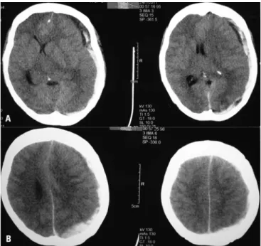
Until pre-intubation sedation, the patient’s level of consciousness was normal, with no abnormal indings on the neurological examination, a score of 15 on the Glasgow coma scale, equal-size and light-reactive pupils, and no focal signs. Two days later, while still sedated, intubated, and under unweanable mechanical ventilation, the patient exhibited a sudden hypertensive crisis with vomiting and the appearance of mydriasis in the left eye. he hypertension was immediately treated, an antiemetic was administered, and the analgesic doses were increased. Cranial CT was performed (Figure 2), which revealed the presence of an acute subdural hematoma and a parietal-occipital intracerebral hemorrhage on the left side. he patient was immediately subjected to emergency neurosurgical treatment.
he patient exhibited profuse bleeding with an onset at the beginning of surgery and lasting throughout the transoperative period, accompanied by hemodynamic instability that was diicult to manage despite luid replacement, blood component transfusion, and the use of vasoactive drugs. Following two trepanations, the hypertensive hygroma and hematoma, which displayed both acute and chronic components, were drained. Notably, the original neurosurgical plan had been to perform a decompressive craniotomy and to treat the acute subdural hematoma; however, the anesthetist had to interrupt the procedure because of progressive and refractory hemodynamic deterioration despite aggressive management. herefore, only the two trepanations were performed to drain the subdural collection.
Subsequently, the patient exhibited spontaneous bleeding at other sites, such as the mouth, nose, and vagina (although the coagulation tests had been normal up to that point), with persistence of hemolytic anemia and refractory shock. Four days after neurosurgery, the patient presented with bilateral mydriasis and nonreactive pupils. Sedation was discontinued, and her Glasgow score was 3. he maximal LDH level was 2,357 U/L, being 1,143 U/L at 20 days after delivery. he maximal total bilirubin concentration was 12.23 mg/dL. he maximal direct bilirubin was 8.29 mg/dL, being 1.43 at 20 days after delivery, when the AST was 118 U/L, and the ALT was 192 U/L. he minimal hemoglobin concentration was 4.6 g/dL and occurred ive days after delivery, whereas it had risen to 5.8 g/dL on the day neurological deterioration occurred. he minimal platelet count was 54,000/mm3,
Table 1 - Laboratory tests performed during hospitalization
Test/reference value Day 1 Day 3 Day 5 Day 11 Day 17 Day 22
Hemoglobin (11.1-16.1 g/dL) 10.6 8.5 4.6 10.0 5.8 7.6
Hematocrit (35-47%) 31.4 24.8 13.6 30.6 16.8 22.3
Total white blood cells (3,600-11,000/mm3) 24,800 33,900 19,600 22,200 20,600 9,400
Platelets (150,000-450,000/mm3) 232 176 183 59 207 159
Lactic dehydrogenase (207-414 U/L) 1,249 2,357 2,101 1,220 1,262 1,143
Aspartate transaminase (5-46 U/L) 150 141 135 --- --- 118
Alanine transaminase (3-50 U/L) 152 92 74 --- --- 192
Total/direct bilirubin (up to 1.2/0.4 mg/dL) 6.31/4.29 6.61/4.47 11.85/7.2 8.81/5.61 6.27/4.31 4.43/2.6
Alkaline phosphatase (50-136 U/L) 314 --- --- 80 ---
---Prothrombin time (PT) 1.00 1.79 1.22 0.95 1.12 0.9
Partial thromboplastin time (16-34 s) 51.5 76.2 54.2 27.1 29.4 29.5
Albumin (3.5-5.5 g/dL) --- --- 2.5 2.4 ---
---Creatinine (0.6-1.3 mg/dL) 2.88 3.67 2.79 1.76 2.13 2.01
Urea (10-45 mg/dL) 94 137 177 202 143 120
Sodium (135-147 mEq/L) 144 132 140 168 138 142
Calcium (8.8-11 mg/dL) 10.89 7.6 8.2 9.6 8.2
---Lactic acid (0.5-1.6 mmol/L) 3.7 1.6 --- 1.7 ---
---Uric acid (1.5-6.0 mg/dL) 7.0 7.9 9.7 --- ---
---Fibrinogen (150-350 mg/dL)
Non-coagulating: >10 times the
upper limit
--- 178 --- ---
---D-dimers (15-147 ng/mL) 4.4 --- --- --- ---
--- not performed on that day.
Figure 1 - Chest computed tomography scan showing a large left pleural effusion and a left collapsed lung.
Figure 2 - Cranial computed tomography scan showing an acute subdural hematoma with a midline shift and a subcortical hemorrhage in the left occipital lobe.
measured and found to be normal (2.1 to 2.4 mg/dL) throughout the hospitalization.
he diagnosis of brain death could not be established because the patient was absolutely unresponsive to stimuli and exhibited a complete absence of brain stem relexes upon neurological examination, due to the refractory hemodynamic instability that had set in combined with the clinical and neurological deterioration despite the luid resuscitation, use of vasopressors, and blood component transfusion. he patient died ive days after surgery, which was 22 days after the delivery.
DISCUSSION
Preeclampsia is characterized by hypertension, proteinuria, and edema. It afects approximately 5 to 7% of pregnant women, 65% of whom might progress to HELLP syndrome. For these reasons, preeclampsia remains a signiicant cause of maternal morbidity and mortality. Typically, preeclampsia occurs during the second or third trimester of pregnancy, but occasionally, the disease has been detected before gestational week 20. Its consequences include hypertensive crises, kidney failure, liver rupture, neurological complications such as seizures and stroke, and increased perinatal morbidity and mortality. he pathogenesis of preeclampsia is believed to be associated with placental ischemia, endothelial dysfunction, and cytotoxic and genetic factors. Severe preeclampsia is deined by systolic arterial pressure levels that are persistently ≥160 mmHg or diastolic arterial pressure ≥110 mmHg, massive proteinuria (4+ in a dipstick, or >2.0 g/24 hours), or the presence of clinical (epigastric pain, nausea, and vomiting) and laboratory (platelet count <50,000/mm3, creatine kinase >200 U/L,
LDH >1,400 U/L, AST >150 U/L, ALT >100 U/l, uric acid >7.8 mg/dL, and serum creatinine >1.2 mg/dL) manifestations.(4)
AFLP is a rare and potentially fatal disorder that might occur at the third trimester of pregnancy, involves microvesicular fatty iniltration of hepatocytes, and has a mortality rate of up to 70%. In most studies, the estimated incidence is 1:10,000-15,000 pregnancies, with mortality of 10 to 20%. AFLP occurs more often in multiple pregnancies, male fetuses, and nulliparous women. AFLP is associated with inherited defects of the mitochondrial fatty acid beta-oxidation, as well as with long-chain 3-hydroxyacyl-CoA dehydrogenase deiciency (LCHAD), including the G 1529C mutation.(3) he earliest symptoms
and itch. Approximately one third of patients exhibit signs of preeclampsia at the onset or during the progression of the disease. he diagnosis is usually clinical and is based on the laboratory and compatible imaging indings, including increased values for AST, ALT, bilirubin, uric acid, creatinine, leukocytes, clotting, and electrolytes, as well as blood glucose (hypoglycemia) disorders and lactic acidosis. he present case exhibited clinical and laboratory features compatible with AFLP, HELLP syndrome, and severe preeclampsia. As the clinical manifestations of AFLP prevailed at the disease onset, treatment was directed against that condition and was thus limited to intensive clinical support care, which includes immediate delivery following maternal stabilization because there is no speciic treatment for this disease.
HELLP syndrome is a severe form of preeclampsia that usually afects 4 to 12% of women with preeclampsia. his syndrome is associated with microthrombi, thrombocytopenia, and clotting disorders and has a poor prognosis. HELLP syndrome may appear from the second trimester of pregnancy up to several days after delivery; in one third of cases, the syndrome appears after childbirth.(3) HELLP syndrome is associated with
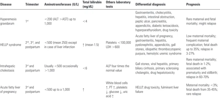
vasoconstriction, increased vascular tonus, platelet aggregation, and an altered thromboxane/prostacyclin ratio. HELLP syndrome is partially caused by activation of the complement and consequent coagulation cascade, resulting in endothelial and microvascular injury in multiple organs, microangiopathic hemolytic anemia, periportal and liver necrosis with increased liver enzymes, and thrombocytopenia. Both AFLP and HELLP syndrome afect the liver, but HELLP syndrome is more common (1:5,000 births). AFLP tends to occur later in the pregnancy and causes reduced liver synthetic function, resulting in hypoglycemia, increased ammonia levels, and clotting disorders. Disseminated intravascular coagulation is common in both conditions; however, the AST and ALT disorders and the increased LDH are more typical of HELLP syndrome, whereas the bilirubin increase is greater in AFLP (Table 2).(5)
he risk of stroke during pregnancy is low; however, when stroke does occur, the morbidity and mortality of its complications are high. Recent studies reported an association between stroke during pregnancy and a history of migraine, gestational diabetes, preeclampsia, and eclampsia,(6) which
most likely indicates physiopathological mechanisms other than those associated with stroke that is unrelated with pregnancy.
Table 2 - Clinical features of the liver diseases during pregnancy
Disease Trimester Aminotransferases (U/L) Total bilirubin (mg/dL)
Others laboratory
tests Differential diagnosis Prognosis
Hyperemesis
gravidarum 1
st <200 (ALT >AST) up to
1,000 <4
Gastroenteritis, cholecystitis, hepatitis, intestinal obstruction, peptic ulcer, pancreatitis, appendicitis, diabetic ketoacidosis, hyperparathyroidism, drug toxicity
Rare maternal and fetal mortality; might relapse
HELLP syndrome 2nd, 3rd, and postpartum
<500 (mean 250) except
in case of liver infarction ↑ (mean 1.5)
Platelets <100,000 LDH >600
Acute fatty liver of pregnancy, gastroenteritis, hepatitis, pyelonephritis, appendicitis, gall stones, idiopathic thrombocytopenic purpura, hemolytic uremic syndrome
Low maternal mortality; frequent maternal complication; fetal death up to 35%; relapse in 3-27%
Intrahepatic cholestasis
3rd and postpartum
Usually <500 occasionally
>1,000 <6
ALP four times the normal value
Gall stones, viral hepatitis, primary biliary cirrhosis, primary sclerosing cholangitis, drug hepatotoxicity
Rare maternal mortality; fetal death in 1-2%; associated with prematurity and stillbirth; relapse in 60-70%
Acute fatty liver of pregnancy
3rd and
postpartum <500 up to a 1,000 ↑
White blood cells ↑, PT ↑, platelets ↓, glucose ↓, uric acid ↑
HELLP, drug toxicity, fulminant liver failure
Maternal mortality <3%; fetal death from 35-45%; rare relapse
Adapted from: Sibai BM. HELLP syndrome. Up to Date. 2013 [cited January 29, 2013]. Available at http://www.uptodate.com/contents/hellp-syndrome.(5) ALT - alanine-aminotransferase;
AST - aspartate-aminotransferase; LDH - lactic dehydrogenase; ALP - alkaline phosphatase; PT - prothrombin time.
of death in women with preeclampsia.(7) In addition,
hypertension acts as an independent cause of stroke because the loss of the self-regulatory mechanisms of the brain blood low leads to vasodilation and brain edema, particularly in the case of individuals without chronic hypertension. Brain ischemia and hemorrhage are almost always accompanied by a blood pressure increase of at least 10% due to alteration of the self-regulatory mechanisms induced by vasoactive substance release at the injury site.(8)
Cases of spontaneous peripartum acute subdural hematoma and intracerebral hemorrhage have been reported in the literature, but in association with the HELLP syndrome(9) or with thrombocytopenia caused
by idiopathic thrombocytopenic purpura.(10) No case of
spontaneous acute subdural hematoma associated with AFLP could be found in the literature.
he present patient exhibited an unfavorable progression partially due to the late diagnosis of preeclampsia. In this regard, it is noteworthy that the hypertension peaks irst appeared a few days after the delivery and that the patient had undergone regular
prenatal care. In addition, the late diagnosis of fetal death contributed to the fatal outcome. Indeed, the fetal movements had ceased two days prior to hospital admission, and the patient had sought medical care from the very onset of the symptoms, thus indicating a delay in the pre-hospital and emergency care. Retrospectively, one might wonder whether plasmapheresis might have modiied the clinical progression, as hemolysis remained throughout the disease course. Plasmapheresis is indicated for patients with persistent HELLP syndrome,(11) but
there are no reports on the use of this modality for AFLP.
CONCLUSION
REFERENCES
1. Selo-Ojeme DO, Omosaiye M, Battacharjee P, Kadir RA. Risk factors for obstetric admissions to the intensive care unit in a tertiary hospital: a case-control study. Arch Gynecol Obstet. 2005;272(3):207-10.
2. Allford SL, Hunt BJ, Rose P, Machin SJ; Haemostasis and Thrombosis Task Force, British Committee for Standards in Haematology. Guidelines on the diagnosis and management of the thrombotic microangiopathic haemolytic anaemias. Br J Haematol. 2003;120(4):556-73.
3. Rahman TM, Wendon J. Severe hepatic dysfunction in pregnancy. QJM. 2002;95(6):343-57.
4. Freitas F, Martins-Costa SH, Ramos JG, Magalhães JA. Rotinas em obstetrícia. 5ª ed. Porto Alegre: Artmed; 2006. p. 389-423.
5. Sibai BM. HELLP syndrome. Up to Date. 2013 [cited 2013 Jan 29]. Available in: http://www.uptodate.com/contents/hellp-syndrome.
6. Scott CA, Bewley S, Rudd A, Spark P, Kurinczuk JJ, Brocklehurst P, et al. Incidence, risk factors, management, and outcomes of stroke in pregnancy. Obst Gynecol. 2012;120(2 Pt 1):318-24.
7. Gogarten W. Preeclampsia and anaesthesia. Curr Opin Anaesthesiol. 2009;22(3):347-51. Review.
8. Slama M, Modeliar SS. Hypertension in the intensive care unit. Curr Opin Cardiol. 2006;21(4):279-87. Review.
9. Yokota H, Miyamoto K, Yokoyama K, Noguchi H, Uyama K, Oku M. Spontaneous acute subdural haematoma and intracerebral haemorrhage in patient with HELLP syndrome: case report. Acta Neurochir (Wien). 2009;151(12):1689-92.
10. Pandey M, Saraswat N, Vajifdar H, Chaudhary L. Subdural haematoma in pregnancy-induced idiopathic thrombocytopenia: Conservative management. Indian J Anaesth. 2010;54(5):470-1.
11. von Baeyer H. Plasmapheresis in thrombotic microangiopathy-associated syndromes: review of outcome data derived from clinical trials and open studies. Ther Apher. 2002;6(4):320-8.
RESUMO
Pré-eclâmpsia, síndrome HELLP (hemólise, elevação de en-zimas hepáticas e plaquetopenia) e fígado gorduroso agudo da gestação são as principais causas de microangiopatia trombótica e disfunção hepática grave durante a gestação, representando um spectrum do mesmo processo patológico. Relatou-se aqui o caso de uma gestante com 35 semanas internada em unidade de terapia intensiva no pós-operatório imediato de cesariana por morte fetal, com náuseas, vômitos e icterícia. Diagnosticaram-se pré-eclâmpsia pós-parto e fígado gorduroso agudo da gestação. Houve evolução tardia com hematoma subdural agudo e
hemorragia intracerebral, sendo realizado tratamento neuroci-rúrgico. A paciente foi a óbito por anemia hemolítica refratária, com sangramento espontâneo em múltiplos órgãos. Pré-eclâmp-sia, síndrome HELLP e fígado gorduroso agudo da gestação são processos patológicos que podem se sobrepor e se associar a complicações potencialmente fatais, como a hemorragia intra-craniana aqui descrita. Sua detecção e diagnóstico precoces são fundamentais para a instituição de manejo adequado e sucesso do tratamento.