SUMMARY
Objective: To investigate the association between non-alcoholic fatty liver disease (NAFLD) and liver function/injury markers with components of metabolic syndrome (MS) in class III obese individuals. Methods: he study population consisted of 144 patients with class III obesity (body mass index [BMI] ≥ 40 kg/ m²). MS was diagnosed according to the National Cholesterol Education Program – Adult Treatment Panel III (NCEP ATP III) criteria, by determining the lipid proile, blood glucose, and basal insulin. Liver function/injury markers were also quantiied. Insulin resistance (IR) was measured by HOMA-IR and NAFLD diagnosis was established by magnetic resonance imaging (MRI). Statistical calculations were performed by SPSS version 13.0. he association was assessed by the Mann-Whitney and Chi-square tests, with a level of signiicance set at 5%.
Results: here was a signiicant association between the diagnosis of MS and NAFLD (χ2 = 6.84, p = 0.01). As for the diagnostic components of MS, there was a positive and
signiicant association between HDL-C (p = 0.05), waist circumference (p < 0.05), and hypertension (χ2 = 4.195, p = 0.041) with NAFLD. HOMA-IR (p < 0.001) also showed a
positive association with liver disease. Conclusion: A positive and signiicant association between NAFLD and components of metabolic syndrome in class III obese individuals was observed, suggesting the need and importance of monitoring these components in NAFLD screening.
Keywords: Fatty liver; metabolic syndrome X; obesity.
©2012 Elsevier Editora Ltda. All rights reserved.
Study conducted at the Micronutrient Research Center, Instituto de Nutrição Josué de Castro, Universidade Federal do Rio de Janeiro, and Clínica Cirúrgica Carlos Saboya, Rio de Janeiro, RJ, Brazil
Submitted on: 05/10/2011
Approved on: 02/19/2012
Correspondence to:
Gabriela Villaça Chaves Av. Pau-Brasil, 211-Bl. J Ilha da Cidade Universitária Rio de Janeiro – RJ, Brazil CEP: 21949-900 Fax: (21) 2280-8343 [email protected]
Conlict of interest: None.
Association between non-alcoholic fatty liver disease and liver
function/injury markers with metabolic syndrome components in
class III obese individuals
GABRIELA VILLAÇA CHAVES1, DAIANE SPITZDE SOUZA2, SILVIA ELAINE PEREIRA3, CARLOS JOSÉ SABOYA4, WILZA ARANTES FERREIRA PERES5
1 PhD in Internal Medicine; Nutritionist, Instituto Nacional de Câncer (INCA), Rio de Janeiro, RJ, Brazil 2 Specialist in Clinical Nutrition; Resident in Oncological Nutrition, INCA, Rio de Janeiro, RJ, Brazil 3 PhD in Internal Medicine; Nutritionist, Clínica Cirúrgica Carlos Saboya, Rio de Janeiro, RJ, Brazil 4 PhD; Surgeon, Clínica Cirúrgica Carlos Saboya, Rio de Janeiro, RJ Brazil
INTRODUCTION
he non-alcoholic fatty liver disease (NAFLD) is charac-terized by accumulation of fat in the liver when it exceeds 5-10% of its weight1. In addition to leading to major
histo-pathological alterations, it may be associated with elevated liver enzymes and abnormal liver function, ranging from steatosis to steatohepatitis, ibrosis, and cirrhosis2.
Although diagnosed worldwide, there are variations in prevalence, reaching about 20-30% in western countries3.
In the United States, where 25% of the adult population is obese, the disease occurs in more than two-thirds of these individuals and in more than 90% of class III obese indi-viduals4. It is estimated that 2% to 3% of the population has
nonalcoholic steatohepatitis (NASH)3.
Approximately 74% to 90% of patients who undergo liver biopsy show alterations due to triacylglycerol ac-cumulation2,5. he disease is highly prevalent (88.7%) in
obese patients undergoing bariatric surgery6, and the
like-lihood of developing steatohepatitis is increased in class III obesity, with 15% to 20% of these patients diagnosed with NASH7.
Recent studies have shown increased prevalence and higher incidence of cardiovascular disease (CVD) in in-dividuals with NAFLD. hese studies have shown hepatic steatosis as an independent risk factor for the development of this disease8,9.
Metabolic syndrome (MS), which involves the combi-nation of risk factors for CVD such as insulin resistance (IR), abdominal fat, dyslipidemia, glucose intolerance, and hypertension, has oten been associated with more severe liver abnormalities10.
OBJECTIVE
To investigate the association between NAFLD and liver function/injury markers with the components of MS in in-dividuals with class III obesity treated at a private clinic in the city of Rio de Janeiro, Brazil.
METHODS
he study included 144 individuals with class III obesity, of both genders, with a mean age of 36.5 (11.7) years, from a private clinic in the city of Rio de Janeiro in the period from January to December 2006, representing ap-proximately 60% of the total annual attendance. Pregnant women, nursing mothers, individuals with liver disease other than NAFLD (positive serology for hepatitis B and C), with daily intake of more than 20 grams of ethanol, and those using hepatotoxic drugs were excluded from the study. NAFLD diagnosis was achieved by magnetic reso-nance imaging assessment.
he class III obesity classiication was based on the World Health Organization (WHO) criteria (1998)11,
deined by body mass index (BMI) ≥ 40 kg/m² for the
diagnosis of this class of obesity. BMI calculation was per-formed using the anthropometric measurements weight (kg) and height (m²)12. Blood pressure (BP) and waist
cir-cumference (WC) were also measured. WC was measured with the patient standing with the abdomen relaxed, arms at the sides of the body and feet side by side, using an inex-tensible tape. he tape surrounded the individual’s largest abdominal sagittal diameter, as individuals with class III obesity have what is called an abdominal “apron”13.
For biochemical evaluation, a blood sample was ob-tained by venipuncture, ater a 12-hour fast. he lipid pro-ile, blood glucose, and basal insulin levels were evaluated. Basal insulin was quantiied by reversed phase high per-formance liquid chromatography (RP-HPLC). Addition-ally, the following markers of liver function (albumin, total bilirubin [TB] and activated prothrombin time [APT] – the latter described in seconds above the control) and liver injury (aspartate aminotransferase [AST], alanine amino-transferase [ALT], and gamma glutamyl transpeptidase [GGT]) were evaluated.
IR was identiied by HOMA-IR14, obtained by the
for-mula: HOMA-IR = fasting insulin (mU/L) × fasting glu-cose (mmol/L)/22.5. he receiver operating characteris-tic curve (ROC) was used for the identiication of IR. To determine the gold standard for implementing the ROC curve, with subsequent identiication of the value of high-est IR sensitivity and speciicity in this sample, reference values in the literature for healthy adult subjects were used15-18, thus obtaining a value > 4.0 as cutof point.
he diagnosis of MS was performed according to the National Cholesterol Education Program – Adult Treat-ment Panel III19 (NCEP - ATP III), which deines MS by
the presence of at least three of the following components: WC ≥ 102 cm in men and ≥ 88 cm in women, HDL-c ≤ 40 mg/dL in men and ≤ 50 mg / dL in women, triglyc-erides ≥ 150 mg/dL, fasting glucose ≥ 110 mg / dL, and blood pressure ≥ 130/85 mmHg.
Statistical calculations were carried out using SPSS, re-lease 13.0. he comparison of numerical and continuous variables (age, WC, lipid proile, blood glucose, insulin, and liver function/injury tests) between groups with and without NAFLD was performed by the Mann-Whitney test. Associations between categorical variables (presence or absence of hypertension (SAH), NAFLD, IR and MS) were performed using the chi-square (χ2) test. he
coef-icient of the proportion was performed to measure the degree of association between the categorical variables NAFLD and metabolic syndrome. he ROC curve was used to identify the most accurate value of HOMA-IR to diagnose IR. he signiicance level was set at 5%.
Presence of MS Absence of MS
p-value χ2 Coeficient of contingency
n % n %
Presence of NAFLD 57 81.4 45 61.6
0.01 6.84 0.21
Absence of NAFLD 13 18.6 29 38.4
NAFLD, non-alcoholic fatty liver disease; MS, metabolic syndrome.
Table 1 – Association between the presence of NAFLD and diagnosis of MS
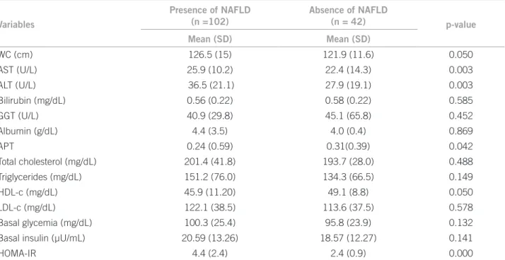
Table 2 – Comparison between the means (SD) of biochemical and anthropometric variables between the individuals with and without NAFLD
Variables
Presence of NAFLD (n =102)
Absence of NAFLD
(n = 42) p-value
Mean (SD) Mean (SD)
WC (cm) 126.5 (15) 121.9 (11.6) 0.050
AST (U/L) 25.9 (10.2) 22.4 (14.3) 0.003
ALT (U/L) 36.5 (21.1) 27.9 (19.1) 0.003
Bilirubin (mg/dL) 0.56 (0.22) 0.58 (0.22) 0.585
GGT (U/L) 40.9 (29.8) 45.1 (65.8) 0.452
Albumin (g/dL) 4.4 (3.5) 4.0 (0.4) 0.869
APT 0.24 (0.59) 0.31(0.39) 0.042
Total cholesterol (mg/dL) 201.4 (41.8) 193.7 (28.0) 0.488
Triglycerides (mg/dL) 151.2 (76.0) 134.3 (66.5) 0.149
HDL-c (mg/dL) 45.9 (11.20) 49.1 (8.8) 0.050
LDL-c (mg/dL) 122.1 (38.5) 113.6 (37.5) 0.578
Basal glycemia (mg/dL) 100.3 (25.4) 95.8 (23.9) 0.132
Basal insulin (µU/mL) 20.59 (13.26) 18.57 (12.27) 0.141
HOMA-IR 4.4 (2.4) 2.4 (0.9) 0.000
SD, standard deviation; NAFLD, non-alcoholic fatty liver disease; WC, waist circumference; AST, aspartate aminotransferase; ALT, alanine aminotransferase; GGT, gamma glutamyl transpeptidase; APT, seconds above the control.
RESULTS
he sample consisted of 144 subjects, of which 43 (29.4%) were males and 101 (70.6%) were females. he mean age of subjects was 36.5 (11.7) years, ranging from 19 to 64 years. here was no signiicant diference between the mean age (p = 0.08) and BMI (p = 0.16) according to gender. he prevalence of NAFLD in the study group was 71% with a positive diagnosis in 75.0% and 69.3% of men and women, respectively.
Of the 144 class III obese patients studied 49% had metabolic syndrome, and 81.4% of those with NAFLD also had the diagnosis of MS, showing a signiicant as-sociation between MS and NAFLD (p = 0.01) (Table 1). Table 2 shows a comparative analysis between the groups with and without a diagnosis of NAFLD. he mean HDL-c was signiicantly lower in patients with NAFLD. Regard-ing the anthropometric component of MS, the mean WC was signiicantly higher in subjects diagnosed with NAFLD. he means of AST and ALT levels were sig-niicantly higher, and the APT means were sigsig-niicantly
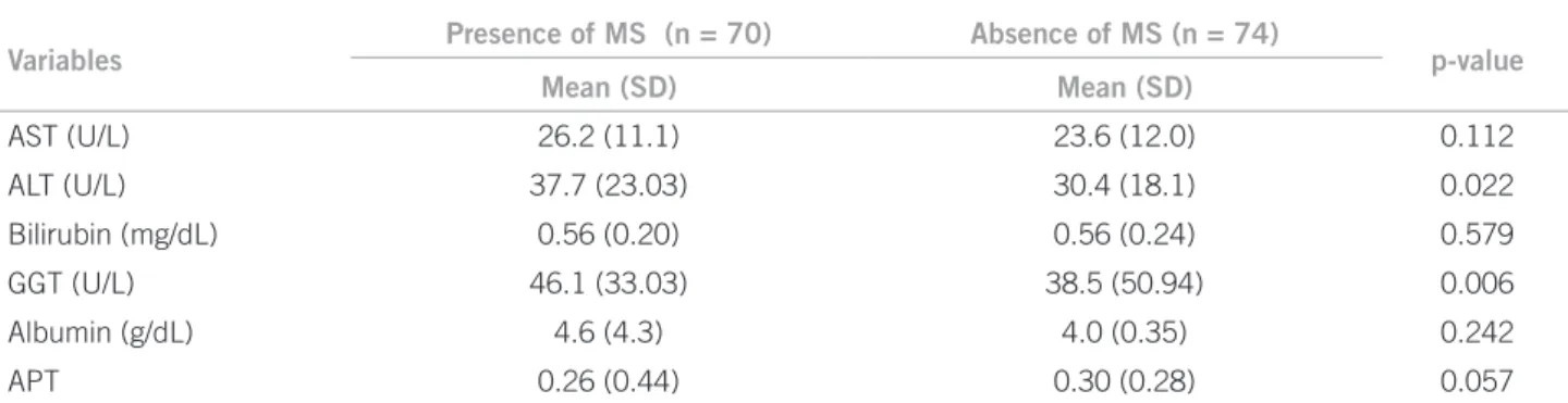
lower in this group. Considering the markers of liver function and injury in individuals with or without a di-agnosis of MS, signiicantly higher means of liver injury markers, ALT and GGT, were observed in subjects who had MS (Table 3).
When association tests for categorical variables were applied, the only component of the lipid proile that was associated with NAFLD was HDL-c, which was below the amount considered adequate by the NCEP-ATP III in 83.0% of patients with liver disease (p = 0.047, χ2 = 4.13).
When analyzing the HOMA-IR, it was observed that 75.5% of subjects had insulin resistance according to this parameter, with only 15% of individuals not presenting the disease (p < 0.001, χ2 = 5.641). Furthermore, patients
with NAFLD had a signiicantly higher mean of this index.
Finally, systemic arterial hypertension (SAH) was also associated with the presence of NAFLD (p = 0.041, χ2 = 4.195), with 57% of individuals who had a diagnosis
Variables
Presence of MS (n = 70) Absence of MS (n = 74)
p-value
Mean (SD) Mean (SD)
AST (U/L) 26.2 (11.1) 23.6 (12.0) 0.112
ALT (U/L) 37.7 (23.03) 30.4 (18.1) 0.022
Bilirubin (mg/dL) 0.56 (0.20) 0.56 (0.24) 0.579
GGT (U/L) 46.1 (33.03) 38.5 (50.94) 0.006
Albumin (g/dL) 4.6 (4.3) 4.0 (0.35) 0.242
APT 0.26 (0.44) 0.30 (0.28) 0.057
SD, standard deviation; MS, metabolic syndrome; AST, aspartate aminotransferase; ALT, alanine aminotransferase; GGT, gamma glutamyl transpeptidase; APT, seconds above the control.
Table 3 – Comparison between the means (SD) of the biochemical variables of liver function/injury between individuals with and without MS
DISCUSSION
MS was diagnosed in 81.4% of individuals afected by NAFLD. his association conirms the indings by Ferreira et al.20 and Soler et al.21, who found a higher prevalence of
MS in individuals with NAFLD. he growing recognition of the association between NAFLD and MS has been stim-ulating the interest in the possible role of this liver disease in the risk for the development of cardiovascular disease22.
he diagnostic criteria of MS that were associated with NAFLD were WC, HDL-c, and blood pressure. Bitencourt et al.6 showed a similar trend when evaluating the clinical
and histological features of NAFLD in obese patients un-dergoing bariatric surgery, in which more than 50% of the cases had diagnostic criteria for MS.
Several studies have shown that the accumulation of body fat in the abdominal region, regardless of the indi-vidual’s total body fat content, is an independent predic-tive factor for fat accumulation in hepatocytes and, there-fore, crucial in the pathogenesis of NAFLD23,24. he WC is
considered more sensitive to metabolic alterations and/or cardiovascular morbimortality than the simple increase in body weight measured by BMI21.
In the present study, the mean WC was signiicantly higher in the group with the disease. his association is ex-plained by the lipolytic nature of visceral fat, due to lower insulin sensitivity and higher concentration of β-receptors in this region, and its proximity to the portal system25.
Vis-ceral fat is drained directly into the portal system, expos-ing the liver to large amounts of free fatty acids, which in-creases the hepatic synthesis of triglycerides and may also decrease its ability to secrete them, resulting in accumula-tion in hepatocytes26.
he importance of monitoring the WC in individuals with NAFLD was described in the study by Yoo27, which
suggested that this component can be used in the screen-ing of NAFLD in Korean adults by means of speciic values for the screening of the disease.
he only lipid fraction and diagnostic factor of MS that was associated with the presence of the disease was
HDL-c, which showed a signiicantly lower mean in sub-jects with NAFLD. Generally, hypertriglyceridemia and low HDL-c are the lipid fraction disorders most oten as-sociated with the presence of steatosis28. Boza et al.29
ob-served signiicantly lower mean HDL-c levels in class III obese individuals with NAFLD, when compared with the group without the disease, which is the only lipid fraction variable that was associated with NAFLD diagnosis. In the study by Dixon et al.30, which evaluated possible
predic-tors of NAFLD in these individuals, no correlation was observed between any lipid fraction with more advanced stages of the disease. However, a weak negative correlation was observed between levels of HDL-c and the degree of simple steatosis, graded according to the lobular paren-chyma involvement. he authors suggest that dyslipidemia may have a greater impact on the disease in class I or class II obesity and a lower inluence in class III.
Marchesini et al.31, who studied the components of
MS in 304 individuals with NAFLD, found that over 90% of patients with some degree of liver disease have at least one component of this syndrome, with approximately one third of individuals having all components. Moreover, they observed a higher prevalence of disease in diabetic indi-viduals, being associated to 20% to 75% of cases. However, this prevalence seems to be more related to the IR than to the hyperglycemia.
In the present study, a signiicant association between IR, as indicated by the HOMA-IR, and the presence of NAFLD was observed. Probably due to the accumulation of abdominal fat, metabolic abnormalities such as these are very prevalent in these individuals21, which is in
agree-ment with studies that suggest that NAFLD is a compo-nent of MS, associated with visceral adiposity and IR23,32.
IR, both in the liver and adipose tissue, has been strongly associated with NAFLD33,34, as IR has been shown
to increase with disease severity35. Compared with control
intrinsic defect of the disease and the lower response to in-sulin in adipocytes stimulates tissue lipolysis, contributing to the progressive accumulation of lipids in hepatocytes through an increased low of free fatty acids in the liver36.
he storage of lipids can reach toxic levels and exacerbate the production of reactive oxygen species in the liver, stimulating the proliferation of macrophages and TNF-α, which also interfere with insulin sensitivity37. hus,
abnor-mal lipid peroxidation results in direct liver damage, with inlammation and even ibrosis21.
A limitation of the present study was the method used to assess IR. he hyperinsulinemic euglycemic clamp tech-nique is considered the gold standard for IR evaluation, as it allows the evaluation of insulin sensitivity in both liver and peripheral tissues. However, this method is not very practical and it is high-cost to be used in population-based studies or in clinical practice38. To date, there is no IR
labo-ratory method that can meet all of the following criteria for universal acceptance and use: suiciently precise measures so that IR can be compared between individuals, measures that can be obtained independently from the glucose from which it is obtained, data collection within the physiologi-cal range of insulin action and low cost and possibility of use in clinical practice.
HOMA is a simple and low cost method for the evalu-ation of IR but it has some limitevalu-ations. In this model, IR measurement is performed for total body surface, con-sidering that insulin sensitivity would be the same in the liver and peripheral tissues. here is some criticism re-garding the speciicity of the techniques used to evaluate basal insulin, which can be corrected by using speciic and standardized methodologies that do not sufer inluence of pro-insulin levels. he proportionality between insu-linemia and the degree of IR is also debatable39. Despite its
limitations, this has been the method most oten used to assess insulin resistance in population studies.
Liver damage can be identiied through liver damage markers: AST, ALT, and GGT. In the present study, the means of AST and ALT, although within the reference values, were signiicantly higher in those with NAFLD. Furthermore, higher mean levels of ALT and GGT were observed in individuals with MS.
he present study did not assess the severity of NAFLD, which can justify the fact that no diference was found in the means of the other liver functions and injury tests in individuals with and without MS, as well as explain the fact that the mean values of liver function tests are within the normal range in the group with the disease, as more severe alterations at liver function and injury tests are observed only in individuals with advanced liver disease. However, data published in the “hird national health and nutrition survey” showed a signiicant association between high concentrations of ALT and insulin resistance, diabetes
mellitus type 2, and MS40. Moreover, Koller et al.41
suggest-ed that the markers of liver injury may be indicators for the screening of individuals with MS or its components. How-ever, further studies should be performed to determine the association between MS and NAFLD severity.
CONCLUSION
In the present study, the association between the diag-nosis of NAFLD and MS was observed. MS components cause metabolic alterations, such as insulin resistance and oxidative stress that may contribute to the progression and worsening of liver disease. herefore, the determination and monitoring of these components are of crucial impor-tance for the screening of NAFLD.
REFERENCES
1. Festi D, Colecchia A, Sacco T, Bondi M, Roda E, Marchesini G. Hepatic ste-atosis in obese patients: clinical aspects and prognostic signiicance. Obes Rev. 2004;5:27-42.
2. Clain DJ, Lekowitch JH. Fatty liver disease in morbid obesity. Gastroenterol Clin North Am. 1987;16:239-52.
3. Bellentani S, Scaglioni F, Marino M, Bedogni G. Epidemiology of non-alcohol-ic fatty liver disease. Dig Dis. 2010;28:155-61.
4. Ruhl CE, Everhart JE. Epidemiology of non-alcoholic fatty liver. Clin Liver Dis. 2004;8:501-19.
5. Luyckx FH, Desaive C, hiry A, Dewé W, Scheen AJ, Gielen JE, et al. Liver abnormalities in severely obese subjects: efect of drastic weight loss ater gas-troplasty. Int J Obes Relat Metab Disord. 1998;22:222-6.
6. Bitencourt AGV, Cotrim HP, Alves E, Almeida AM, Barbosa DBV, Santos AS, et al. Doença hepática gordurosa não alcoólica: características clínicas e his-tológicas em obesos graves submetidos à cirurgia bariátrica. Acta Gastroen-terol Latinoam. 2007;37:224-30.
7. Scheen AJ, Luyckx FH. Obesity and liver disease. Best Pract Res Clin Endocri-nol Metab. 2002;16:703-16.
8. Adams LA, Lymp JF, St Sauver J, Sanderson SO, lindor KD, Feldstein A, et al. he natural history of nonalcoholic fatty liver disease: a population-based cohort study. Gastroenterology. 2005;129:113- 21.
9. Targher G, Bertolini L, Padovani R, Poli F, Scala L, Tessari R, et al.Increased prevalence of cardiovascular disease in type 2 diabetic patients with non-alco-holic fatty liver disease. Diabet Med. 2006;23:403-9.
10. Gupte P, Amarapurkar D, Agal S, Baijal R, Kulshrestha P, Pramanik S, et al. Non-alcoholic steatohepatitis in type 2 diabetes mellitus. J Gastroenterol Hep-atol. 2004;19:854-8.
11. World Health Organization. Obesity: preventing and managing the global epi-demic. Report of a WHO consultation on obesity. World Health Organ Tech Rep Ser. 1998;894:1-253.
12 Cuppari L, Sampaio, LR, Baxmann A, Kamimura MA. Avaliação nutricional. In: Cuppari L. Guias de medicina ambulatorial e hospitalar. Nutrição clínica no adulto. UNIFESP. São Paulo: Manole; 2002. p. 89-127.
13. Empana JP, Ducimetieri P, Charles MA, Jouven X. Sagittal abdominal diameter and risk of sudden death in asymptomatic middle-aged men: the Paris pro-spective study I. Circulation. 2004;110:2781-5.
14. Matthews DR, Hosker JP, Rudenski AS, Naylor BA, Treacher DF, Turner RC. Homeostasis model assessment: insulin resistance and beta-cell function from fasting plasma glucose and insulin concentrations in man. Diabetologia. 1985;28:412-9.
15. Geloneze B, Repetto EM, Geloneze SR, Tambascia MA, Ermetice MN. he threshold value for insulin resistance (HOMA-IR) in an admixtured popula-tion IR in the Brazilian metabolic syndrome study. Diabetes Res Clin Prac. 2006;72:219-20.
16. Buccini GS, Wolthal DL. Valores de corte para índices de insulinorresisten-cia, insulinosensibilidad e insulinosecreción derivados de la fórmula HOMA y del programa HOMA2. Interpretación de los datos. Rev Argent Endocrinol Metab. 2008;45:3-21.
17. Bonora E, Kiechl S, Willeit J, Oberhollenzer F, Egger G, Targher G, et al. Preva-lence of insulin resistance in metabolic disorders: the Bruneck study diabetes. 1998;47:1643-9.
19. he third report of the National Cholesterol Education Program (NCEP). Ex-pert panel on detection. Evaluation and treatment of high blood cholesterol in adults (Adult treatment panel III). JAMA. 2001;285:2486-97.
20. Ferreira VSG, Pernambuco RB, Lopes EP, Morais CN, Rodrigues MC, Arruda MJ, et al.Frequência e fatores de risco associados à doença hepática gordurosa não alcoólica em pacientes com diabetes melito tipo 2. Arq Bras Endocrinol Metab. 2010;54:362-8.
21. Soler GLN, Silva AWSM, Silva VCG, Teixeira RJ. Doença hepática gordurosa não-alcoólica: associação com síndrome metabólica e fatores de risco cardio-vascular. Rev SOCERJ. 2008;21:94-100.
22. Targher G, Arcaro G. Non-alcoholic fatty liver disease and increased risk of cardiovascular disease. Atherosclerosis. 2007;191:235-40.
23. Marceau P, Biron S, Hould FS, Marceau S, Simard S, hung SN, et al.Liver pathology and the metabolic syndrome X in severe obesity. J Clin Endocrinol Metab. 1999;84:1513-7.
24. Stranges S, Dorn JM, Muti P, Freudeheim JL, Farinaro E, Russel M, et al. Body
fat distribution, relative weight, and liver enzyme levels: a population-based study. Hepatology. 2004;39:754-63.
25. Arner P. Diferences in lipolysis between human subcutaneous and omental adipose tissues. Ann Med. 1995;27:435-8.
26. Björntorp, P. Do stress reactions cause abdominal obesity and comorbidities? Obes Rev. 2001;2:73-86.
27. Yoo HJ, Park MS, Lee CH, Yang SJ, Kim TN, Lim KI, et al. Cutof points of
abdominal obesity indices in screening for non-alcoholic fatty liver disease in Asians.Liver Int.2010;30:1189-96.
28. Angelico F, Del Ben M, Conti R, Francioso S, Feole K, Maccioni D, et al. Non-alcoholic fatty liver syndrome: a hepatic consequence of common metabolic diseases. J Gastroenterol Hepatol. 2003;18:588-94.
29. Boza C, Riquelme A, Ibañez L, Duarte I, Norero E, Viviani P, et al.Predictors of nonalcoholic steatohepatitis (NASH) in obese patients undergoing gastric bypass. Obes Surg. 2005;15:1148-53.
30. Dixon JB, Bhathal PS, O’Brien PE. Nonalcoholic fatty liver disease: predictors of nonalcoholic steatohepatitis and liver ibrosis in the severely obese. Gastro-enterology. 2001;121:91-100.
31. Marchesini G, Bugianesi E, Forlani G, Cerrelli F, Lenzi M, Manini, et al. Non-alcoholic fatty liver, steatohepatitis, and the metabolic syndrome. Hepatology. 2003;37:917-23.
32. Luyckx FH, Lefebvre PJ, Scheen AJ. Non-alcoholic steatohepatitis: associa-tion with obesity and insulin resistance, and inluence of weight loss. Diabetes Metab. 2000;26:98-106.
33. Seppälä-Lindroos A, Vehkavaara S, Häkkinen AM, Goto T, Westerbacka J, So-vijärvi A, et al. Fat accumulation in the liver is associated with defects in insu-lin suppression of glucose production and serum free fatty acids independent of obesity in normal men. J Clin Endocrinol Metab. 2002;87:3023-8. 34. Bugianesi E, Pagotto U, Manini R, Vanni E, Gastaldelli A, Lasio R, et al.Plasma
adiponectin in nonalcoholic fatty liver is related to hepatic insulin resistance and hepatic fat content, not to liver disease severity. J Clin Endocrinol Metab. 2005;90:3498-504.
35. Angelico F, Del Ben M, Conti R, Francioso S, Feole K, Fiorello, Set al. Insulin resistance, the metabolic syndrome, and nonalcoholic fatty liver disease J Clin Endocrinol Metab. 2005;90:1578-82.
36. Utzschneider KM, Kahn SE. Review: the role of insulin resistance in nonalco-holic fatty liver disease. J Clin Endocrinol Metab. 2006; 91:4753-61. 37. Neuschwander-Tetri BA, Caldwell SH. Nonalcoholic steatohepatitis:
sum-mary of an AASLD single topic conference. Hepatology. 2003;37:1202-19. 38. Ruano M, Silvestre V, Castro R, García-Lescún MC, Aguirregoicoa E, Marco
A, et al. HOMA, QUICKI and MFfm to measure insulin resistance in morbid obesity. Obes Surg. 2006;16:549-53.
39. Geloneze B, Tambascia MA. Avaliação laboratorial e diagnóstico da resistência insulínica. Arq Bras Endocrinol Metab. 2006;50:208-15.
40. Liangpunsakul S, Chalasani N. Unexplained elevations in alanine amino-transferase in individuals with the metabolic syndrome: results from the third national health and nutrition survey (NHANES III). Am J Med Sci. 2005;329:111-6.