DOI: 10.1590/1518-8345.0782.2693 www.eerp.usp.br/rlae
Santos CT, Almeida MA, Lucena AF. The Nursing Diagnosis of risk for pressure ulcer: content validation. Rev.
Latino-Am. Enfermagem. 2016;24:e2693. [Access ___ __ ____]; Available in: ____________________. DOI:
http://dx.doi.org/10.1590/1518-8345.0782.2693 month day year URL
The Nursing Diagnosis of risk for pressure ulcer: content validation
1Cássia Teixeira dos Santos
2Miriam de Abreu Almeida
3Amália de Fátima Lucena
3Objective: to validate the content of the new nursing diagnosis, termed risk for pressure ulcer.
Method: the content validation with a sample made up of 24 nurses who were specialists in skin
care from six different hospitals in the South and Southeast of Brazil. Data collection took place
electronically, through an instrument constructed using the SurveyMonkey program, containing
a title, deinition, and 19 risk factors for the nursing diagnosis. The data were analyzed using
Fehring’s method and descriptive statistics. The project was approved by a Research Ethics
Committee. Results: title, deinition and seven risk factors were validated as “very important”:
physical immobilization, pressure, surface friction, shearing forces, skin moisture, alteration in
sensation and malnutrition. Among the other risk factors, 11 were validated as “important”:
dehydration, obesity, anemia, decrease in serum albumin level, prematurity, aging, smoking,
edema, impaired circulation, and decrease in oxygenation and in tissue perfusion. The risk factor
of hyperthermia was discarded. Conclusion: the content validation of these components of the
nursing diagnosis corroborated the importance of the same, being able to facilitate the nurse’s
clinical reasoning and guiding clinical practice in the preventive care for pressure ulcers.
Descriptors: Pressure Ulcer; Nursing Diagnosis; Nursing Process; Risk Factors; Nursing.
1Paper extrated from Master’s Thesis “Development and content validation of nursing diagnosis risk of ulcer pressure”, presented to Escola de Enfermagem, Universidade Federal do Rio Grande do Sul, Porto Alegre, RS, Brasil. Supported by Fundo de Incentivo à Pesquisa e Eventos (Fipe), Hospital de Clínicas de Porto Alegre, process # 130034.
2 RN, Hospital de Clínicas de Porto Alegre, Porto Alegre, RS, Brazil. Profeasor, Centro Universitário Metodista de Porto Alegre, Porto Alegre, RS,
Brazil.
Rev. Latino-Am. Enfermagem 2016;24:e2693
Introduction
Pressure Ulcers (PU) are lesions in the skin and/or
underlying tissue, usually over a bony prominence, as a
result of pressure or pressure in combination with shear
and/or friction(1).
International studies indicate rates of prevalence
of pressure ulcers in American hospitals at around
12.3% among inpatients in clinical care units and
22% in intensive care units(2). In Sweden, one General
Hospital had a prevalence rate of PU of 23%(3) while
in Switzerland, the prevalence of PU of 26.5% was
identiied among hospitalized children(4).
These data evidence that PU remain a frequent
health problem with far-reaching effects, as they
increase the risk of developing other health issues
such as infections, osteomyelitis, septic arthritis and
sepsis. PU cause patients great physical and emotional
suffering, reducing their independence in daily activities
and compromising their process of rehabilitation, and,
consequently, having a negative impact on their quality
of life(5).
In addition to this, it is known that the inancial
costs are high for the health systems, associated
with the acquiring of material for treating PU and
their complications; these lead to more prolonged
hospitalizations, with a need for more time spent on
the care provided to patients with PU. These costs vary
from US$2,000 to US$70,000 per wound, considering
an annual average for the hospital varying from
US$400,000 to US$700,000(5).
In the light of this, preventing PU is shown to
be essential, as it can impact positively on reducing
the prevalence and incidence of this health issue and
its complications and, therefore, reduces the costs
of treating these. In this perspective and in the light
of the absence of a Nursing Diagnosis (ND) clearly
naming and deining the situation of risk for PU in the
NANDA International (NANDA-I) Taxonomy II, Brazilian
nurses undertook a study(6) which contributed to the
development of the ND of Risk for pressure ulcer. It
was located in Domain 11- Safety/Protection, Class 2-
Physical injury, recently published in the 2015 – 2017
edition of this diagnostic classiication system(7). The content validation of the new ND was undertaken
from when it was irst constructed(6), in order to evidence the reliability and degree of agreement in relation to the
components which structure it; that is, title, deinition
and risk factors(8-10). The studies on nursing diagnosis
content validation are essential sources in searching for
evidence and in the reduction of the probability of errors
in the nurse’s diagnostic process and decision-making
process. As a result, in the present study, the objective
was to validate the content of the components of the
Nursing Diagnosis Risk for pressure ulcer (title, deinition
and risk factors), in accordance with specialists’ opinion.
Method
This is a Diagnostic Content Validation (DCV) study
of the components of the ND of Risk for pressure ulcer,
through the opinion of specialists(11). The sample was
made up of 24 nurses, members of skin and wound
care study groups, from ive hospitals in the southern
region of Brazil, and one in the southeastern region.
The specialists were selected according to the following
inclusion criteria: to participate or have participated in a
skin and wound care study group for, at least, one year;
to have had clinical practice in skin care, particularly in
care for patients at risk for PU, for at least one year;
to use a PU prevention and treatment protocol with
application of the Braden scale as the instrument for
predicting risk; and to respond to the instrument within
the time period established of 60 days. Those nurses
who met the inclusion criteria but who were absent
from work during the period of the study due to holiday,
absence and/or leave were excluded from the study.
For data collection, the SurveyMonkey program
was used, available free of charge on the Internet,
in which was created a questionnaire with a link
generated automatically, which was sent by email to
the study participants. The responses were stored on
the program’s database. The irst part of the instrument
contained data on the specialists’ characterization and
professional and academic proile. The second part
of the instrument focused on data of the DCV of the
ND Risk for pressure ulcer, and contained the title
and deinition of this new ND, in which the specialists were to place an “X” on a ive point Likert-type scale,
covering one of the following possibilities: 1 - strongly
disagree; 2 - disagree; 3 - do not know; 4 – agree,
and 5 - strongly agree. Following that, the instrument
presented the risk factors which made up the ND with
their respective conceptual deinitions and, in addition, a ive point Likert scale in which the specialists were
to mark one of the following alternatives: 1- does not
indicate risk for PU; 2 - indicates little risk for PU; 3 -
indicates moderate risk for PU; 4 - indicates high risk for
PU and 5 - indicates a very high risk for PU. Along with
the data collection instrument, the respondents were
also sent an informative pamphlet on how to ill out the
instrument and return it to the researcher, and on the
ethical aspects of the study. The return of the illed-out
instrument was taken as acceptance to participate in the
The analysis of the variables related to the sample’s
characterization was undertaken through descriptive
statistics on the SurveyMonkey program and using the
IBM Statistical Package for the Social Sciences (SPSS)
program, version 18.0.
The analysis related to the ND’s DCV was also
statistical, taking into account the score attributed by
the specialists to each one of its components and, based
on that, the weighted average of the same indicated on
the Likert scale with variation between 1 and 5 points,
where: 1=0; 2=0.25; 3=0.50; 4=0.75 and 5=1(11).
Any component (title, deinition, risk factor) which
received a mean greater or equal to 0.80 was considered
“very important”; those with a mean below 0.80, but above 0.50, as “important”; and those with a mean
equal to or less than 0.50 were discarded, as they were
not considered important for this ND in the specialists’
opinion(11). The project was approved by the Research
Ethics Committee, under Protocol 13-0034.
Results
The study involved the participation of 24 specialist
nurses, the large majority of whom were female
(95.8%), with work in the area of nursing for a median
time of 63.5 (20.75 – 183) months and with a median
time of participation in study groups on skin and wounds
of 48 (16.5 – 72) months. The academic title of the
majority was specialist (58.3%), with them currently
working in clinical care (54.2%). It was ascertained that
they participated in events on the issue of prevention
and treatment of PU, besides publication in annals and
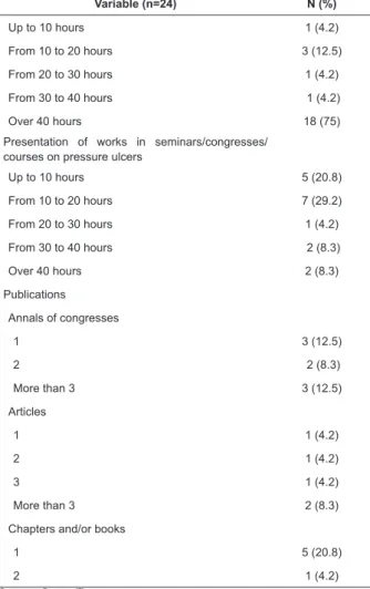
scientiic articles (Table 1).
Table 1 – Characterization of the sample of specialist
nurses (n=24). Porto Alegre, RS, Brazil, 2014.
Variable (n=24) N (%)
Title
Ph.D 2 (8.3)
M.A 4 (16.7)
Specialist 14 (58.3)
Graduate degree 4 (16.7)
Area of work
Clinical care 13 (54.2)
Teaching of nursing 1 (4.2)
Nursing management 5 (20.8)
Other 5 (20.8)
Length of work in the area of nursing (in months)* 63,5 (20.75-183)
Length of participation in skin groups (in months)* 48 (16.5-72)
Participation in courses on pressure ulcers/skin lesions
Variable (n=24) N (%)
Up to 10 hours 1 (4.2)
From 10 to 20 hours 3 (12.5)
From 20 to 30 hours 1 (4.2)
From 30 to 40 hours 1 (4.2)
Over 40 hours 18 (75)
Presentation of works in seminars/congresses/ courses on pressure ulcers
Up to 10 hours 5 (20.8)
From 10 to 20 hours 7 (29.2)
From 20 to 30 hours 1 (4.2)
From 30 to 40 hours 2 (8.3)
Over 40 hours 2 (8.3)
Publications
Annals of congresses
1 3 (12.5)
2 2 (8.3)
More than 3 3 (12.5)
Articles
1 1 (4.2)
2 1 (4.2)
3 1 (4.2)
More than 3 2 (8.3)
Chapters and/or books
1 5 (20.8)
2 1 (4.2)
Source: Santos(7). *median (25%-75%)
The content validation of the ND Risk for pressure
ulcer included the analysis of the title, deinition and 19
risk factors which make up the same.
The title and the deinition were validated with a mean of ≥ 0.80 (Table 2).
Table 2 – Title and deinition validated by specialists for
the ND Risk for pressure ulcer. Porto Alegre, RS, Brazil,
2014.
Components of the ND Risk for pressure ulcer Mean
Title – Risk for pressure ulcer 0.92
Deinition - Risk of tissue damage in the skin and
underlying tissue, as a result of compression of the soft tissues generally over a bony prominence, during a time period capable of causing local ischemia and, consequently, necrosis
0.87
Source: Santos(7).
Nineteen risk factors for the ND Risk for pressure
ulcer were submitted to DCV. Seven (56.8%) were
validated as “very important”, with a mean of ≥ 0.80
(Table 3).
(continue...)
Rev. Latino-Am. Enfermagem 2016;24:e2693
Table 3 – Risk factors validated as “very important” for
the ND Risk for pressure ulcer. Porto Alegre, RS, Brazil,
2014.
Risk factors for pressure ulcer validated as
“very important” Mean
Physical immobilization 0.97
Pressure 0.90
Shearing forces 0.90
Surface friction 0.89
Skin moisture 0.88
Malnutrition 0.84
Alteration in sensation 0.82
Source: Santos(7).
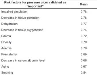
Eleven (57.8%) risk factors were validated as
“important”, with a mean of > 0.5 and < 0.8 (Table 4).
Table 4 - Risk factors validated as “important” for the ND
Risk for pressure ulcer. Porto Alegre/RS, 2014.
Risk factors for pressure ulcer validated as
“important” Mean
Impaired circulation 0.78
Decrease in tissue perfusion 0.78
Dehydration 0.77
Decrease in tissue oxygenation 0.74
Edema 0.72
Obesity 0.70
Anemia 0.70
Prematurity 0.69
Decrease in serum albumin level 0.68
Aging 0.67
Smoking 0.54
Source: Santos(7).
Only the risk factor of hyperthermia was discarded,
as it received a mean of ≤ 0.50.
Discussion
The DCV(11) has been recognized by NANDA-I(7) as
an important method for reining the ND, with a level
of evidence of 2.3, as it requires nurses’ specialized
opinion regarding the components of a nursing
diagnosis. These studies seek to ascertain the reliability
of the ND in practice, as well as considering its validity
in relation to the degree of agreement regarding the
components which structure it: title, deinition, deining
characteristics (signs and symptoms), related factors
(etiology/cause) and risk factors. This type of study has
been used both for developing new NDs and for reining
those already existing, with a view to greater accuracy(7).
Among the limitations of validation studies is the
initial difficulty for defining the inclusion criteria for
specialists, as there is no consensus in the literature
in relation to the ideal number for the sample,
besides the difficulty of finding nurse specialists in
the areas of interest for investigation(8-10). However,
in the present study, the choice of the specialists
was based on their academic background and, in
particular, on the clinical experiences of the nurses
who make up the different hospitals’ study groups
on care of the skin and tissues, so as to favor an
accurate judgment on the components of the ND Risk
for pressure ulcer. In addition to this, the diversity in
the origin of the specialists extended the reliability
of the data evaluated from different perspectives,
showing there to exist, among the specialists,
convergent opinions related to the physiopathology
and risk factors for PU.
The data referent to the validation of the title of the
diagnosis “Risk for pressure ulcer” and of its deinition “risk of cell damage in the skin and underlying tissue
as a result of the compression of soft tissues, generally
over a bony prominence, during a period of time capable
of causing local ischemia and, consequently, necrosis”
obtained means of > 80 points, that is to say, they were
considered on the Likert scale as “strongly agree”.
This score referent to the title of the ND
demonstrated agreement among the specialists,
in addition to the same covering in a clear way the
essential axes of an ND in accordance with NANDA-I(7):
1 - focus of the diagnosis (in this case PU); 2 - subject of
the diagnosis (when not made clear, this automatically
comes to be the individual); 3 – judgment (combined
in the diagnostic concept; in this case PU) and 7 -
situation of the diagnosis (covered by the risk category).
Similarly, the deinition of the ND also presented a
mean which showed agreement among the specialists,
which demonstrates clarity and objectivity, based in the
physiopathology and etiology of the PU.
The importance of a speciic ND, with a clear title and deinition regarding the risk of PU, has been
evidenced by studies(12-14) which have demonstrated
that this clinical situation is common, both in patients
at home and in hospitals, which justiied the ND’s
development and inclusion in a diagnostic terminology,
which supports the nurse in her management of the
process of preventive care for this health issue.
Seven risk factors (37%) were validated by the
forces, surface friction, skin moisture, malnutrition and
alteration in sensations.
The risk factor of physical immobilization was
validated with a mean of 0.97, this being the highest score
and agreement among the specialists, demonstrating
this to be one of the principal factors increasing the
patient’s vulnerability to PU. It is known that reduced
mobility increases the probability of greater time of
pressure on the skin, favoring tissue ischemia and the
occurrence of surface friction and shearing forces, with
consequent possibility of breaking the skin and initiating
ulceration(15). Corroborating this idea, one transversal
and exploratory study, with 43 older adults at risk of
PU, hospitalized in clinical units of a Brazilian hospital,
indicated – on the “activity” subscale of the Braden
scale – that approximately 39.5% of these patients
were bedridden or conined to a chair; on the subscale of “mobility”, 60% of the patients were totally immobile or signiicantly limited, which explains these older adults’
risk of developing PU(16).
The risk factors related to pressure and shearing
forces were validated with a mean of 0.90, and surface
friction with a mean of 0.89. These external forces do
not act in isolation, and cause the reduction of supply
of blood to the skin and tissues. When associated
with the patient’s intrinsic factors (such as immobility,
malnutrition and low tissue perfusion and oxygenation),
they cause breaking of the skin due to ischemia,
gradually increasing the development of the PU unless
a prevention intervention is made. The repositioning
of the patient, the use of polyurethane mattresses or
air mattresses, the use of protective dressings on bony
prominences and the constant assessment of humidity
are examples of preventive interventions for PU(17).
The risk factor of skin moisture received a mean of
0.88, reafirming its importance for the development of
PU. The exposure of the skin to humidity, principally to
urine and feces, associated with abrasive forces such as
surface friction and shearing forces, predisposes to an
increase in irritation, causing maceration and ulceration
and – once the PU is installed – the prognosis is negative
regarding healing(14).
The risk factor of malnutrition was validated with
a mean of 0.84. Under conditions of weight loss, the
musculature becomes hypertrophic, and the thin
panniculus causes a break in the skin. With deiciency in
nutrients, change also takes place in tissue healing, in
the inlammatory reaction, and in the immune function
when exposed to pressure. Poor nutrition can also
be associated with low weight, indicated by the Body
Mass Index (BMI <20), which favors the development
of PU over the bony prominence, associated with
pressure(13,18-19).
The risk factor of alteration in sensations was
validated with a mean of 0.82. Reduction in sensation
occurs due to illnesses which trigger this form of
harm, such as neurological ones, or through the use
of analgesics and sedatives which, besides reducing
sensation to physical stimulus, harm mobility. This is
due to reduction of the normal stimulus to pain, leading
the patient not to relieve prolonged pressure(19).
Eleven risk factors were considered “important” by
the specialists, with means between 50 and 80 points:
impaired circulation, decrease in tissue perfusion,
dehydration, decrease in tissue oxygenation, edema,
obesity, anemia, prematurity, decrease in serum albumin
level, aging and smoking.
The risk factor of impaired circulation was validated
with a mean of 0.78. Impairment in the peripheral
circulation leads to reduction in local capillary pressure,
with a negative impact on the nutrition of the tissues
due to the deicient peripheral blood supply, with a
probability of hypoxia, anoxia, and tissue ischemia.
Peripheral vasoconstriction can be related to peripheral
cardiovascular diseases, kidney diseases, anemia,
arterial hypertension, diabetes mellitus, kidney and
respiratory failure, concomitant infections, orthopedic
lesions and use of medications, among other factors(20).
In the light of the numerous illnesses related to the
circulation, this risk factor deserves attention and has
been described in research on PU(12, 21), principally among
older adults, whose circulatory system is impaired by
the characteristics of senescence.
The risk factor of dehydration was validated with
a mean of 0.77. Dehydration impairs the vital functions
of circulation, reducing the oxygenation of the tissues.
It is known, furthermore, that deicit in ingesting liquids
causes reduction in the skin turgor, this becoming
increasingly fragile, which, coupled with the forces of
abrasion (friction, pressure and shearing), increases the
risk of ulceration(18).
The risk factor of decrease in tissue oxygenation
was validated with a mean of 0.74, and decrease in
tissue perfusion with 0.78. Decrease in tissue perfusion
and oxygenation reduces the rate of metabolism and
energy of the tissue, predisposing to hypoxemia and
organic dysfunction. Studies indicate that, in this
situation, the patient is more predisposed to PU because
of the deicit in perfusion and oxygenation, which can
occur in situations such as trauma, loss of blood, and
infection(22).
The risk factor of edema was validated with a mean
of 0.72. Edema is abnormal accumulation of liquid, in
which there is increase in vascular permeability, and
reduction in lymphatic drainage and, due to this, the
Rev. Latino-Am. Enfermagem 2016;24:e2693
in nutrients. When the tissue luid increases and leaks
outside the cells, the pressure on the blood vessels
increases and, therefore, the blood low and oxygenation
of the tissues reduce, favoring ulceration(19).
The risk factor of obesity was validated with a mean
of 0.70. In obesity, there is the formation of adipose
tissue, which reduces the vascularization of the skin
surface, which can favor ischemia in the tissues and
the development of PU, when some area of the body is
subjected to pressure. Associated with this, the obese
individual may have other comorbidities such as diabetes
mellitus, making her still more vulnerable to PU(13,18-19).
The risk factor of decrease in serum albumin level
was validated with a mean of 0.68. Albumin is the most
abundant protein in the plasma, used for determination
of nutritional status. In low concentration, it causes
changes in oncotic pressure and the formation of
edema, which compromises the diffusion of oxygen and
nutrients to the tissues, predisposing to hypoxia and cell
death(13,18).
The risk factor of anemia was validated with a mean
of 0.70. This consists of the reduction in the quantity of
hemoglobin in the blood stream, which is responsible for
transporting oxygen to cells and tissues. The reduction
of oxygen for the ibroblasts, cells responsible for
healing of the tissues, reduces the formation of collagen
and increases the tissue’s susceptibility by precipitating
ischemia and necrosis(13,19). Supporting this, one study
which described the proile of patients with PU, in a public
hospital in São Paulo, indicated – among other factors –
that the result of the laboratory tests presented a mean
albumin of 2.7, glycemia of 169.7, hemoglobin of 9.5,
leukocytes of 14.888 and C-reactive Protein of 79.2(22).
These data, related to the levels of serum albumin and
hemoglobin, demonstrated the inluence of the same in
increasing the risk of PU.
The risk factor of prematurity was validated with a
mean of 0.69. It is known that the skin of a premature
child (age between the 20th and 37th week of gestation)
is fragile and that the physiological systems are not
completely formed. There are deiciencies in oxygenation
and vascularization of the skin and tissues, as well as in
the integrity of the skin, with any break or ulceration
being able to lead to systemic infection and increase in
morbidity. In addition to this, hospitalized newborns in
the critical units often require mechanical ventilation,
cardiological monitoring and nutritional support, which
hinders changing their position, favoring the increase of
pressure and shearing on more vulnerable areas and,
consequently, the development of PU(23).
The risk factor of aging was validated with a mean of
0.67. It is known that the elderly population is considered
at risk, due to its presenting decline in biological, psychic
and social functions, as well as developing chronic
degenerative diseases which cause prolonged periods
of hospitalization and, later, of rehabilitation. As age
advances, the skin becomes drier; as a consequence
of the reduction in sweat and sebaceous glands, there
is a reduction in vascularization and in properties such
as perception of pain and the inlammatory response,
besides there being hemodynamic changes and muscular
atrophy, which causes the bony structures to become
more prominent(24). When these factors are associated
with the morbid conditions and with other risk factors
(such as changes in mobility, nutrition, and anal and
urinary incontinence) the predisposition to developing
PU increases.
The risk factor of smoking was validated with a
mean of 0.54. The nicotine present in cigarettes causes
vasoconstriction, and, because of this, impedes the blood
low from occurring normally, hindering oxygenation and
tissue perfusion, which favors necrosis and ulceration.
In one case control study, undertaken in the United
Kingdom, the cutaneous reactive response was assessed,
after the installation of pressures in the sacral region, so
as to identify the differences in the reactivity of blood
low in a group of individuals who were smokers and
non-smokers, demonstrating that the smokers had a greater
probability of forming tissue ischemia in comparison with
non-smokers(25). This datum strengthens the fact that PU
is strongly related to the vascular risk factors brought by
tobacco, corroborating what has been validated by the
specialists in the present study.
The risk factor of hyperthermia, although not
validated by the specialists as important, as it presented
a mean of < 0.50, is found described in the literature as a factor which favors the compromising of the body’s
metabolism, the instability of enzymatic functions, and
the alteration of the metabolic pathways dependent
on oxygen, causing reduction in the oxygenation of
the tissues. This, associated with other concomitant
factors such as immobility, malnutrition or obesity and
extremes of age (prematurity or aging), makes the risk
of PU imminent(18).
The results obtained in this study were sent to
the Diagnosis Development Committee (DDC) of the
NANDA-I, responsible for analyzing proposals for
new diagnoses for this taxonomy, and were approved
and published in its most recent edition(6) with some
modiications such as maintaining the risk factor of
hyperthermia.
Conclusion
The DCV of the new Nursing Diagnosis Risk
demonstrated that its title, its deinition and 18 of the
19 risk factors raised were considered to be important
components of this ND.
It is known that PU begin silently and are like
icebergs; very dangerous below the surface, but
unobtrusive on the surface. Thus, a speciic and accurate ND for this clinical situation, with a clear deinition and well-deined risk factors for this health issue, will assist
the nurse in the process of clinical judgment, as well
as supporting her in selecting preventive interventions
which allow a favorable result, that is, the non
development of the lesion.
The short time for elaborating and submitting this
ND to the NANDA-I DDC is considered to be a limiting
factor for the study, bearing in mind that this taxonomy
is updated every two years. Nevertheless, this ND’s
importance for the teaching of nursing is emphasized,
given that its elements could contribute to the
construction of logical reasoning regarding this clinical
situation, as well as leading to further research such as
the application of the same in real care environments,
with results for qualifying the care.
It is also understood that the classiication systems
with standardized language, such as NANDA-I, are
instruments which favor the qualiication of the nursing
process, assist in clinical reasoning, and enable the
better practice of nursing in the ambit of direct patient
care, and in the communication, recording and managing
of the care; and that, for this, it is necessary to reine
and develop new elements such as the ND validated in
this study.
References
1. National Pressure Ulcer Advisory Panel (NPUAP),
European Pressure Ulcer Advisory Panel (EPUAP) and
Pan Paciic Pressure Injury Alliance (PPPIA). Prevention
and Treatment of Pressure Ulcers: Quick Reference
Guide. Emily Haesler (Ed.). Cambridge Media: Perth,
Australia; 2014.
2. Van gilder C, Amlung S, Harrison P, Meyer S. Results of
the 2008-2009 international pressure ulcer prevalence
survey and a 3-year, acute care, unit–speciic analysis.
Ostomy Wound Manage. 2009;55(11):39-45.
3. Leijon S, Bergh I, Terstappen K. Pressure ulcer
prevalence, use of measures, and mortality risk in an
acute care populations: a quality improvement project.
Wound Ostomy Continence Nurs. J. 2013;40(5):469-74.
4. Schlüer AB, Schols JM, Halfens RJ. Risk and associated
factors of pressure ulcers in hospitalized children over
1 year of age. J Spec Pediatr Nurs. 2014;19(1):80-9.
5. Carson D, Emmons K, Falone W, Preston AM.
Development of pressure ulcer program across
a university health system. J Nurs Care Qual.
2012;27(1):20-7.
6. Heardman TH, Kamitsuru S. (Eds.). NANDA
International Nursing Diagnoses: Deinitions & Classiication, 2015-2017. Oxford: Wiley Blackwell; 2014.
7. Santos CT. Desenvolvimento e validação de conteúdo
do diagnóstico de enfermagem Risco de úlcera por
pressão [dissertação de mestrado]. Porto Alegre (RS):
Escola de Enfermagem da Universidade Federal do Rio
Grande do Sul; 2014.
8. Arreguy-Sena C, Carvalho EC. Risco para trauma
vascular: proposta do diagnóstico e validação por
peritos. Rev Bras Enferm. 2008;62(1):71-8.
9. Capellari C, Almeida MA. Nursing diagnosis ineffective
protection: content validation in patients under
hemodialysis. Rev Gaúcha Enferm. 2008;29(3):415-22.
10. Juchem BC, Almeida MA, Lucena AF. Novos
diagnósticos de enfermagem em imagenologia:
submissão à NANDA International. Rev Bras Enferm.
2010;63(3):480-6.
11. Fehring R. Methods to validate nursing diagnosis.
Heart Lung.1987;16(6):625-629.
12. Lucena AF, Santos CT, Pereira AGS, Almeida MA, Dias
VLM, Friedrich MA. Clinical proile and Nursing Diagnosis of Patients at Risk of Pressure Ulcers. Rev. Latino-Am.
Enfermagem. 2011;19(3):523-30.
13. Sibbald RG, Goodman L, Norton L, Krasner DL,
Ayello EA. Prevention and Treatment of Pressure Ulcers.
Skin Ther Lett. 2012;17(8):4-7.
14. Zambonato BP, Assis MCS, Beghetto MG.
Associação das subescalas de Braden com o risco do
desenvolvimento de úlcera por pressão. Rev Gaúcha
Enferm. 2013;34(1):21-8.
15. Peterson MJ, Gravenstein N, Schwab WK, Van
Oostrom JH, Caruso LJ. Patient repositioning and
pressure ulcer risk: monitoring interface pressures of
at-risk patients. J Rehabil Res Dev. 2013;50(4):477-88.
16. França SPS, Melo JS, Araújo LS. Risco de
desenvolvimento de úlcera por pressão em idosos. Rev
Enferm UFPE. 2013;7(1):755-62.
17. Lahmann NA, Kottner J. Relation between pressure,
friction and pressure ulcer categories: A secondary data
analysis of hospital patients using CHAID methods. Int J
Nurs Stud. 2011;48(12):1487-94.
18. Barrientos C, Urbina L, Ourcilleón A, Pérez C. Efectos
de la implementación de un protocolo de prevención de
úlceras por presión en pacientes en estado crítico de
salud. Rev Chil Med Intensiv. 2005;20(1):12-20.
19. Agrawal K, Chauhan N. Pressure ulcers: Back to the
Rev. Latino-Am. Enfermagem 2016;24:e2705
Received: Mar. 18th 2015 Accepted: Aug. 2nd 2015
Copyright © 2016 Revista Latino-Americana de Enfermagem This is an Open Access article distributed under the terms of the Creative Commons (CC BY).
This license lets others distribute, remix, tweak, and build upon your work, even commercially, as long as they credit you for the original creation. This is the most accommodating of licenses offered. Recommended for maximum dissemination and use of licensed materials.
Corresponding Author: Amália de Fátima Lucena
Universidade Federal do Rio Grande do Sul. Escola de Enfermagem Rua São Manoel, 963
Bairro: Rio Branco
CEP: 90620-110, Porto Alegre, RS, Brasil E-mail: [email protected]
20. Duque HP, Menoita E, Simões A, Nunes A, Mendanha
M, Matias A. Manual de boas práticas - úlceras de
pressão: uma abordagem estratégica. Coimbra:
Formasau - Formação e Saúde; 2009.
21. Apold J, Rydrych D. Preventing device-related
pressure ulcers using data to guide statewide change. J
Nurs Care Qual. 2012;27(1):28-34.
22. Chacon JMF, Blanes L, Hochman B, Ferreira LM.
Prevalence of pressure ulcers among the elderly living
in long-stay institutions in São Paulo. São Paulo Med J.
2009;127(4):211-5.
23. Rodrigues CAS. Avaliação da integridade cutânea
do recém-nascido prematuro [trabalho de conclusão de
curso especialização]. Rio de Janeiro: Hospital Federal
de Bonsucesso; 2011.
24. Linck CL, Crossetti MGO. Fragilidade no idoso: o que
vem sendo produzido pela enfermagem. Rev Gaúcha
Enferm. 2011;32(2):385-93.
25. Nobre M, Voegeli D, Clough GFA comparison of
cutaneous vascular responses to transient pressure
loading smokers and no smokers. J Rehabil Res Dev.