Phage Therapy as an Approach to Prevent
Vibrio anguillarum
Infections in Fish
Larvae Production
Yolanda J. Silva1, Liliana Costa1, Carla Pereira1, Cristiana Mateus1,
Aˆ ngela Cunha1, Ricardo Calado1, Newton C. M. Gomes1, Miguel A. Pardo2, Igor Hernandez2, Adelaide Almeida1*
1.CESAM and Department of Biology, University of Aveiro, Campus Universita´rio de Santiago, 11 3810-193 Aveiro, Portugal,2.Food Research Division AZTI–Tecnalia, Bizkaiko Teknologi Parkea, Astondo Bidea, 609 Eraikina, 48160 Derio, Spain
Abstract
Fish larvae in aquaculture have high mortality rates due to pathogenic bacteria, especially theVibriospecies, and ineffective prophylactic strategies. Vaccination is not feasible in larvae and antibiotics have reduced efficacy against multidrug resistant bacteria. A novel approach to controllingVibrioinfections in aquaculture is needed. The potential of phage therapy to combat vibriosis in fish larvae production has not yet been examined. We describe the isolation and characterization of two bacteriophages capable of infecting pathogenicVibrioand their application to prevent bacterial infection in fish larvae. Two groups of zebrafish larvae were infected with V. anguillarum(,106 CFU mL21) and one was later treated with a phage lysate (,108 PFU mL21). A third group was only added with phages. A
fourth group received neither bacteria nor phages (fish control). Larvae mortality, after 72 h, in the infected and treated group was similar to normal levels and significantly lower than that of the infected but not treated group, indicating that phage treatment was effective. Thus, directly supplying phages to the culture water could be an effective and inexpensive approach toward reducing the negative impact of vibriosis in larviculture.
Introduction
Aquaculture industries frequently suffer heavy financial losses that threaten their growth and sustainability, due mainly to uncontrolled microbial diseases [1,2,3]. Several factors may contribute to disease outbreaks, such as unfavorable
OPEN ACCESS
Citation:Silva YJ, Costa L, Pereira C, Mateus C, Cunha Aˆ, et al. (2014) Phage Therapy as an Approach to PreventVibrio anguillarumInfections in Fish Larvae Production. PLoS ONE 9(12): e114197. doi:10.1371/journal.pone.0114197
Editor:Krystyna Da˛browska, Institute of Immunology and Experimental Therapy, Polish Academy of Sciences, Poland
Received:March 28, 2014
Accepted:November 2, 2014
Published:December 2, 2014
Copyright:ß2014 Silva et al. This is an open-access article distributed under the terms of the
Creative Commons Attribution License, which permits unrestricted use, distribution, and repro-duction in any medium, provided the original author and source are credited.
Data Availability:The authors confirm that all data underlying the findings are fully available without restriction. Relevant data are available within the paper. Additional data are available upon request to authors Adelaide Almeida for Laboratory of Applied and Environmental Microbiology of University of Aveiro, Aveiro, Portugal, and Igor Hernandez for Food Research Division AZTI-Tecnalia, Derio, Spain.
Funding:This work was supported by FEDER COMPETE (Competitiveness Factors Operational Program) and by FCT (Portuguese Foundation for Science and Technology), under the research project FCOMP-01-0124-FEDER-013934; and Center for Environmental and Marine Studies (CESAM) for funding (Project Pest-C/MAR/LA0017/ 2013). Financial support to Y. J. Silva, C. Pereira, and L. Costa was provided by FCT in the form of PhD grants (SFRH/BD/65147/2009 and SFRH/BD/ 76414/2011) and a BI grant (BI/UI88/5319/2011), respectively. The funders had no role in study design, data collection and analysis, decision to publish, or preparation of the manuscript.
environmental conditions, overfeeding, high water temperature, fast bacterial growth, infrequent water renewal, and improper removal of wounded and dead fish from the farming area.
Bacteria are the main pathogenic agents in the aquaculture industry. Vibriosis is the primary disease of marine and estuarine fish in both natural and commercial production systems throughout the world, but it may also occur in freshwater fish [4,5,6,7,8,9]. This bacterial infection causes significant mortality in fish, up to 100% mortality in infected facilities, and is responsible for most of the current disease outbreaks in fish farming plants. Vibriosis is caused by species from the genera Vibrio (i.e.,V. anguillarum, V. vulnificus, V. alginolyticus, V. parahaemo-lyticus andV. salmonicida) andPhotobacterium(i.e.,P. damselaesubsp.damselae, formerly Vibrio damselae) [5,6].
The regular use of artificial feed supplemented with antibiotics in an effort to prevent the spread of diseases and to control infections in aquaculture systems, has resulted in the development of resistant strains that render antibiotic treatments for infection ineffective. In fact, in the marine environment, most (,90%) bacterial strains are resistant to more than one antibiotic and,20% are resistant to at least five antibiotics [10,11]. The observed resistances limit the applicability of antibiotics as treatment against marine pathogens. Although commercial vaccines against vibriosis are available for fish, vaccination is not an option for fish larvae as it is unfeasible to handle large numbers of these small-sized and frail organisms. Moreover, fish larvae do not have the ability to develop specific immunity [12], which provides an added advantage of reducing the possibility of phage particle removal from the circulatory system by the host defense system [13].
Alternative strategies must be developed to control fish diseases in aquaculture. These strategies should reduce the risk of developing and spreading microbial resistance, and be reasonably inexpensive and more environmentally friendly. Phage therapy is a proven eco-friendly alternative approach to prevent and control pathogenic bacteria in aquaculture [4,14,15,16,17,18,19,20]. The use of phages to prevent infection or to inactivate different fish pathogenic bacteria is well documented [15,18,21,22,23,24,25]. Experimental results with marine animal models have demonstrated the therapeutic efficacy of phage therapy against infectious diseases caused by Pseudomonas aeruginosa, Photobacterium damselae subsp. piscicida, Enterococcus seriolicida, Aeromonas salmonicida, Vibrio harveyi, Vibrio parahaemolyticus,Vibrio anguillarum, Pseudomonas plecoglossicida, andLactococcus garvieae[4,14,15,16,17,18,19,20]. Some animal models include the yellowtail (Seriola quinqueradiata), larval stages of shrimp (Penaeus monodon), Ayu (Plecoglossus altivelis), Atlantic salmon (Salmo salar), rainbow trout
(Oncorhynchus mykiss), seabass (Dicentrarchus labrax), and seabream (Sparus aurata) [4,14,15,16,17,18,19,20].
The success of phage therapy to control pathogenic bacteria of fish depends on virus survival in aquaculture water and their ability to inactivate a broad range of fish pathogens. The phage burst size (number of phages produce by each host cell) and the latent period (time elapsed from virus entry into the cell until the first progeny are released) are also important factors to consider when phages are selected. Phages with high burst sizes and short latent periods are more effective to inactivate bacteria; however, great burst sizes are associated with a long latent period [26] which makes the selection for phage therapy difficult.
In aquaculture, phage therapy can be applied as a preventive approach against bacterial infections during larvae production, before releasing them in the aquaculture tanks, thereby improving the overall production of adult fish and the sustainability of fish farming. During the intensive rearing of marine larvae, various forms of interactions between bacteria and biologic surfaces may occur [27], resulting in the formation of indigenous microbiota that can be beneficial or pathogenic for the animal. In aquaculture, fish larvae are maintained in
incubators with hatching eggs and debris, resulting in a 1000-fold increase in bacterial counts of the culture water throughout hatching [28]. Marine fish larvae begin drinking before the yolk sac is consumed and thus bacteria enter the digestive tract before active feeding starts [27]. Older larvae may also ingest bacteria by grazing on suspended particles and egg debris [29,30,31].
In larval cultures, phages can be supplied in the feed, using infected bacteria as a vehicle or by direct release into the culture water [1,15]. The use of bacteria infected with phages as carriers can be seen as a protective method to insure that phage particles are delivered directly to the organ infected without suffering any damage. However, Nakai et al (1999) demonstrated that this strategy did not enhance the protective effect. When they administrated Lactococcus garvieae infected with phages to treat the infection, the curative effect of the phage was not influenced, but the results did not differ from those when phages were directly administrated. The later strategy is inexpensive, flexible, and requires no specific equipment, but the antimicrobial effects are assumed to depend on phage stability in the medium and their ability to arrive at the infected tissues (i.e., intestine) by passive diffusion. Consequently, to develop an effective, safe and controlled phage therapy protocol to be used in larviculture, detailed information is needed on the properties and behavior of the selected phage. The host range of the phage, the phage time of permanence in the water, its latent period, the burst size, lytic potential, its avoidance of lysogenic induction and conversion, and the potential development of host resistance are crucial factors that must be considered.
as a biologic model system because it is a well understood, easily observable, and testable organism [32].
Experimental Procedures
Ethics Statement
Sewage water was collected in the EEIS9 lift station of the Aveiro Network Sewage (Portugal) with the permission of Sistema Multimunicipal de Saneamento da Ria de Aveiro (SIMRIA). (http://www.simria.pt/index.php). The field studies were done with permission of the fish farm Corte das Freiras of Aveiro (Portugal) (https://www.racius.com/corte-das-freiras-aquicultura-lda/).
Thein-vivo studies were carried out in strict accordance with the
recommendations of European Commission (2003/65/CE and 2007/526/CE) and the Spanish legislation (RD 1201/2005). Protocols were designed to comply with the European policy on the ‘‘3 Rs’’(Reduce, Refine, and Replace) in animal experimentation. All of the protocols used for the challenges were supervised by a Food Research Division AZTI-Tecnalia veterinarian and approved by the ethics committee of the competent authority (Animal Experimentation Ethical
Committee, Bizkaiko Foru Aldundia - Diputacio´n Foral de Bizkaia). Embryos were collected directly from the breeding tanks shortly after fertilization and stored up to the test in optimal conditions. Larvae were not intentionally sacrificed during the experiment, and final sacrifice was done according with the standard procedures, using cold treatment.
Bacterial Strains
Three bacterial strains, Vibrio parahaemolyticus, Vibrio anguillarum, and
Aeromonas salmonicida, previously isolated from the aquaculture system Corte das Freiras in Ria de Aveiro (an estuarine system located in the north-western coast of Portugal - 80449W, 409399N) were used in this study [33,34]. The other 7 strains used in this study were obtained either from the American Type Culture
Collection (ATCC): Photobacterium damselae subsp. damselae(ATCC 33539),
Photobacterium damselae subsp. piscicida (ATCC 29690), Vibrio fischeri (ATCC
49387), Aeromonas hydrophila (ATCC 7966), or previously isolated from Ria de
Aveiro: Pseudomonas aeruginosa, Pseudomonas fluorescens andPseudomonas
putida, [35].
Bacteria and Phage Culture and Titration
Except when annotated, bacteria were plated on Tryptic Soy Agar (TSA; Merck, Darmstadt, Germany) medium and incubated for 24 h at 25
˚
C. Fresh cultures of the host strains, Vibrio parahaemolyticus,Vibrio anguillarum, were maintained on TSA at 4˚
C. When necessary, bacteria were grown in Tryptic Soy Broth (TSB; Merck, Darmstadt, Germany) overnight at 25˚
C (O.D.600of 0.8, corresponding toThe phage titer was determined using the double agar layer method, with TSA as base agar and soft TSA (0.6% agarose) as soft agar. Bacteria were incubated for 12 h at 25
˚
C. The spot test was used to study bacterial susceptibility, using TSA as the culture medium [36]. An aliquot of 30 mL of the phage lysate was placed onto the surface of the bacterial overlay and the occurrence of lytic plaques was verified after 12 h of incubation at 25˚
C.Phage Isolation and Purification
Sewage water from a lift station of the sewage network of Aveiro, Portugal (station EEIS9 of SIMRIA Multi Sanitation System of Ria de Aveiro) was filtered through
0.45 mm pore size polycarbonate membranes (Millipore, Bedford, MA, USA). The
filtrate was added to double-concentrated TSB medium with 1 mL of fresh culture of the host, V. parahaemolyticusand V. anguillarum, to produceV. para-haemolyticusandV. anguillarumphages. The mixtures were incubated at 25
˚
C for18 h at 80 rpm, and then filtered through a 0.2 mm membrane (Millipore).
Chloroform (final volume of 1%) was added to the supernatants and phage concentration was determined as described before. Plates were incubated at 25
˚
C and examined for the presence of lytic plaques after 12 h.One single plaque was removed from the agar, diluted in TSB, and then chloroform (final volume of 1%) was added to eliminate bacteria. The sample was centrifuged and the supernatant was used as a phage source for a second isolation procedure. Three successive single-plaque isolation cycles were performed to obtain pure phage stocks for both bacteria. All lysates were centrifuged at 10,000 g for 10 min at 4
˚
C, to remove intact bacteria or bacterial debris. The phage stock was stored at 4˚
C and 1% chloroform (final volume) was added. The phage produced onV. parahaemolyticuswas designated as VP-2 and the phage produced on V. anguillarum as VA-1.Phage Host Range Determination
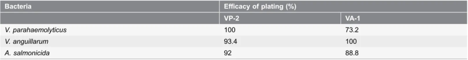
Bacterial susceptibility to both bacteriophages was assayed for the 10 pathogenic bacterial strains (Table 1). The hosts tested included species from generaVibrio, Aeromonas, Photobacteriumand Pseudomonas that include the main pathogenic bacteria of fish [34,37]. The spot test was used as an initial approach for the detection of bacterial infection [36] and the efficiency of plating was determined for the bacteria with positive spot tests (occurrence of lytic plaques) by the double-layer agar method. The plating efficiency for each host was calculated by
comparison with an efficacy of 100% for the phages, VP-2 and VA-1, using V.
parahaemolyticus and V. anguillarum as hosts, respectively. Three independent experiments were performed for each phage.
Phage Nucleic Acid Isolation
for 2 h at 10
˚
C. Five hundred microliters of SM buffer were added to the pellet and was let to rest for 2 h. The suspension was then treated with DNase I and RNase A at 37˚
C for 20 min to remove any free nucleic acids contamination. Nucleic acid extraction from phage particles was performed as described by Griffiths et al. [38]. Extraction was performed by the addition of 0.5 mL of hexadecyltrimethylammonium bromide (Sigma Aldrich, St. Louis, MO, USA) extraction buffer and 0.5 mL of phenol-chloroform-isoamyl alcohol (25:24:1; pH 8.0; Sigma Aldrich) to the sample. The sample was lysed for 30 s in a FastPrep FP120 (BIO 101/Savant) at 5.5 ms21and centrifuged (16,000 g) for 5 min at 4˚
C. The supernatant was pipetted into a clean vial, mixed 1:1 with chloroform-isoamyl alcohol (Sigma Aldrich) and centrifuged (16,000 g) for 5 min at 4˚
C. The supernatant was removed to a clean tube and the nucleic acids were precipitated with two volumes of 30% (wt/v) polyethylene glycol 6000 – 1.6 M NaCl for 2 h at 25˚
C. This mixture was centrifuged (18,000 g) at 4˚
C for 10 min, the pellet washed in ice cold 70% (v/v) ethanol, centrifuged (18.000 g) at 4˚
C for 10 min and then the pellet was air dried prior to re-suspension in 30 mL Tris-EDTA buffer. Nucleic acid yield was quantified in the Qubit 2.0 Fluorometer (Invitrogen, Carlsbad, CA, USA). The nucleic acid was then digested with DNase I (Ambion, Austin, TX, USA), and RNase I (Sigma Aldrich) separately, as described by the manufacturers. DNase I was inactivated by heating at 80˚
C for 5 min whereas RNase A was inactivated by EDTA (final concentration 20 mM) and Proteinase K (final concentration of 50 mg mL21; Bioron) addition and incubation at 56˚
C for 1 h. Five microliters of nucleic acids of each reaction was then loaded onto a agarose gel and separated by electrophoresis (0.8% agarose gel electrophoresis at 80 V for 45 min), using XXL DNA Ladder GeneOn (25 kb DNA ladder) as marker and observed in a Molecular Imager (Chemi Doc XRS+, BioRad, Hercules,CA, USA). To determine whether phages VP-2 and VA-1 are single or double-stranded DNA viruses, 10 ng of DNA was incubated with 20 U of S1nuclease (MBI Fermentas, Portugal) at 37
˚
C for 1 h as described by Sambrook et al [39]. The resulting product was electrophoresed through 0.8% agarose gel at 80 V for 40 min.Table 1.Efficiency of plating (%) of phages VP-2 and VA-1 on different bacteria.
Bacteria Efficacy of plating (%)
VP-2 VA-1
V. parahaemolyticus 100 73.2
V. anguillarum 93.4 100
A. salmonicida 92 88.8
Phage Survival Determination
Phage survival was tested in marine water collected on three different dates, in February, March, and May 2013, at the Aquaculture Corte das Freiras (Aveiro, Portugal). At each date, 50 mL of water (salinity 18–21 practical salinity units; pH 7.6–7.7) was filtered sequentially through 0.45-mm and 0.22-mm pore-size membranes (Millipore) and then sterilized in an autoclave (121
˚
C for 20 min). Phage lysates were added (estimated final concentration 107 PFU mL21) and incubated at 25˚
C without shaking. The phage titer was determined as described above, at time zero and at intervals of 12 h until day 1, 24 h until day 5, 48 h until day 9, 72 h until day 12, 120 h until day 45, and 240 h until the end of the experiment (day 185). Three independent experiments were performed for each phage.One-Step Growth Assays
The one-step growth curves of phages VP-2 (on V. parahaemolyticusand on V.
anguillarum) and VA-1 (on V. anguillarum) were performed according to the method of Almeida [40]. Briefly, mid-exponential host bacterial cultures of V. parahaemolyticus and V. anguillarum were adjusted to an optical density51 at 600 nm (,109 CFU mL21) and 10 mL phage lysate was added to 10 mL bacterial culture (multiplicity of infection - MOI of 0.001). Phages were allowed to adsorb for 5 min at room temperature and the mixture was centrifuged (10,000 g for 5 min). The pellet was re-suspended in 10 mL TSB at 25
˚
C and serially diluted to 1024. Samples (1 mL) were obtained at 10 to 20 min intervals and the phage titration was done by the double agar layer method. The burst size (number of phages produced by infected bacteria) was determined dividing the average number of lysis plaques at the stationary phase by the average number of lysis plaques at the latent phase.Phage Therapy Assays
Preliminary studies of phage therapy were performed to select the best phage to inactivate V. anguillarum. VP-2 and VA-1 phages were tested using their natural bacteria as host, V. parahaemolyticusand V. anguillarum, respectively. The phage VP-2 was also tested on V. anguillarum.
Killing curves
Optimal MOI was determined using VP-2 phage on its natural host, V.
sampling time (0, 2, 4, 6, 8, 10, 12, 14, 16, 18, 24, and 36 h) and bacteria and phages were counted as described before. Three independent experiments were performed for each MOI and the results were averaged.
Phage therapy experiments with phages VA-1 and VP-2 usingV. anguillarumas the host were performed only at MOI 100 under the previously described conditions.
Prophage Detection in Host Bacteria Before and After Phage Therapy
To assess lysogeny among the isolated bacteria and to check for the occurrence of lysogenic induction after phage therapy, the mitomycin C (a known agent of prophage induction) test was applied. To detect if the natural bacteria can harbour prophages in their genome, fresh bacterial cultures of V. parahaemo-lyticus and V. anguillarum were used. To detect if phage-treated bacteria can incorporate the phages in their DNA, bacteria/phages suspensions (V.
parahaemolyticus/VP-2 and V. anguillarum/VP-2 and V. anguillarum/VA-1), at a MOI of 100, were collected after 48 h of incubation at 25
˚
C were used. The bacterial cultures were centrifuged at 12,000 g for 10 min to collect the pellet of bacteria and remove free phages. The pellets were resuspended in fresh TSB medium. Each bacterial culture was then split equally into two Eppendorf microtubes (1 ml in each): one was added of mitomycin C (final concentration,1 mg mL21, Sigma-Aldrich) and the other was used as control (no added of
mitomycin C). The cultures were incubated overnight at 25
˚
C without shaking and then centrifuged (10,000 g, 10 min) (ThermoHeraeus Pico, Hanau,Germany) Supernatants were checked for the presence of phages by the spot test. The absence of a clear zone after inducing stress indicates that the bacteria contained no prophages in their genome. Three independent assays were performed for each condition.
Determination of phage-resistant mutants
AV. anguillarumculture (final concentration 105 CFU mL21) and a phage lysate (final concentration 107 PFU mL21) were inoculated in TSB and incubated at 25
˚
C without agitation for 24 hours (test sample). One control sample was included, the bacterial control, without phages, and was incubated exactly as the test sample. Aliquots of experimental samples and control were removed and plated by incorporation on TSA plates. Plates were incubated at 25˚
C for 24 h. To determine the mutation frequencies per cell per generation, ten isolated colonies were picked out from the TSA plates of the test sample, inoculated into ten tubes with TSB, grown with agitation until approximate concentration ofPhage Therapy in Zebrafish Larvae
As previously stated, all of the protocols used for the challenges were supervised by a veterinarian and approved by the ethics committee to comply with the European policy on the ‘‘3 Rs’’, to obtain reliable scientific information using the lowest number of animals possible.
Zebrafish (Danio rerio) larvae were selected as model organisms for testing the efficacy of phage therapy againstVibrioinfections. Due to its low salt requirement for growth and the success in previous experiences infecting zebrafish [42],V. anguillarum was selected as the candidate to infect zebrafish larvae. Bacteria were grown in TSB overnight and diluted 1:5 in fresh TSB. After 30 min at 27
˚
C, cellswere centrifuged and washed twice with SEW (CaCl2 66.2 mM, MgSO4*7H2O
13.4 mM, NaHCO321 mM, KCl 3.8 mM, and NaCl 0.5%, all from Merck). The
bacterial suspension was adjusted to,107 CFU mL21with SEW. Four groups of zebrafish larvae at 5 d post-fecundation, with each group comprising 3 sets of 20 specimens, were selected by haphazard sampling for the present study (a total of 4 groups 63 sets of 20 specimens each5240 larvae were used). Each set of 20
specimens of the 4 groups was treated separately, corresponding to 3 independent samples per condition. The 3 sets of the first group were infected with V. anguillarum and treated with VP-2 phage (test group, larvae+Vibrio+phages), the
3 sets of the second group were infected with the bacterium but not treated with the phage (bacterium control, larvae+Vibrio), and the 3 sets of the third group were not infected with the bacterium but phages were added (phage control, larvae+phages). To the 3 sets of the fourth group neither bacteria nor phages were
added (fish control). Each set of larvae was suspended in 3.0 mL SEW prior to inoculation (if they were inoculated). When required (test group and bacterium control), 250 mL bacterial culture was added (final concentration of
106 CFU mL21) and all groups were incubated at 27
˚
C with shaking (160 rpm). One hour after infection, 200 mL phage solution of VP-2 (,108 PFU mL21) was added (MOI 100, test group and phage control). Samples were incubated at 27˚
C with shaking (160 rpm) and samples were obtained at 1, 5, 24, 48, and 72 h post infection. Control groups were treated the same way, but instead of phage lysate,200 mL (phage control), 250 mL (bacterium control), or 450mL (no-treated
group) of SEW were added. Fish mortality in each of the 3 sets of the 4 groups was determined by visual inspection of all fish (inspecting for dead fish) every 24 h. Bacterial and phage concentration was determined as described before.
Statistical Analysis
Statistical analysis was performed using SPSS (SPSS 20.0 for Windows, SPSS Inc., Chicago, IL, USA). Significant differences among the different MOI tested using
the phage VP-2 and the host V. parahaemolyticusand between both phages VA-1
(difference between the concentration obtained in the control and the
concentration obtained in the test sample) for the different times of each of the three independent assays. Significant differences between samples of larvae fish experiments were also determined. The groups infected with V. anguillarum treated with phage were compared with the non–treated infected fish (samples with bacteria but without phages) and with the controls (samples with phages but without bacteria and samples without bacteria and without phages. Normal distributions were assessed using the Kolmogorov-Smirnov test and homogeneity of variances was assessed by Levene’s test. A value of p,0.05 was considered statistically significant.
Results
Presence of Prophages in the Bacterial Host
No phages were detected in the supernatant of cultures ofV. anguillarum andV. parahaemolyticus or in the mixture of bacteria and phages after treatment with mitomycin C, demonstrating the absence of inducible prophages in the two strains. Phages presented lytic cycles with no evidence of lysogeny induction. However, more specific tests, such as search of integrase genes to evaluate the development of lysogeny are needed.
Phage Host Range
Phage VP-2 infectedV. anguillarumandA. salmonicidawith an efficiency of 93% and 92% respectively, but was not effective against A. hydrophila, P. damselae subsp. damselae, P. damselae subsp.piscicida, V. fischeri, P. aeruginosa, P. fluorescens or P. putida. (Table 1). Phage VA-1 infected V. parahaemolyticus and A. salmonicidawith an efficiency of approximately 73% and 89%, respectively, but was not effective against the other tested strains (Table 1). The lysis plaques of phages VP-2 and of VA-1 were clear with a diameter of about 2.0 mm.
VP-2 and VA-1 Phage Characterization
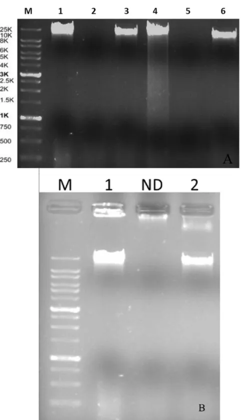
Nucleic Acid Characterization
Nucleic acid lysates with 357 ng mL21and 210 ng mL21, respectively for phages VP-2 and VA-1 were obtained. Enzyme digestion with DNase I and RNase A revealed that both phages are DNA phages (Figure 1A). Analysis of the enzyme digestion with S1 Nuclease revealed that phages VP-2 and VA-1 were double-stranded DNA viruses (Figure 1B).
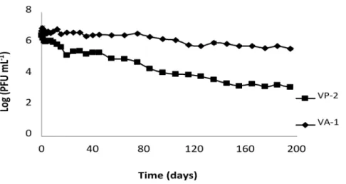
Phage Survival
Afterwards, the phage titer decreased slightly until day 35 and then more rapidly to day 195 (Figure 2).
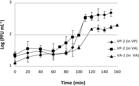
Burst Size and Latent Period
The VP-2 phage one-step growth experiments revealed that lysis occurred after 90 min of incubation when using V. parahaemolyticus as the host. Each infected bacterium produced approximately 15 phages (Figure 3). Phage VP-2 one-step
growth experiments performed using V. anguillarum as the phage host revealed
that lysis occurred at 100 min and that each infected bacterium produced an average of 10 phages (Figure 3). When the phage one-step growth experiments were performed using the VA-1 phage on V. anguillarum, lysis occurred at 100 min and each infected bacterium produced an average of 6 phages (Figure 3).
Kill Curves
VP-2 Phage on V. parahaemolyticus
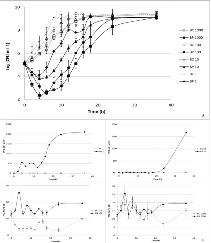
At a multiplicity of infection (MOI) of 1, the maximum bacterial inactivation was 3.0 log colony-forming units (CFU) per mL achieved after 6 h of phage therapy. Phage therapy efficiency at MOI 1 was significantly lower than that observed at MOI 10, 100, and 1000 (p,0.05) at the different times (Figure 4A). Increasing the MOI to a value of 10 enhanced the maximum rate of inactivation to 3.6 log CFU mL21after 8 h of incubation. After 6 h, the inactivation rate was already 3.4 log CFU mL21 and was, in general, significantly different from that observed for
Figure 2. VP-2 and VA-1 phage survival in aquaculture marine water.Both phages had an initial concentration of 7 log PFU mL21. Values represent the mean of three independent experiments; error bars
(overlapped by symbols) represent the standard deviation.
doi:10.1371/journal.pone.0114197.g002
acid.Agarose gel (0.8%) showing electrophoretic patterns of S1 Nuclease restriction digestion of phage DNA(B). Lane M: XXL DNA Ladder GeneOn; Lane 1: VP-2 DNA and Lane 2: VA-1 DNA. ND – Not determined.
MOI 100 and 1000 (p,0.05). At a MOI of 100, the highest inactivation rate (4.1 log CFU mL21) was achieved after 8 h of phage therapy, and after 6 h it was 3.7
log CFU mL21. At the highest MOI value (1000), the maximum value of
inactivation was 4.7 log CFU mL21 after 10 h of phage therapy and 4.0 log CFU mL21after 6 h. Phage inactivation at MOI 100 was only different from that of MOI 1000 after 6 h of phage therapy (p,0.05;Figure 4A).
No decrease in phage survival was observed during the 36 h of the experiments for the VP-2 phage alone and for the phage in the presence of its host (Figure 4B). While the phage alone remained almost constant throughout (p.0.05), a significant increase (p,0.05) in 1.0 log PFU mL21was observed for the MOI of 1 (p,0.05) after 36 h when the phage was incubated in the presence of its host. Increasing the MOI to 10 also significantly increased the rate of phage survival by 0.6 log PFU mL21 after the same period of time (p,0.05). For the highest MOI values (100 and 1000), only a slight increase in phage survival (less than 0.3 log PFU mL21) was recorded (p.0.05; Figure 4B).
VP-2 Phage on V. anguillarum
At a MOI of 100, the maximum bacterial inactivation was 5.0 log CFU mL21
achieved after 8 h of phage therapy. After 6 h of incubation, however, the rate of inactivation was already 3.8 log CFU mL21 (p,0.05; Figure 5A). No decrease in phage survival was observed during the 24 h of the experiment for the VP-2 phage alone or in presence of V. anguillarum as the host (Figure 5B). While the phage control remained nearly constant over time (p.0.05), a significant increase (p,0.05) of 1.9 log PFU mL21was observed when the phage was incubated in the presence of its host (p,0.05; Figure 5B).
Figure 3. VP-2 and VA-1 phage one-step growth experiment in the presence ofV. parahaemolyticus
andV. anguillarumas hosts.PFU per mL are shown at different time points. Values represent the mean of three independent experiments; error bars represent the standard deviation.
VA-1 Phage on V. anguillarum
The maximum rate of bacterial inactivation was 3.9 log CFU mL21achieved after 6 h of phage therapy, at a MOI of 100 (p,0.05). At 8 h of phage therapy, the inactivation rate remained quite high (2.5 log CFU mL21; Figure 6A).
No decrease in phage survival occurred during the experiment for either the VA-1 phage alone or the phage in the presence of V. anguillarum as a host (Figure 6B). While phage control remained nearly constant during the entire testing period (p.0.05), a significant increase (p,0.05) of 1.4 log PFU mL21was observed when the phage was incubated in the presence of its host after 24 h of phage therapy (p,0.05; Figure 6B).
Frequency of phage-resistant mutants
The frequency of emergence of resistant mutants was 8.461024(Table 2).
Mutant colonies were visible only after 48 h but in the control sample colonies were observed after 24 h. The number of colonies after 48 and 24 h, in the sample and in the control, respectively, was constant up to 6 days.
Phage Therapy in Infected Larvae
After 72 h, the group infected with V. anguillarum treated with phage had mortality rates similar (p.0.05) to those of fish control and the group treated
BC – Bacterial control; BP – Bacteria plus phage. (B) Phage concentration (PFU mL21): PC – Phage control, and PB – Phage plus bacteria. Values
represent the mean of three independent experiments; error bars represent the standard deviation.
doi:10.1371/journal.pone.0114197.g004
Figure 5. Inactivation ofV. anguillarumby the VP-2 phage at a MOI of 100 during the 24 h experiment.Bacteria and phage were inoculated in TSB and incubated at 25˚C. Phage and bacteria, had an initial concentration of 107PFU mL21and 105CFU mL21. For each MOI, two control samples were included, the bacterial control and the phage control, respectively, without phages and without bacteria. Both controls were incubated exactly as the test samples. (A) Bacterial concentration (Log CFU mL21): BC – Bacterial control; BP – Bacteria plus phage. (B) Phage concentration (PFU mL21): PC – Phage
control, and PB – Phage plus bacteria. Values represent the mean of three independent experiments; error bars represent the standard deviation.
with only the phage but without exposure to Vibrio (mean below 3%, Table 3). The mortality rate was much lower (p,0.05) than that observed for the non– treated infected fish (17%; Table 3). The larvae that were exposed to only phage were not affected during the experiment.
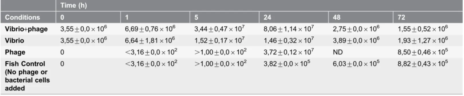
Bacterial concentrations in infected and treated larvae group samples increased until 24 h of incubation but after 48 h decreased by,1.5 log CFU mL21. At 72 h, the number of bacteria increased, reaching values similar to those observed at the beginning of the experiment, but there was a clear shift in the colony type on the TSA medium. The colonies detected at 24, 48 and 72 h were not consistent with the Vibrio style colonies on TSA. The pattern of variation of the bacterial concentrations in samples corresponding to fish larvae infected with bacteria but not treated with phage was similar to that of the infected and treated samples (Table 4). The number of bacteria in the samples of the phage control group and in the fish control samples increased by 6 log CFU mL21during the experiment,
Figure 6. Inactivation ofV. anguillarumby the VA-1 phage at a MOI of 100 during the 24-h experiment.Bacteria and phage were inoculated in TSB (final volume 50 mL) and incubated at 25˚C. Phage and bacteria, had an initial concentration of 107PFU mL21and 105CFU mL21. For each MOI, two
control samples were included, the bacterial control and the phage control, respectively, without phages and without bacteria. Both controls were incubated exactly as the test samples. (A) Bacterial concentration (Log CFU mL-1): BC – Bacterial control; BP – Bacteria plus phage. (B) Phage concentration: PC – Phage control, and PB – Phage plus bacteria. Values represent the mean of three independent experiments; error bars represent the standard deviation.
doi:10.1371/journal.pone.0114197.g006
Table 2.Frequencies ofV. anguillarumspontaneous VP-2 phage-resistant mutants.
Control sample (CFU mL21) Sample treated with phages (CFU mL21) Frequency of mutants
Days of incubation
1 2.64¡0.476108 0 0
2 2.64¡0.406108 2.28¡0.126105 8.4061024
3 2.64¡0.406108 2.22¡0.166105 8.3561024
4 2.75¡0.386108 2.22¡0.166105 8.2061024
5 2.99¡0.746108 2.17¡0.176105 8.1761024
6 2.76¡0.276108 2.39¡0.326105 8.1161024
reaching values similar to those obtained in the infected and treated samples and in the bacterial control samples (Table 4).
Discussion
In recent years, the potential of phage therapy to control bacterial infections in aquaculture has generated great expectations [4,14,15,16,17,18,19,20]. To permit its commercial application, however, an accurate evaluation of this approach is needed for the development of effective, safe, and controlled
protocols. Although studies have addressed the use of phages to control vibriosis in aquaculture [4,5,20], the present study is the first to examine the potential to control vibriosis in fish larvae production. The selection of the appropriate bacteriophage, the phage delivery method, and the life stage (eggs, larvae, juveniles, or adult fish) during which phage therapy is applied are key factors in the success of phage-mediated control of Vibrio in aquaculture.
The criteria required to select phages for phage therapy in aquaculture include i) host range; ii) latent period; iii) burst size; iv) survival in the environment; and
Table 3.Fish mortality after 72 h of treatment at 27˚C.
Sample Mortality
Assay 1 Assay 2 Assay 3 Sum of all assays
Average percentage of all assays
(n520) (n520) (n520) (n560) (n560)
Vibrio+phage 0 0 1 1 2¡3a
Vibrio 3 4 3 10 17¡3a,b
Phage 0 0 0 0 0b
Control (non-Vibrioand non-phages)
0 1 1 2 3¡3b
Values represent the Mean¡Standard Deviation of three samples.
asignificant difference with Vibrio samples (samples with bacteria but without phages),
bsignificant difference with phage samples (with phages but without bacteria) and with control samples (non-Vibrio and non-phages) (p
,0.05). doi:10.1371/journal.pone.0114197.t003
Table 4.Concentration (CFU mL21) of viable bacteria during the 72 h experiment.
Time (h)
Conditions 0 1 5 24 48 72
Vibrio+phage 3,55¡0,06106 6,69¡0,766106 3,44¡0,476107 8,06¡1,146107 2,75¡0,06106 1,55¡0,526106 Vibrio 3,55¡0,06106 6,64¡1,816106 1,52¡0,176107 1,46¡0,326107 3,89¡0,06106 1,93¡1,276106 Phage 0 ,3,16¡0,06102 .1,00¡0,06102 3,72¡0,126107 ND 8,50¡0,466105 Fish Control
(No phage or bacterial cells added
0 ,3,16¡0,06102 .1,00¡0,06102 3,82¡0,06105 6,03¡0,06105 8,82¡0,436105
ND – Not determined. Values represent the Mean¡Standard Deviation of three samples.
NOTE: After 48 h, there is a shift in the major bacterial type, with the new colony types not matchingVibriocolonies.
v) efficiency of bacterial inactivation. The two dsDNA phages tested in this study (isolated on V. parahaemolyticusand on V. anguillarum): i) infect the same three hosts (V. parahaemolyticus, V. anguillarum, and A. salmonicida), presenting high efficiency to inactivate the both pathogenic Vibrio species tested; ii) present long periods of survival in marine aquaculture water; and iii) have similar latent periods and burst sizes. Nonetheless, phage VP-2 showed a higher efficiency to
inactivate V. anguillarum than phage VA-1 (maximum ofV. anguillarum
inactivation of 5.0 log CFU mL21for VP-2 and of 3.9 log CFU mL21for VA-1), even higher than that obtained when this phage was used to inactivate its own host (V. parahaemolyticus; 4.2 log CFU mL21 maximum inactivation).
The results of this study strongly suggest that the VP-2 phage can protect fish larvae againstVibrioinfections. Although infecting fish withV. anguillarumin the absence of the phage resulted in low fish mortality (17%), the survival of infected fish larvae in the presence of the phage increased significantly, reaching values similar to those obtained in the control treatment (fish control). Addition of the phage lysate to the fish larvae without bacterial challenge did not decrease larvae fish survival. The mortality in this group was similar to that of controls (fish control). Although phage VP-2 produced no observable effects in larval fish (morphological alterations or mortality), it is necessary to evaluate this phage (whole genome sequencing) for the presence of genes encoding toxins and antibiotic resistance. The phage lysate could also potentially contain residual bacterial antigens or endotoxins [39]. In this study, fish larvae experimentally treated only with the phage preparation (phage lysate at ,109 PFU mL-1diluted 10-fold) did not show any negative effect on fish health, therefore, it is likely that the VP-2 phage lysate has few or no toxins. Moreover, as infecting fish with V. anguillarum in the absence of the phage resulted in low fish mortality (17%), future experiments using several concentrations of bacteria at a MOI of 100 in order to get a higher fish killing rate or to reach a plateau, would be very useful to confirm the potential of phage therapy to inactivate pathogenic bacteria in fish.
Phage VP-2 significantly improved the survival of larval fish infected with V. anguillarum, but killing curves assays demonstrated that, 8 h after phage addition, some bacteria remained viable and could regrow, as reported in previous studies [43,36]. A big fraction of the remained bacteria are phage-resistant mutants (frequency of phage-resistant mutants 8.461024) but it has been shown that
during treatment the phage titer increased despite bacterial numbers have remained constant or increased, thus suggesting that cell turnover of these resistant bacteria is likely occurring to generate the phage. Future microscopic studies should look at the bacterial cells to characterize their morphological diversity, in order to understand what is occurring during phage therapy.
Levin and Bull [49] also proposed that phages decrease the bacterial level enough to be eliminated by the fish immune system by acquired response, but this is not the case in fish larvae because they are unable to develop specific acquired immunity [12]. Further studies on this topic are needed.
Whether or not it is possible to supply phages to cultured fish by
intraperitoneal or intramuscular administration [9,15,16], research on bacter-iophage treatment in aquaculture has mainly focused on oral administration [5,9,15,16,17,18]. Oral administration can affect the phage viability due to the harsh conditions of the gastrointestinal tract [15,16]. In fish larviculture, where mass mortality is commonly associated with the actions of opportunistic bacteria, phages cannot be supplied by oral administration and must be directly released into the culture water. Consequently, phage survival in aquaculture water must be high to reach the specific site of infection, the intestine. As the phage VP-2 is able to survive long periods in marine aquaculture water (at least 5 months), the release of the phage directly into the water allows the phage to control not only the bacteria inside the larvae fish, but also avoid colonization on fish larvae skin. In the case of infections byV. anguillarum, avoiding colonization on the fish skin can play a key role in preventing disease development [50]. In addition, the survival of VP-2 in aquaculture water allows for the use of phage therapy as a prophylactic measure to prevent infections, as phages can inactivate pathogenic bacteria in the water without affecting the beneficial bacterial community (e.g., probiotics). In fact, in larval fish assays, the phage VP-2 controlledV. anguillarumgrowth but did not affect other bacteria that grew during the experiment. After 24 h of treatment, the concentration of V. anguillarum began to decrease, but at 48 and 72 h the bacterial density increased. At 48 and 72 h, however, there was a shift in the major bacterial type, with the new colony types not matching Vibrio colonies. Other bacteria already present in the larvae may have grown in the saline embryo water (SEW) and masked the decrease in theVibrioconcentration. Actually, the growth of these bacteria was also observed in the control group with added phage but not infected with bacteria and in the control with no bacteria or phage added. The bacterial colonies that do not present the Vibrio type were, most likely,
Conclusions
Overall, our findings suggest that phage therapy is a suitable alternative approach against vibriosis in aquaculture, with phage administration directly to the culture water as a promising method for treatment of fish larvae. Further studies need to be performed in the field and in a real scale, at a semi-intensive fish farm, where environmental conditions are known to vary considerably at different time scales, to evaluate the potential of this approach under environmental/seasonal variation. As MOI of 10 and even of 1 are enough to efficiently inactivate the pathogenic bacteria, phage lysates with about 109 PFU mL-1 can be diluted 10 to 100 times. In this way, small scale production (5 L) of phages is enough to produce sufficient material for purification. The resulting suspensions can be scaled-up, using pilot stirred bioreactors working with culture volumes of 10 to 300 L, to increase batch size. As phage solutions at 107–108 PFU mL-1 are enough, with 5 L of stock suspension 50–500 L of work suspension can be easily obtained. About 50 L of phage solution at 107–108 PFU mL-1 are enough for a treatment at real scale. As bacterial infection occurred at concentrations around 105 CFU mL21, phage suspensions with 107–108 PFU mL21 allows the use of MOI of 100 which has been shown efficient rates of fish pathogenic bacterial inactivation in previous studies [37,44]. This study provides the first evidence that phage therapy is a feasible alternative approach against vibriosis during fish larvae production in aquaculture systems.
Author Contributions
Conceived and designed the experiments: AA IH YS NG RC. Performed the experiments: YS LC CP CM MP. Analyzed the data: YS LC CP CM MP AC AA IH. Contributed reagents/materials/analysis tools: AA IH. Wrote the paper: YS LC CP CM MP AC AA IH RC NG.
References
1. Almeida A, Cunha A, Gomes NCM, Alves E, Costa L, et al.(2009) Phage therapy and photodynamic therapy: Low environmental impact approaches to inactivate microorganisms in fish farming plants. Marine Drugs 7: 268–313.
2. Shao ZJ (2001) Aquaculture pharmaceuticals and biologicals: current perspectives and future possibilities. Adv Drug Deliver Rev 50: 229–243.
3. Wahli T, Knuesel R, Bernet D, Segner H, Pugovkin D, et al.(2002) Proliferative kidney disease in Switzerland: current state of knowledge. J Fish Dis 25: 491–500.
4. Higuera G, Bastı´as R, Tsertsvadze G, Romero J, Espejo RT (2013) Recently discoveredVibrio anguillarumphages can protect against experimentally induced vibriosis in Atlantic salmon,Salmo salar. Aquaculture 392–395: 128–133.
5. Martı´nez-Dı´az SF, Hipo´lito-Morales A(2013) Efficacy of phage therapy to prevent mortality during the vibriosis of brine shrimp. Aquaculture 400: 120–124.
7. Noya M, Magarinos B, Lamas J(1995) La administracion intraperitoneal y oral de glucano afecta al sistema immune no especifico y a la resistencia de la dorada,Sparus aurata, a la pasteurelosis. In Proceedings of the Fifth National Congress of Aquaculture, Sant Carles de la Rapita, Spain. pp. 734– 738.
8. Toranzo AE, Barreiro S, Casal JF, Figueras A, Magarin˜os B, et al.(1991) Pasteurellosis in gilthead seabream,Sparus aurata: first report in Spain. Aquaculture 99: 1–15.
9. Verner-Jeffreys DW, Algoet M, Pond MJ, Virdee HK, Bagwell NJ, et al.(2007) Furunculosis in Atlantic salmon (Salmo salarL.) is not readily controllable by bacteriophage therapy. Aquaculture 270: 475–484.
10. Martinez JL (2003) Recent Advances in Marine Biotechnology. Molecular Genetics of Marine Organisms; Fingerman, N., Eds.; Science Publishers, Inc: Enfield, New Hampshire, USA. Vol. 10.
11. Pasqualina L, Gabriella C, Eleonora M, Renata Z, Santi D(2011) Susceptibility to antibiotics ofVibrio
spp. andPhotobacterium damselaessp. piscicida strains isolated from Italian aquaculture farms. New Microbiol 34: 53–63.
12. Vadstein O(1997) The use of immunostimulation in marine larviculture: possibilities and challenges. Aquaculture 155: 401–417.
13. Duckworth DH, Gulig PA(2002) Bacteriophages: potential treatment for bacterial infections. BioDrugs 16: 57–62.
14. Karunasagar I, Shivu MM, Girisha SK, Krohne G(2007) Biocontrol of pathogens in shrimp hatcheries using bacteriophages. Aquaculture 268: 288–292.
15. Nakai T, Sugimoto R, Park K, Matsuoka S, Mori K, Nishioka T, et al.(1999) Protective effects of bacteriophage on experimentalLactococcus garvieaeinfection in yellowtail. Dis Aquat Org 37: 33–41.
16. Nakai T, Park SC(2002) Bacteriophage therapy of infectious diseases in aquaculture. Res Microbiol 153: 13–18.
17. Park SC, Shimamura I, Fukunaga M, Mori KI, Nakai T(2000) Isolation of bacteriophages specific to a fish pathogen,Pseudomonas plecoglossicida, as a candidate for disease control. Appl Environ Microbiol 66: 1416–1422.
18. Park S, Nakai T (2003) Bacteriophage control of Pseudomonas plecoglossicida infection in ayu Plecoglossus altivelis. Dis Aquat Org 53: 33–39.
19. Shivu MM, Rajeeva BC, Girisha SK, Karunasagar I, Krohne G, et al. (2007) Molecular characterization ofVibrio harveyibacteriophages isolated from aquaculture environments along the coast of India. Environ Microbiol 9: 322–331.
20. Vinod MG, Shivu MM, Umesha KR, Rajeeva BC, Krohne G, et al.(2006) Isolation ofVibrio harveyi
bacteriophage with a potential for biocontrol of luminous vibriosis in hatchery environments. Aquaculture 255: 117–124.
21. Crothers-Stomps C, Høj L, Bourne DG, Hall MR, Owens L (2010) Isolation of lytic bacteriophage againstVibrio harveyi. J Appl Microbiol 108: 1744–50.
22. Merino S, Camprubi S, Tomas JM(1990) Isolation and characterization of bacteriophage PM2 from
Aeromonas hydrophila. FEMS Microbiol Lett 68: 239–244.
23. Munro J, Oakey J, Bromage E, Owens L(2003) Experimental bacteriophage-mediated virulence in strains ofVibrio harveyi. Dis Aquat Org 54: 187–194.
24. Stevenson RMW, Airdrie DW (1984) Isolation of Yersinia ruckeri bacteriophages. Appl Environ Microbiol 47: 1201–1205.
25. Wu JL, Lin HM, Jan L, Hsu YL, Chang LH (1981) Biological control of fish pathogen,Aeromonas hydrophila, by bacteriophage AH1. Fish Pathol 15: 271–276.
26. Abedon ST, Herschler TD, Stopar D(2001) Bacteriophage latent-period evolution as a response to resource availability. Appl Env Microbiol 13: 4233–4241
27. Olafsen JA(2001) Interactions between fish larvae and bacteria in marine aquaculture. Aquaculture 200: 223–247.
29. Beveridge MCM, Sikdar PK, Frerichs GN, Millar S(1991) The ingestion of bacteria in suspension by the common carpCyprinus carpioL. J Fish Biol 39: 825–831.
30. Olafsen JA(1984) Ingestion of bacteria by codGadus morhuaL. larvae. In: The Propagation of Cod,
Gadus morhuaL. Institute of Marine Research, Bergen. pp. 627–643.
31. Olafsen JA, Hansen GH(1992) Intact antigen uptake by intestinal epithelial cells of marine fish larvae. J Fish Biol 40: 141–156.
32. Laanto E, Bamford JKH, Laakso J, Sundberg LR(2012) Phage-driven loss of virulence in a fish pathogenic bacterium. PLoS One 7: e53157.
33. Almeida A, Alcaˆntara F (1992) Bacterial colonization of seston particles in brackish waters (Ria de Aveiro, Portugal). Mar Ecol Prog Ser 89: 165–173.
34. Pereira C, Silva YJ, Santos AL, Gomes NCM, Cunha A, et al.(2011) Bacteriophages with potential for inactivation of fish pathogenic bacteria: survival, host specificity and effect on bacterial community structure. Marine Drugs 9: 2236–2255.
35. Louvado A, Coelho FJRC, Domingues P, Gomes NCM, Santos AL, et al. (2012) Isolation of surfactant-resistant pseudomonads from the estuarine surface microlayer. J Microbiol Biotech 22: 283– 291.
36. Vieira A, Silva YJ, Cunha A, Gomes NCM, Ackermann HW, et al.(2012) Phage therapy to control multidrug-resistantPseudomonas aeruginosaskin infections:in vitroandex vivoexperiments. Eur J Clin Microbiol Infect Dis 31: 3241–3249.
37. Mateus C, Costa C, Silva Y, Cunha A, Almeida A (2014) Efficiency of phage cocktails in the inactivation of Vibrio in aquaculture. Aquaculture 424: 167–173
38. Griffiths RI, Whiteley AS, O’Donnell AG, Bailey MJ(2000) Rapid method for coextraction of DNA and RNA from natural environments for analysis of ribosomal DNA and rRNA-based microbial community composition. Appl Environ Microbiol 66: 5488–5491.
39. Sambrook J, Fritsch EF, Maniatis T(1989) Molecular Cloning: A Laboratory Manual, 2nd edn. Cold Spring Harbor, NY, USA: Cold Spring Harbor Laboratory Press.
40. Almeida A(1994) Morfologia e Infecc¸a˜o Bacteriofa´gica. Relato´rio de uma Aula Pra´tica para Prestac¸a˜o de Provas de Apida˜o Pedago´gica e Capacidade Cientı´fica. Universidade de Aveiro.
41. Filippov AA, Sergueev KV, He Y, Huang X-Z, Gnade BT, et al. (2011) Bacteriophage-Resistant Mutants in Yersinia pestis: Identification of Phage Receptors and Attenuation for Mice. PLoS ONE 6(9): e25486.
42. Oyarbide U, Rainieri S, Pardo MA(2012) Zebrafish (Danio rerio) Larvae as a system to test the efficacy of polysaccharides as immunostimulants. Zebrafish 9: 74–84.
43. Barrow P, Lovell M, Berchieri Jr A (1998) Use of lytic bacteriophage for control of experimental
Escherichia colisepticemia and meningitis in chickens and calves. Clin Diagn Lab Immunol 5: 294–298.
44. Silva YJ, Costa L, Pereira C, Cunha A, Calado R, et al.(2014) Influence of environmental variables in the efficiency of phage therapy in aquaculture. Microb Biotechnol 7: 401–413.
45. Anonymous(1983) Phage therapy. Lancet 2: 1287–1288.
46. Capparelli R, Nocerino N, Lanzetta R, Silipo A, Amoresano A, et al.(2010) Bacteriophage-resistant
Staphylococcus aureusmutant confers broad immunity against staphylococcal infection in mice. PLoS One 5: e11720.
47. Filippov AA, Sergueev KV, He Y, Huang XZ, Gnade BT, et al.(2011) Bacteriophage-resistant mutants inYersinia pestis: identification of phage receptors and attenuation for mice. PLoS One 6: e25486.
48. Wagner PL, Waldor MK(2002) Bacteriophage control of bacterial virulence. Infect Immun 70: 3985– 3993.
49. Levin BR, Bull JJ(2004) Population and evolutionary dynamics of phage therapy. Nat Rev Microbiol 2: 166–73.
51. Lan CC, Love DR (2012) Molecular Characterisation of Bacterial Community Structure along the Intestinal Tract of Zebrafish (Danio rerio): A Pilot Study. ISRN Microbiol 14: 590385.