Rev Odontol UNESP. 2017 Nov-Dec; 46(6): 362-367 © 2017 - ISSN 1807-2577
ORIGINAL ARTICLE
Doi: http://dx.doi.org/10.1590/1807-2577.06517
Micro-CT analysis of illing ability and porosity
of root-end illing materials
Análise em micro-CT da capacidade de preenchimento e porosidade de materiais retrobturadores
Mário TANOMARU-FILHO
a*, Camila Galletti ESPIR
a, Katia Cristina Toledo NOGUEIRA
a,
Fernanda Ferrari Esteves TORRES
a, Juliane Maria GUERREIRO-TANOMARU
aaFaculdade de Odontologia de Araraquara, UNESP – Universidade Estadual Paulista, Araraquara, SP, Brasil
Resumo
Introdução: Capacidade de preenchimento da cavidade retrógrada e porosidade são propriedades importantes
de materiais retrobturadores e podem ser avaliadas por meio de microtomografia computadorizada (micro-CT).
Objetivo: Avaliar a capacidade de preenchimento e porosidade de materiais retrobturadores por meio de micro-CT.
Material e método: Cavidades com 1 mm de diâmetro e 3 mm de altura foram preparadas em dentina bovina
utilizando pontas ultrassônicas (CVD No. 6.1107-6) e foram preenchidas com Mineral Trióxido Agregado (MTA), Sealer 26 (S26) e cimento de óxido de zinco e eugenol (OZE). As cavidades foram escaneadas em micro-CT antes e após o preenchimento. A capacidade de preenchimento foi calculada com base na porcentagem em volume, das cavidades preenchidas. O número e porcentagem dos poros fechados foram avaliados em toda extensão da cavidade preenchida (total) e por terços (cervical, médio e apical) por meio de análises bi e tridimensionais. Os dados de preenchimento foram submetidos aos testes estatísticos ANOVA e Tukey e a porosidade aos testes de Kruskall-Wallis e Dunn, com nível de significância de 5%. Resultado: S26 e OZE apresentaram maior capacidade de preenchimento que o MTA (p<0,05). S26 mostrou maior porosidade total (em número e porcentagem) (p<0,05). Em todos os terços, após as análises 2D e 3D, a porosidade foi maior para S26 em comparação ao MTA e OZE (p<0,05). Conclusão: Embora Sealer 26 tenha apresentado maior porosidade, o material foi associado a uma adequada capacidade de preenchimento. A análise em micro-CT mostrou ausência de correlação entre capacidade de preenchimento e porosidade.
Descritores: Microtomografia por Raio-X; porosidade; materiais dentários; endodontia.
Abstract
Introduction: Filling ability of retrograde cavity and porosity are important properties for root-end illing materials
and may be evaluated by using microcomputed tomography (micro-CT). Objective: To evaluate illing ability and porosity of root-end illing materials using microcomputed tomography (micro-CT). Material and method: Cavities with 1 mm internal diameter and 3 mm depth were prepared in bovine dentin sections by using ultrasonic tips (CVD No. 6.1107-6), and illed by Mineral Trioxide Aggregate (MTA); Sealer 26 (S26) and zinc oxide and eugenol cement (ZOE). Before and ater illing, cavities were scanned by using micro-CT (SkyScan 1176). Filling and porosity were analyzed by using CTAn sotware. Filling ability was calculated based on volumetric percentage of the illed cavity. he number and percentage of closed pores were measured throughout entire extension of the illed cavity (total) and in each third (cervical, middle and apical), by using bi and tridimensional analyses. he illing data were submitted to ANOVA and Tukey statistical tests, and porosity data to Kruskall-Wallis and Dunn tests, at a 5% signiicance level.
Result: S26 and ZOE presented higher illing ability than MTA (p<0.05). S26 showed the highest total porosity (number
and percentage) (p<0.05). In all thirds ater 2D and 3D analyses, porosity was higher for S26 in comparison to MTA and ZOE (p<0.05). Conclusion: Although Sealer 26 presented more porosity, the material was associated with a great illing ability. Micro-CT analysis showed no correlation between illing ability and porosity.
Descriptors: X-Ray Microtomography; porosity; dental materials; endodontics.
INTRODUCTION
Microcomputed tomography (micro-CT) is a non-destructive tool used for diferent studies in Endodontics as analysis of root canal illing1, illing ability of the reparative materials2 and the evaluation of interface material/dentin associated with diferent
he porosity of a material may be afected by physicochemical properties. Cements with absence of macropores and lower porosity may present less penetration of oral luids, bacteria and bacterial toxins into root canal5. Pores in sealers appear to be originated from the air trapped in the mass of cement during handling manipulation6. Porosity may be analyzed by means of microscopy, using porosimetry by mercury intrusion7, or by using microcomputed tomography8,9. De Souza et al.10, using a 3D model, quantitatively evaluated the degree of porosity of calcium-silicate based materials analyzing the images obtained by microcomputed tomography. 3D models were evaluated and the porosity parameters of each material were obtained by comparison with standard porosity values of Biodentine®. his tool allows a tridimensional mensuration (with volumetric results – mm3) of failures. Surface area, volumetric analysis, the amount of pores and speciic characteristics of these structures can be assessed.
Clinically, these physical properties of root-end illing materials are relevant. High porosity values for endodontic materials may afects its physical properties7, besides increasing the leakage5. A better sealing can be obtained using a material presenting low disintegration and solubility11 and it may be directly related to leakage12. Also, proper sealing may be related to complete illing of the cavity. Analyzing physicochemical (i.e solubility, pH, illing ability) and biological (i.e biocompatibility, bioactivity) characteristics to the root-end illing material may improve the success of the treatment2,4,7,12.
Mineral Trioxide Aggregate (MTA) is a biocompatible calcium silicate-based material with capacity to induce repair by mineralized tissue13. However, the luid consistency makes it diicult to insert MTA into the root-end cavity, and can harm illing and sealing14. Sealer 26 (S26) is a resin-based endodontic sealer, composed of bismuth oxide, calcium hydroxide and epoxy resin, and requires higher powder/resin proportion to favor insertion into root-end cavities13. It has excellent sealing properties when used as root-end illing material15, and presents interface adaptation similar to calcium silicate-based cements16. Zinc oxide and Eugenol cement (ZOE) may also be used with higher powder/liquid proportion17, favoring its insertion into root-end cavities.
he aim of this study was to evaluate the illing ability and porosity of root-end illing materials by using microcomputed tomography. he null hypothesis was that there is no diference between the materials, and the properties of porosity and illing ability are associated.
MATERIAL AND METHOD
Sample Preparation
Bovine teeth were used in this study. he coronal portion was removed. he root portion was sectioned into 5mm slices, using an Isomet 1000 (Buehler Ltd., Lake Bluf, IL, USA) machine. On dentine surface of each slice, root-end cavities with standardized dimensions (1.5 mm diameter and 3 mm deep) were prepared by making intermittent forward-backward movements18 using a high-speed 2137 bur (KG SORENSEN, Cotia, SP, Brazil). Ultrasonic tip CVD No 6.1107-6 (CVD-Vale, São José dos Campos/SP, Brazil) coupled to an ultrasonic device CVDentus (CVD-Vale, São José
dos Campos/SP, Brazil) was used on the dentinal walls. In order to maintain the same position during the scans, the samples were ixed in culture plates with silicone. Each well of the culture plate was illed with a standard amount of silicone and the samples were placed into this material, at all stages.
Sample Scanning
Ater cavity preparation, samples with empty cavities were subjected to microtomographic scannings (Micro-CT SkyScan 1176, Bruker micro-CT Kontich, Belgium). he scanning procedure was performed with 50 kV X-ray tube voltages and 800 µA anode current; aluminum ilter of 0.5; isotropic voxel of 9 µm; and an evolution cycle of 360°. To standardize the position of samples, specimens were ixed in culture plates with silicone, as already described.
Filling Ability
he prepared cavities were then randomly divided into three groups, according to the material. MTA Angelus (Angelus, Londrina, PR, Brazil) was manipulated by a same operator previously calibrated using powder/liquid proportion of 1g/330µL. S26 (Dentsply, Petropolis, RJ, Brazil) was prepared in a thicker consistency than used as endodontic sealer, in a 4:1 powder/resin proportion (14). ZOE cement (S.S.White Art. Dent. Ltda., Rio de Janeiro, RJ, Brazil) was used in the proportion of 1 g zinc oxide to 0.2 mL eugenol. he cavities were illed with the materials by using a condenser (Trinity, São Paulo, SP, Brazil). Samples were maintained at 37ºC and 100% humidity for three times the material setting time. Ater this period, each illed sample was subjected to another microtomographic scanning, as previously described.
Filling Ability Analysis
he scanned images were reconstructed by using NRecon sotware (V1.6.4,7; SkyScan, Belgium). he correction parameters smoothing, beam hardening and ring artefacts were carefully adjusted and maintained for all the periods, using values of 7, 20% and 10, respectively. hese image parameters were used for all materials, to standardize the analysis process (scanning, reconstruction and analyzing criteria). Ater reconstruction, images were analyzed by using CTAn sotware (V1.11.8; SkyScan, Belgium). To obtain the total volume of material in mm3, the area of interest the region of interest (ROI) was deined by previous tests and a calibrated operator made all the analyzes of each sample, excluding the dentin. he binary value (threshold) was adjusted to assure the analysis of the material and the total volume of the material in mm3 from quantitative analysis was obtained. Filling was determined by calculating the subtracting the initial (empty cavities) from the inal (illed cavities) assessed volume of each sample. he results were then converted into percentages based on the initial volume. 3D models from each specimen were carefully obtained by using CTVol sotware (V2.0, SkyScan, Belgium) to analyze qualitatively the data.
Porosity Analysis
(V1.11.8; SkyScan, Belgium). he porosity parameters for each sample were measured and number and percentage of closed pores were obtained. Total porosity throughout the entire material extent was determined. he limits of each sample were deined by using the “top” and “bottom” tool (Figure 1A). A circular VOI, measuring average 0.75 x 0.75, was determined to each analyzes to exclude any possible artefact related to the sample border. hus, approximately 1 mm2 of margin was excluded. he internal portion of the materials was analyzed by the diference in radiopacity. Ater this, using the sotware’s porosity tool, the bidimensional total porosity values were obtained, according to number and percentage of closed pores. his feature allows porosity analysis for individual objects in 2D (bidimensional). It has been deined as the area of any spaces fully surrounded by solid, as a percent of the area of solid plus closed pores. Subsequently, by using the 3D (tridimensional) analysis tool, the number and percentage of closed pores were obtained by means of tridimensional data, providing volumetric data. In this case, a closed pore in 3D is a connected assemblage of space voxels that is fully surrounded on all sides in 3D by solid voxels. In addition a tridimensional model of the samples was obtained by using the CTVol sotware (V2.0, SkyScan, Belgium).
To obtain more precise and speciic analysis, sections with smaller thickness were also analyzed. he “top” and “bottom” tool
was used to deine 0.5mm sections to evaluate the porosity in the diferent cavity thirds: coronal, middle and apical (Figure 1B). he analysis was performed as previously described. For each third, the number and percentage of closed pores were calculated, and 3D models were obtained.
Data from illing analysis were submitted to ANOVA and the Tukey tests, with 5% signiicance. Porosity data were submitted to the Kruskall-Wallis test, complemented by the Dunn test, with a level of signiicance of 5%.
RESULT
Filling Ability
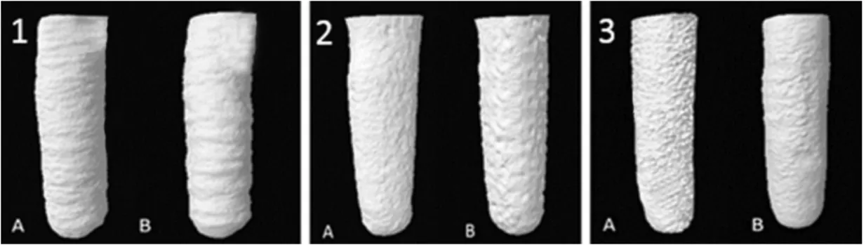
Data obtained for illing volumetric analysis are represented in Table 1. Similar illing ability was observed for S26 and ZOE (p>0.05), and both presented higher values than MTA (p<0.05). 3D model analyses (Figure 2) show similar characteristics for S26 and ZOE, in comparison with MTA.
Porosity
he data obtained for porosity are described in Table 2. 2D and 3D analyses presented similar patterns of comparison among the materials. S26 showed higher values for number and percentage of closed pores considering the total extent or each third analysis (p<0.05). MTA and ZOE cement were similar for all the analyses (p>0.05). Considering absolute values for both tests, the bidimensional analysis showed higher values in percentage and lower values for number of pores, while the tridimensional analysis revealed higher number of pores and a lower percentage. 3D models porosity models for each third were obtained (Figure 3). he higher porosity observed to S26 was clearly observed in the qualitative analysis in comparison with MTA and ZOE, in all thirds evaluated.
Figure 1. Models used for porosity analysis. (A) Sample analysis for full extent (total porosity); (B) sample analysis for sections (porosity in thirds).
Figure 2. Representative micro-CT tree-dimensional reconstructions of the evaluated samples (1 – MTA; 2 – Sealer 26; 3 – Zinc oxide and eugenol cement). Empty cavity (A) and the cavity illed with each material (B).
Table 1. Mean percentage values (± standard deviation) of cavity illings by diferent root-end illing materials
MTA Sealer 26 ZOE
Mean illing 75.66 (8.854)b 84.46 (1.959)a 84.07 (5.220)a
DISCUSSION
Micro-CT volumetric analysis shows that S26 and ZOE cement presented greater illing ability than MTA. he larger powder to resin/liquid used to manipulate S26 and ZOE cement made it possible to increase their consistency, favoring their insertion into the root-end cavity. By using microcomputed tomography, Cavenago et al.4 evaluated the solubility of MTA with diferent powder-liquid ratio (4:1, 3:1 and 2:1) before and ater immersion in water for 7 days. hey observed that the material manipulated with larger quantity of water promoted greater change in volume. herefore, the diiculty with inserting MTA into the cavity13 cannot be improved by changing the powder/liquid proportion.
For this reason, a better ill can be achieved using materials which allow changes in handling, facilitating insertion into the cavity (i.e. S26 and ZOE cement).
Proper cavity illing with a material presenting low disintegration and solubility may allow a better sealing. Favorable sealing ability for some materials may be related to its dimensional stability and illing, which led to less leakage11. Chittoni et al.12 observed lower bacterial leakage for Sealer 26 in comparison with MTA. Sealer 26 has also demonstrated to prevent bacterial leakage when compared with IRM15. Amoroso-Silva et al.16 demonstrated that Sealer 26, MTA and calcium silicate cements were similar ater analyzing sealing by luid leakage and dentinal adaptation. In the present study, similar illing ability was observed for S26 and ZOE, and both presented higher values than MTA. Torres et al.2 observed that ZOE presented better illing ability than MTA. Dias et al.19 observed that the modiication of a composite resin with small amounts of zinc oxide (ZnO) microparticles signiicantly inhibited
the S. mutans growth on resin surface without signiicant alterations
of its mechanical strength. his illing capacity may be related to the better consistency. he results obtained may suggest this relationship. S26 and ZOE cement presented similar characteristics of illing compared to MTA. he presence of space at the interface of the illing material and the root canal wall can result from deicient adaptation of the illing material to the root dentin20. Despite the excellent properties of MTA, the condensation technique may have some inluence in its sealing ability.
Porosity analysis aims to measure the “failure fraction” and empty spaces, counting the spaces and characterizing their connections. he closed pores represent empty spaces completely surrounded by material, which is diicult to be analyzed by conventional methodologies. he use of microcomputed tomography allows Figure 3. Representative aspect of material porosities observed in the
diferent evaluated thirds for each evaluated material.
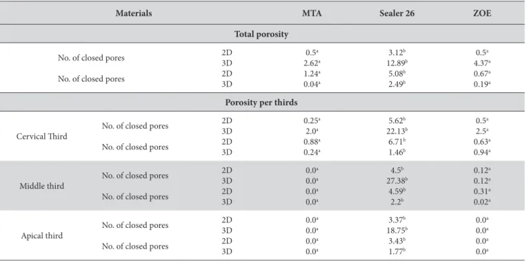
Table 2. Porosity values (total number and percentage of closed pores) in cavities illed with diferent root- end illing materials in 2D and 3D analysis
Materials MTA Sealer 26 ZOE
Total porosity
No. of closed pores
No. of closed pores
2D 3D 2D 3D 0.5a 2.62a 1.24a 0.04a 3.12b 12.89b 5.08b 2.49b 0.5a 4.37a 0.67a 0.19a
Porosity per thirds
Cervical hird
No. of closed pores
No. of closed pores
2D 3D 2D 3D 0.25a 2.0a 0.88a 0.24a 5.62b 22.13b 6.71b 1.46b 0.5a 2.5a 0.63a 0.94a Middle third
No. of closed pores
No. of closed pores
2D 3D 2D 3D 0.0a 0.0a 0.0a 0.0a 4.5b 27.38b 4.59b 2.2b 0.12a 0.12a 0.31a 0.02a Apical third
No. of closed pores
No. of closed pores
2D 3D 2D 3D 0.0a 0.0a 0.0a 0.0a 3.37b 18.75b 3.43b 1.77b 0.0a 0.0a 0.0a 0.0a
evaluation of these faults10. Microtomography has been used to evaluate the porosity of diferent materials8,9. Kerckhofs et al.8 aiming to validate micro-CT as an imaging tool for analysis of pores, pointed out errors inherent to the analyses that may result in an incorrect interpretation. Artifacts might inluence the quality of the images, challenging image analyses, once the images are reconstructed with several independent detector measurements21. As an example, streaking artefacts, which happen generally due to an inconsistency in a single measurement; ring artefacts, which appear due to errors in an individual detector calibration, distortion artefacts, due to the geometry of image reconstruction. In order to minimize these errors, all reconstructions were performed using the artifact reduction tools. De Souza et al.10 proposed a standard to obtain the threshold, using known values for the porosity of each material. herefore, the use of microcomputed tomography for porosity analysis also presents variations that must suggest patterns based on the experiment performed and type of sample.
According to the manufacturer (Bruker-microCT, Kontich, Belgium), the CTAn sotware allows the analysis of porosity of any type of material. In this study, samples were scanned at a high resolution (9um), considered proper for quantifying pores22. During the reconstruction of images, standardized parameters were used in order to decrease artefacts. Analysis of the total sample extent presented more diiculty with deining the parameters, especially the histogram. herefore, analysis of smaller sections (or thirds) is considered important for correct analysis.
As regards the results of this study, the two measurement analyses showed greater porosity for S26 in comparison with MTA and ZOE cement, for both number and percentage of pores. However, the 2D analyses showed higher percentage values, while the 3D presented a higher number of pores. In microscopic 2D images, a
failure may appear as a closed pore, while in the 3D evaluation this pore is considered connected to the external space, which is one advantage of 3D analysis in comparison with conventional methods. herefore, 3D analysis is indicated for this type of porosity analysis. Bidimensional analysis was evaluated to obtain complementary data. In the present study, bidimensional and tridimensional analysis presented correlated results for material porosities.
he analysis of porosity did not show correlation with illing data, since S26 and ZOE cement promoted greater illing. Otherwise, more porosity was observed for S26 when compared with MTA and ZOE cement, rejecting the null hypothesis. he immediate porosity (ater the material set) is related to the material composition and hydration mechanism, especially to water/powder proportion for MTA23. MTA has demonstrated porosity in diferent formulations, especially ater long evaluation periods3. he low porosity observed for MTA in the present study may be related to the diferent methodologies and analysis performed immediately ater cavity illing. Mutal, Gani6, ater a qualitative analysis of pores in endodontic sealers, including zinc oxide and eugenol cements and Sealer 26, observed that the frequency and size of pores were related to the consistency of the material, and was increased for sealers with calcium hydroxide, such as Sealer 26.
CONCLUSION
Based on the methodology used and the results obtained, we could conclude that Sealer 26 and ZOE cement showed better illing ability in comparison to MTA. On the other hand, Sealer 26 presented porosity higher in number and percentage than MTA and zinc oxide and eugenol.
REFERENCES
1. Jung M, Lommel D, Klimek J. The imaging of root canal obturation using micro-CT. Int Endod J. 2005 Sep;38(9):617-26. PMid:16104975. http://dx.doi.org/10.1111/j.1365-2591.2005.00990.x.
2. Torres FFE, Bosso-Martelo R, Espir CG, Cirelli JA, Guerreiro-Tanomaru JM, Tanomaru-Filho M. Evaluation of physicochemical properties of root-end filling materials using conventional and Micro-CT tests. J Appl Oral Sci. 2017 Jul-Aug;25(4):374-80. PMid:28877275. http:// dx.doi.org/10.1590/1678-7757-2016-0454.
3. Gandolfi MG, Parrilli AP, Fini M, Prati C, Dummer PM. 3D micro-CT analysis of the interface voids associated with Thermafil root fillings used with AH Plus or a flowable MTA sealer. Int Endod J. 2013 Mar;46(3):253-63. PMid:23039158. http://dx.doi.org/10.1111/j.1365-2591.2012.02124.x.
4. Cavenago CB, Pereira TC, Duarte MA, Ordinola-Zapata R, Marciano MA, Bramante CM, et al. Influence of powder to-water ratio on radiopacity, setting time, pH, calcium ion release, and a micro-CT volumetric solubility of white mineral trioxide aggregate. Int Endod J. 2014 Feb;47(2):120-6. PMid:23647286. http://dx.doi.org/10.1111/iej.12120.
5. Milutinović-Nikolić AD, Medić VB, Vuković ZM. Porosity of different dental luting cements. Dent Mater. 2007 Jun;23(6):674-8. PMid:16860859. http://dx.doi.org/10.1016/j.dental.2006.06.006.
6. Mutal L, Gani O. Presence of pores and vacuoles in set endodontic sealers. Int Endod J. 2005 Oct;38(10):690-6. PMid:16164682. http:// dx.doi.org/10.1111/j.1365-2591.2005.00988.x.
7. Camilleri J, Grech L, Galea K, Keir D, Fenech M, Formosa L, et al. Porosity and root dentine to material interface assessment of calcium silicate-based root-end filling materials. Clin Oral Investig. 2014;18(5):1437-46. PMid:24100638. http://dx.doi.org/10.1007/s00784-013-1124-y.
9. N’Diaye M, Degeratu C, Bouler JM, Chappard D. Biomaterial porosity determined by fractal dimensions, succolarity and lacunarity on microcomputed tomographic images. Mater Sci Eng C Mater Biol Appl. 2013 May;33(4):2025-30. PMid:23498228. http://dx.doi.org/10.1016/j. msec.2013.01.020.
10. De Souza ET, Nunes Tameirão MD, Roter JM, De Assis JT, De Almeida Neves A, De-Deus GA. Tridimensional quantitative porosity characterization of three set calcium silicate-based repair cements for endodontic use. Microsc Res Tech. 2013 Oct;76(10):1093-8. PMid:23913667. http://dx.doi.org/10.1002/jemt.22270.
11. Carvalho-Júnior JR, Guimarães LFL, Correr-Sobrinho L, Pécora JD, Sousa-Neto MD. Evaluation of solubility, disintegration, and dimensional alterations of a glass ionomer root canal sealer. Braz Dent J. 2003;14(2):114-8. PMid:12964655. http://dx.doi.org/10.1590/ S0103-64402003000200008.
12. Chittoni SB, Martini T, Wagner MH, Da Rosa RA, Cavenago BC, Duarte MA, et al. Back-scattered electron imaging for leakage analysis of four retrofilling materials. Microsc Res Tech. 2012 Jun;75(6):796-800. PMid:22147679. http://dx.doi.org/10.1002/jemt.21128.
13. Tanomaru-Filho M, Luis MR, Leonardo MR, Tanomaru JM, Silva LA. Evaluation of periapical repair following retrograde filling with different root-end filling materials in dog teeth with periapical lesions. Oral Surg Oral Med Oral Pathol Oral Radiol Endod. 2006 Jul;102(1):127-32. PMid:16831685. http://dx.doi.org/10.1016/j.tripleo.2005.09.008.
14. El-Ma’aita AM, Qualtrough AJ, Watts DC. A micro–computed tomography evaluation of Mineral Trioxide Aggregate root canal fillings. J Endod. 2012 May;38(5):670-2. PMid:22515899. http://dx.doi.org/10.1016/j.joen.2012.01.009.
15. Siqueira J Jr, Roccas I, Abad E, Castro A, Gahyva S, Favieri A. Ability of three root-end filling materials to prevent bacterial leakage. J Endod. 2001 Nov;27(11):673-5. PMid:11718135. http://dx.doi.org/10.1097/00004770-200111000-00005.
16. Amoroso-Silva PA, Marciano MA, Guimarães BM, Duarte MA, Sanson AF, Moraes IG. Apical adaptation, sealing ability and push-out bond strength of five root-end filling materials. Braz Oral Res. 2014;28(1):1-6. PMid:25166765. http://dx.doi.org/10.1590/1807-3107BOR-2014. vol28.0043.
17. Sousa-Neto MD, Guimarães LF, Gariba Silva R, Saquy PC, Pécora JD. The influence of different grades of rosins and hydrogenated resins on the powder-liquid ratio of Grossman cements. Braz Dent J. 1998;9(1):11-8. PMid:9835799.
18. Wuchenich G, Meadows D, Torabinejad M. A comparison between two root end preparation techniques in human cadavers. J Endod. 1994 Jun;20(6):279-82. PMid:7931024. http://dx.doi.org/10.1016/S0099-2399(06)80816-2.
19. Dias HB, Bernardi MIB, Ramos MADS, Trevisan TC, Bauab TM, Hernandes AC, et al. Zinc oxide 3D microstructures as an antimicrobial filler content for composite resins. Microsc Res Tech. 2017 Jun;80(6):634-43. PMid:28271628. http://dx.doi.org/10.1002/jemt.22840. 20. Prado M, Simao RA, Gomes BP. A microleakage study of gutta-percha/AH Plus and Resilon/Real self-etch systems after different irrigation
protocols. J Appl Oral Sci. 2014 Jun;22(3):174-9. PMid:25025557. http://dx.doi.org/10.1590/1678-775720130174.
21. Barrett JF, Keat N. Artifacts in CT: recognition and avoidance. Radiographics. 2004 Nov;24(6):1679-91. PMid:15537976. http://dx.doi. org/10.1148/rg.246045065.
22. Mendoza F, Verboven P, Mebatsion HK, Kerckhofs G, Wevers M, Nicolai B. Three-dimensional pore space quantification of apple tissue using X-ray computed microtomography. Planta. 2007 Aug;226(3):559-70. PMid:17361459. http://dx.doi.org/10.1007/s00425-007-0504-4. 23. Camilleri J. Evaluation of selected properties of mineral trioxide aggregate sealer cement. J Endod. 2009 Oct;35(10):1412-7. PMid:19801242.
http://dx.doi.org/10.1016/j.joen.2009.07.008.
CONFLICTS OF INTERESTS
he authors declare no conlicts of interest.
*CORRESPONDING AUTHOR
Mário Tanomaru-Filho, Departamento de Odontologia Restauradora, Faculdade de Odontologia, UNESP – Universidade Estadual Paulista, Rua Humaitá, 1680, Centro, 14801-903 Araraquara - SP, Brasil, e-mail: [email protected]