ABSTRACT
Radiopacity evaluation of Portland and MTA-based
cements by digital radiographic system
Alvaro Henrique BORGES1, Fabio Luiz Miranda PEDRO1, Alex SEMANOFF-SEGUNDO1,
Carlos Eduardo Saraiva MIRANDA2, Jesus Djalma PÉCORA3, Antônio Miranda CRUZ FILHO3
1- DDS, MSc, PhD, Department of Endodontics, Dental School, University of Cuiabá, Cuiabá, MT, Brazil.
2- DDS, MSc, PhD, Department of Restorative Dentistry, Ribeirão Preto Dental School, University of São Paulo, Ribeirão Preto, SP, Brazil. 3- DDS, MSc, PhD, Department of Chemistry, University of Ribeirão Preto, Ribeirão Preto, SP, Brazil.
Corresponding address: Antônio Miranda da Cruz Filho - Praça Pompilio Conceição, casa 10 - Residencial Vila Aliança, Jd. Botânico - 14021-594 - Ribeirão Preto, SP - Brasil - Phone: +55-16-3234-9551 - Fax: +55-16-3602-4792 - e-mail: [email protected]
Received: July 29, 2009 - Modiication: February 16, 2010 - Accepted: May 25, 2010
O
bjective: The aim of the present study was to evaluate the radiopacity of Portland and MTA-based cements using the Digora TM digital radiographic system. Material andMethods: The performed tests followed speciication number 57 from the American National
Standard Institute/American Dental Association (2000) for endodontic sealing materials. The materials were placed in 5 acrylic plates, especially designed for this experiment, along
with a graduated aluminum stepwedge varying from 1 to 10 mm in thickness. The set was
radiographed at a 30 cm focus-object distance and with 0.2 s exposure time. After the
radiographs were taken, the optical laser readings of radiographs were performed by Digora
TM system. Five radiographic density readings were performed for each studied material and for each step of the aluminum scale. Results: White ProRoot MTA (155.99±8.04), gray ProRoot MTA (155.96±16.30) and MTA BIO (143.13±16.94) presented higher radiopacity
values (p<0.05), while white non-structural Portland (119.76±22.34), gray Portland
(109.71±4.90) and white structural Portland (99.59±12.88) presented lower radiopacity
values (p<0.05). Conclusions: It was concluded that MTA-based cements were the only materials presenting radiopacity within the ANSI/ADA speciications.
Key words: endodontics. Dental radiography. Digital radiography. Radiology. Retrograde
obturation. Root canal illing materials.
INTRODUCTION
The role of endodontic sealers is to establish a perfect and hermetic periapical environment seal18.
Ideally, these materials should be biocompatible with periradicular tissues, non-absorbable, adaptable to dentin walls and should present good handling characteristics and no cytotoxicity6,19,22,23.
Mineral trioxide aggregate (MTA)-based cements have been widely investigated for endodontic applications19. The use of MTA as retrofilling
material, in animals, has shown an induction of
lower inlammatory response4. MTA has been also
employed for pulp capping20, in root perforations
reparation18 and as barrier for teeth with open
apexes13.
Although MTA is known for its superiority compared to other retroilling materials, it is more
expensive, limiting its use. Biocompatibility studies comparing MTA and Portland cements have shown similar results22. Most components are similar
for both materials10. Bismuth oxide, which is
responsible for radiopacity, is present in MTA, but not in Portland cement10,12. This material is classiied
as structural or non-structural cement. Structural cement presents high quantities of carbonatic material in its composition, being responsible for material resistance2.
The ideal filling material should present sufficient radiopacity to be distinguished from dental structures and be evaluated inside the cavity24. Studies evaluating radiopacity employ
an aluminum stepwedge, and more recently, digital methods that determine gray values have been proposed3, involving radiograph digitization
pixel gray values25. In this process, these values
are converted into millimeters of aluminium equivalent and related to radiopacity of materials5.
Using a digital radiography system, this study evaluated the radiopacity of Portland and MTA-based cements according to the American National Standard Institute/American Dental Association’s
speciication #57 for endodontic sealing materials1.
MATERIAL AND METhODS
Five acrylic plates (2.2 cm x 4.5 cm x 1 mm) with 6 holes measuring 1 mm in depth and 5 mm of internal diameter were fabricated5. The acrylic
plates were placed onto a glass plate covered by
cellophane paper and each oriice was illed with
one of the tested cements (Figure 1).
For the radiographic exposure, each acrylic plate containing the cements was positioned together with another acrylic plate (1.3 cm x 4.5 cm x 1 mm), which contained a graduated aluminum stepwedge
varying from 1 to 10 mm in thickness, and uniform
steps of 1 mm each1.
The set of plates was built with standardized measurements in a way that they would correspond exactly to the sensor size (phosphor plate), from Digora TM system (Soredex, Orion Corporation,
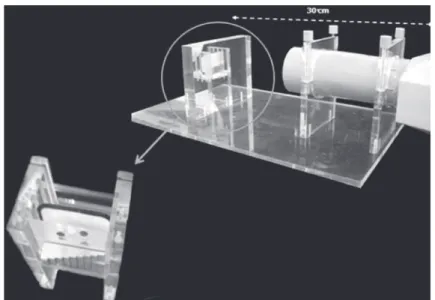
Helsink, Finland), used for data collection. A 70 kVp and 8 mA radiograph machine, Spectro 70X
(Dabi Atlante, Dabi Atlante Indústrias Médico Odontológicas Ltda, Ribeirão Preto, SP, Brazil), was used. The focus-object distance was 30 cm (ANSI/ ADA 2000) and exposure time at 0.2 s, as instructed for digital radiography of phosphor plates, by the manufacturer (Figure 2).
An acrylic positioning device with metallic fastener held sensors and provided an adequate and standardized focus-object distance. The radiograph
machine head was ixed on the same position with
central beam presenting 90°angle of incidence with the acrylic/sensor surface plates set. A rectangular collimator (Dabi Atlante, Dabi Atlante Indústrias Médico Odontológicas Ltda) presenting 3x4 cm aperture reduced possible secondary radiation by being attached to the end of cylinder.
The sensor, after being exposed, was inserted into the laser optical reader of DigoraTM for Windows
5.1 software. As soon as the first image was revealed on screen, parameters suggested by the system were established, allowing to image standardization. The same phosphor plate was used for all exposures to avoid possible differences between plates.
The system performed a radiographic density reading over images of each cement revealed on screen, and also a reading of steps on an aluminum stepwedge, resulting in a numeric value for each reading. This value was written down by the evaluator. After evaluating the 5 acrylic set of plates, 5 measurements for each type of cement and for each step of the aluminum scale were obtained. Mean values of radiographic density and graduated aluminum stepwedge were determined
for each material. Mean values were taken by a
single evaluator previously trained and blinded with regard to the different groups. Intergroup relation analysis was tested using one-way ANOVA (α=0.05). Pairwise multiple comparisons were carried out using the Bonferroni test (α=0.05) in
the cases where the ANOVA test showed signiicant
differences.
RESULTS
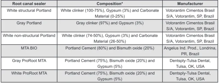
The mean radiographic density values of the cements, in mm Al, are presented in Table 1. MTA-based cements (MTA BIO, gray and white ProRoot MTA) presented the highest radiopacity values
Root canal sealer Composition* Manufacturer
White structural Portland White clinker (100-75%), Gypsum (3%) and Carbonate Material (0-25%)
Votorantim Cimentos Brasil S/A, Votorantim, SP, Brazil
Gray Portland Gray clinker (97%) and Gypsum (3%) Votorantim Cimentos Brasil S/A, Votorantim, SP, Brazil
White non-structural Portland White clinker (74-50%), Gypsum (3%) and Carbonate Material (26-50%)
Votorantim Cimentos Brasil S/A, Votorantim, SP, Brazil
MTA BIO Portland Cement (80%) and Bismuth oxide (20%) Angelus Ind. Prod., Londrina, PR, Brazil
Gray ProRoot MTA Portland Cement (75%), Bismuth oxide (20%) and Gypsum (5%)
Dentsply-Tulsa Dental, Tulsa, OK, USA
White ProRoot MTA Portland Cement (75%), Bismuth oxide (20%) and Gypsum (5%)
Dentsply-Tulsa Dental, Tulsa, OK, USA
Figure 1- Tested materials and compositions *information according to the manufacturers
among the tested materials (p<0.05), overcoming
3 steps from the aluminum stepwedge, which is the minimum radiopacity recommended by the ANSI/
ADA speciication number 571 (Figure 3).
No statistically differences were observed between each other. Portland cements (gray, white structural and white non-structural) presented the
lowest radiopacity values (p<0.05), not reaching
the ANSI/ADA1 (2000) recommendation.
DISCUSSION
Up to present moment, there are no speciic
standards for retrofilling materials to support and reference studies on their physico-chemical properties. Published studies followed standards
proposed by the ANSI/ADA speciication number
Root canal sealer Radiographic
density (mm Al)
White structural Portland 99.59±12.88a
Gray Portland 109.71±4.90a
White non-structural Portland 119.76±22.34a
MTA BIO 143.13±16.94b
Gray ProRoot MTA 155.96±16.30b
White ProRoot MTA 155.99±8.04b
Radiopacity means ± standard deviation of the tested materials and results of ANOVA and Bonferroni test
(α=0.05). Different letters indicate statistically signiicant differences at 5% signiicance level.
Table 1- Radiographic density of the cements (mean ± standard deviation)
Figure 2- Experimental set-up used to ix x-ray machine central beam and the acrylic plates/phosphor plate set at a focus-object distance of 30 cm. In greater magniication, experimental set-up with the aluminium stepwedge, wells for root canal illing materials and radiography digital phosphor plate
Figure 3- Radiopacity of cements in comparison with steps of the aluminium scale. The black color relates to cements
whose radiopacity density (mm Al) fulills the ANSI-ADA speciication number 57 (step 3). The white color refers to cements
571 for endodontic sealing materials11,27, and the
ISO 6876 standard for zinc oxide and eugenol endodontic sealing materials6,15. This equivalence
is based on the fact that, under clinical conditions, retrofilling materials and root filling materials remain in direct contact with periodontal and periapical tissues8,18.
Both ISO and ANSI/ADA have adopted equivalence procedures with an aluminium scale steps, in order to analyze several dental materials radiopacity3. It is known that the radiopacity of 1
mm of dentin is equivalent to 1 mm of aluminum in a graduated stepwedge9. According to the ANSI/
ADA speciication number 571, an endodontic sealing
material should present radiopacity correspondent to at least 3 mm Al.
Digital measurement methods have been proposed by determining gray-tones values, measured in pixels21. These systems can differentiate
all shades of gray on a digital image, while the
naked human eye cannot identify 255 shades, on a non-digitized ilm5. Some studies used direct
methods of analysis5, while others preferred indirect
methods, through scanning images obtained by
occlusal ilms25,26. Besides, digital x-ray ilms provide
reduction in processing time and in number of steps
that could interfere on inal radiograph quality21.
Retroilling materials should present enough
radiopacity to be radiographically distinguished from surrounding structures, such as tooth and alveolar bone, and to reveal empty spaces and inappropriate contours17. Only gray and white ProRoot MTA
cements and MTA BIO, among the studied materials, met the ANSI/ADA recommendations. This fact was expected since ProRoot MTA and MTA BIO are reinforced with 20% bismuth oxide in their composition7,10.However, other studies
reported a lower quantity of bismuth oxide on MTA BIO composition, justifying its lower radiopacity in comparison to ProRoot MTA, corroborating with this
study’s indings6,8,16.
The original formulation of Portland cement did not present bismuth oxide10, determining its low
radiopacity and making impossible to distinguish it
from bone tissue14. Mean values obtained for this
cement were lower than 2 mm Al, not reaching the minimum requirements of the ANSI/ADA1 (2000).
The inadequate radiopacity of Portland cement has been reported8. In order to address this issue,
radiopacity was studied when Portland cement
was associated to different radiopaciiers14. Results
demonstrated that incorporation of a radiopaciier
agent promotes satisfactory radiopacity, being also higher than dentin radiopacity14. However, it should
be further investigated if the cement/radiopaciier
agent mixture does not interfere with the original physicochemical properties and biocompatibility of Portland cements.
CONCLUSIONS
Based on the employed methodology and obtained results, it can be concluded that only
MTA-based cements met the ANSI/ADA speciication
number 571 with respect to radiopacity.
REFERENCES
1- American Dental Association - ANSI/ADA. Speciication 57:
endodontic sealing material. Chicago: ANSI/ADA; 2000. 2- Associação Brasileira de Cimento Portland. Guia básico de utilização do cimento Portland. 7a. ed. São Paulo; 2002.
3- Baksi GB, Sen BH, Eyuboglu TF. Differences in aluminium
equivalent values of endodontic sealer: conventional versus digital radiography. J endod. 2008;34:1101-4.
4- Bernabé PFe, Holland R, Morandi R, Souza V, Nery MJ, Otoboni Filho JA, et al. Comparative study of MTA and other materials in
retroilling of pulpless dogs' teeth. Braz Dent J. 2005;16:149-55.
5- Carvalho JR Jr, Correr-Sobrinho L, Correr AB, Sinhoreti MA,
Consani S, Sousa-Neto MD. Radiopacity of root illing materials
using digital radiography. Int endod J. 2007;40:514-20. 6- Chng HK, Islam I, Yap AUJ, Tong YW, Koh eT. Properties of a
new root-end illing material. J Endod. 2005;31:665-8.
7- Dammaschke T, Gerth HUV, Züchner H, Schäfer E. Chemical and physical surface and bulk material characterization of white
ProRoot MTA and two Portland cements. Dent Mat. 2005;21:731-8.
8- Danesh G, Dammaschke T, Gerth HUV, Zandbiglari T, Schäfer
e. A comparative study of selected properties of ProRoot mineral trioxide aggregate and two Portland cements. Int endod J. 2006;39:213-9.
9- Devito KL, Ortega AI, Haiter-Neto F. Radiopacity of calcium hydroxide cement compared with human tooth structure. J Appl Oral Sci. 2004;12:290-3.
10- estrela C, Bammann LL, estrela CRA, Silva RS, Pécora JD. Antimicrobial and chemical study of MTA, Portland cement, calcium hydroxide paste, Sealapex and Dycal. Braz Dent J. 2000;11:3-9. 11- Fridland M, Rosado R. Mineral trioxide aggregate (MTA) solubility and porosity with different water-to-powder ratios. J endod. 2003;29:814-7.
12- Funteas UR, Wallace JA, Fochtman eW. A comparative analysis of mineral trioxide aggregate and Portland cement. Aust endod J. 2003;29:43-4.
13- Hayashi M, Shimizu A, ebisu S. MTA for obturation of mandibular central incisors with open apices: case report. J endod. 2004;30:120-2.
14- Húngaro Duarte MA, Oliveira el Kadre GD, Vivan RR, Guerreiro Tanomaru JM, Tanomaru Filho M, Moraes IG. Radiopacity of portland cement associated with different radiopacifying agents. J endod. 2009;35:737-40.
15- International Organization for Standardization. ISO 6876: dental root canal sealing materials. 2nd ed. Geneva: ISO; 2001.
16- Islam I, Chng HK, Yap AU. Comparison of the physical and mechanical properties of MTA and Portland cement. J endod. 2006;32:193-7.
17- Laghios CD, Benson BW, Gutmann JL, Cutler CW. Comparative
radiopacity of tetracalcium phosphate and other root-end illing
materials. Int endod J. 2000;33:311-5.
18- Martins GR, Carvalho CAT, Valera MC, Oliveira LD, Buso L, Carvalho AS. Sealing ability of castor oil polymer as a root-end
illing material. J Appl Oral Sci. 2009;17:220-3.
19- Oliveira MG, Xavier CB, Demarco FF, Pinheiro ALB, Costa AT, Pozza DH. Comparative chemical study of MTA and Portland cements. Braz Dent J. 2007;18:3-7.
20- Queiroz AM, Assed S, Leonardo MR, Nelson-Filho P, Silva LAB. MTA and calcium hydroxide for pulp capping. J Appl Oral Sci. 2005;13:126-30.
21- Rasimick BJ, Shah RP, Musikant BL, Deutsch AS. Radiopacity of endodontic materials on ilm and a digital sensor. J Endod.
2007;33:1098-101.
22- Ribeiro DA, Matsumoto MA, Duarte MAH, Marques MeA, Salvadori DMF. In vitro biocompatibility tests of two commercial types of mineral trioxide aggregate. Braz Oral Res. 2005;19:183-7. 23- Saidon J, He J, Zhu Q, Safavi K, Spångberg LSW. Cell and tissue reactions to mineral trioxide aggregate and Portland cement. Oral Surg Oral Med Oral Pathol Oral Radiol endod. 2003;95:483-9. 24- Tagger M, Katz A. A standard for radiopacity of root-end
(retrograde) illing materials is urgently needed. Int Endod J.
2004;37:260-4.
25- Tanomaru-Filho M, Jorge eG, Guerreiro Tanomaru JM,
Gonçalves M. Radiopacity evaluation of new root canal illing
materials by digitalization of images. J endod. 2007;33:249-51. 26- Tanomaru-Filho M, Silva GF, Duarte MAH, Gonçalves M,
Tanomaru JM. Radiopacity evaluation of root-end illing materials
by digitization of images. J Appl Oral Sci. 2008;16:376-9.
27- Wiltbank KB, Schwartz SA, William G, Schindler WG. Effect of