Rosangela da Silveira CorrêaI Ruffo Freitas-JuniorII
João Emílio PeixotoIII Danielle Cristina Netto RodriguesII
Maria Eugênia Fonseca LemosI Cíntia Melazo DiasIV
Rubemar de Souza FerreiraV Rosemar Macedo Souza RahalII
I Rede Goiana de Pesquisa em Mastologia. Centro Regional de Ciências Nucleares do Centro-Oeste. Comissão Nacional de Energia Nuclear. Abadia de Goiás, GO, Brasil
II Rede Goiana de Pesquisa em Mastologia. Programa de Mastologia. Universidade Federal de Goiás. Goiânia, GO, Brasil III Rede Goiana de Pesquisa em Mastologia.
Instituto Nacional de Câncer. Rio de Janeiro, RJ, Brasil
IV Rede Goiana de Pesquisa em Mastologia. Diretoria de Radioproteção e Segurança. Comissão Nacional de Energia Nuclear. Brasília, DF, Brasil
V Diretoria de Radioproteção e Segurança. Comissão Nacional de Energia Nuclear. Rio de Janeiro, RJ, Brasil
Correspondence:
Rosangela da Silveira Corrêa
Comissão Nacional de Energia Nuclear, Centro Regional de Ciências Nucleares do Centro-Oeste
BR-060 km 174,5, Zona Rural, Parque Estadual Telma Ortegal
Received: 10/26/2011 Approved: 4/17/2012
Article available from: www.scielo.br/rsp
Effectiveness of a quality
control program in
mammography for the Brazilian
National Health System
ABSTRACT
OBJECTIVE: To assess the effectiveness of a quality control program in
mammography services of the Brazilian National Health System (SUS).
METHODS: A prospective study using temporal analysis of a health
surveillance action was conducted. A total of 35 service providers that had mammography equipment in operation and regularly performed exams between 2007 and 2009 in the state of Goiás, Central-Western Brazil, participated in this study. Services were assessed during three site visits by performance testing of mammography equipment, fi lm processors, and other materials, and image quality and entrance surface dose in a phantom were also assessed. Each service was scored according to the percentage of tests that conformed to standards.
RESULTS: The mean percentage for compliance among the participating
service providers were 64.1% (13.3%) in the fi rst visit, 68.4% ( 15.9%) in the second, and 77.1% (13.3%) in the third (p < 0.001). The main improvements resulted from adjustments to the breast compression force, the automatic exposure control system, and the alignment of the compression paddle. The doses measured were within the conformity range in 80% of the services assessed.
CONCLUSIONS: The implementation of this program in the mammography
services was effective at improving the operational parameters of the mammography machines, although 40% of the services did not reach the acceptable level of 70%. This result indicates the need to continue this health surveillance action.
DESCRIPTORS: Mammography, standards. Effectiveness. Evaluation of
a OIEA. Organismo Internacional de Energía Atómica. Control de calidad en mamografía. Protocolo elaborado en el marco de dos proyectos regionales ARCAL/OIEA. Viena; 2006. (IAEA-TECDOC-1517). [cited 2011 Sept 12] Available from: http://www.pub.iaea.org/MTCD/ publications/PDF/te_1517s_web.pdf
b Wolf CJM, Perry NM, editors. European guidelines for quality assurance in mammography screening. Luxembourg: European Commission, Europe Against Cancer Programme; 1993.
c Ministério da Saúde (BR), Agência Nacional de Vigilância Sanitária. Portaria/MS/SVS nº 453, de 1º de junho de 1998. Aprova o Regulamento Técnico que estabelece as diretrizes básicas de proteção radiológica em radiodiagnóstico médico e odontológico, dispõe sobre o uso dos raios-x diagnósticos em todo o território nacional e dá outras providências. Diario Ofi cial Uniao. 2 jun 1998. [cited 2011 Sept 18]. Available from: http://www.anvisa.gov.br/legis/portarias/453_98.htm
d Ministério da Saúde (BR). Instituto Nacional de Câncer (INCA). Controle do câncer de mama: documento de consenso. Rio de Janeiro; 2004. [cited 2011 Sept 12]. Available from: http://www1.inca.gov.br/publicacoes/Consensointegra.pdf
e Ministério da Saúde (BR). Cadastro Nacional de Estabelecimentos de Saúde – CNES. Brasília, DF; 2007. [cited 2011 Sept 12]. Available from: http://cnes.datasus.gov.br
The main objective of mammography as a screening method for breast cancer is to reduce the mortality rate through the increase of cases detected at early stages of the disease.5 Randomized trials show statistically significant reduction in mortality rates for women undergoing mammography screening.9,14,16,17 However, screening effectiveness is directly related to the quality and performance of equipment, materials and procedures employed.8 As a result, reference documents were drawn up for the implementation of quality control programs in mammography.6,a,b
The Brazilian College of Radiology created in 1992 the Quality Certifi cation Program in Mammography on a voluntary basis, in order to initiate an action plan for quality control in mammography in Brazil.10 Radiological protection guidelines in medical and dental radiodiagnosis were established by the Brazilian Ministry of Health, in 1998.c
The National Cancer Institute (INCA) recommended in 2004 the creation of quality control guidelines to the Brazilian National Health System (SUS), as part of the criteria for accreditation and monitoring of mammography services.d This recommendation was aimed at implementing quality control programs in the accredited network. Thus, the services became responsible for conducting periodic performance tests in mammography units, fi lm processors and other materials, as well as verifying the image quality and the radiation dose applied to patients.
Screening programs for breast cancer are aimed at improving public health. Quality control actions for achieving satisfactory imaging by exposing patients to acceptable radiation doses constitute a major compo-nent of such programs. Thus, the objective of this study was to evaluate the effectiveness of the quality control program in mammography for continuous monitoring of services that integrate the SUS network.
METHODS
A prospective temporal analysis of a health surveil-lance action was carried out. The unity of the research
INTRODUCTION
was the mammography service, and data collection was performed by site visits in three periods: 2007, 2008 and 2009. Service providers that had equip-ment with conventional processing system (screen/ fi lm) and performed mammograms through the SUS from July 2007 to March 2009, in the state of Goiás, Central-Western Brazil, were included in this research. Data collection was divided into two stages: service provider registration and performance assessment of mammography units, fi lm processors and other materials, as well as evaluation of image quality and radiation doses applied.
Of the 45 mammography service providers caring for SUS patients in the state of Goiás between 2007 and 2009, 35 participated in this study. The following service providers were excluded: two were banned from performing mammography on the SUS, four were closed (during the research period) for not having the technical conditions required, two were not involved in all stages of the research, and two were excluded at the end because they replaced their conventional image processing for a digital system.
An evaluation report was sent to each service provider after the visits, containing the measured and the accep-tance value (or value range) for each parameter. Upon delivery of the report, the service provider received a deadline from the health surveillance organ of the State of Goiás for the completion of nonconformities.
Identifi cation of services was performed using data from the National Registry of Health Care Services (CNES/DATASUS/MS).e Background information on the equipment and materials used in mammography was collected. Participating services were coded by the acronym GO (state of Goiás), followed by a three-fi gure numerical sequence starting at the code GO001, to preserve the mammography services identity.
Optical De
nsity
Contrast scale (Contrast scale = 0.60 ± 0.06)
Fibers 4 ( ĭ 0.75 mm)
Masses 4 (0.75 mm)
Microcalcification groups 4 (0.25 mm)
Metallic grids = 4 (12 pl/mm) I. Contraste Index
4
3
2
12 11 10 9 8 1
Step
Optical density between 1.30 and 1.80 Disks (low contrast) 7 (1.5% of contrast)
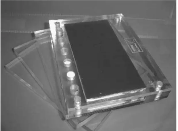
Figure 1. A. Mammographic phantom ; B. Image of the breast phantom produced in the mammography services.
carried out according to the technical regulation by MS/ SVS,c the European Guidelines for Quality Assurance in Breast Cancer Screening and Diagnosis15 and the Protocol on Quality Assurance in Mammography of the International Atomic Energy Agency (IAEA).a Such documents provided the measurement processes used in the evaluations of performance parameters and their acceptance limits.
Radiation dose and image quality were assessed through a mammographic phantom3 (Figure 1A). It simulates a 5.3 cm compressed breast made of 50% fat and 50% fi broglandular tissue. It consists of three acrylic sheets (10 mm x 120 mm x 160 mm) and one acrylic plate (20 mm x 120 mm x 160 mm), which contains a wax insert (5 mm x 70 mm x 140 mm) and a fi ve-step scale that produces areas of different optical densities, used for the assessment of image contrast (Figure 1B). The wax insert contains four metal grids for evaluating the spatial resolution of the image, fi ve groups of Al2O3 microspheres simulating microcalcifi cations, eight polyester disks simulating low contrast areas, six nylon strands simulating fi brous tissues and fi ve spherical nylon caps simulating tumor masses.
The images were generated under the same conditions in each mammography unit, with the phantom posi-tioned on the breast support platform, aligned with the cassete on the edge of the chest wall and the sensor for automatic exposure control (AEC) activated and positioned under the phantom’s main body. The strain selected for the exposure was held constant at 28 kV at all mammography services. After exposure, the fi lm was developed at the service’s processor, in normal use conditions (Figure 1B).
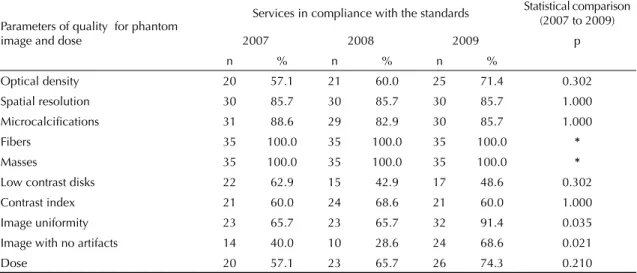
Tabela 2. Assessment and statistical comparison between mammography services, according to parameters of quality for phantom image and dose in compliance with the standards. Goiás, Central-Western Brazil, 2007 to 2009.
Parameters of quality for phantom image and dose
Services in compliance with the standards Statistical comparison (2007 to 2009)
2007 2008 2009 p
n % n % n %
Optical density 20 57.1 21 60.0 25 71.4 0.302 Spatial resolution 30 85.7 30 85.7 30 85.7 1.000 Microcalcifi cations 31 88.6 29 82.9 30 85.7 1.000
Fibers 35 100.0 35 100.0 35 100.0 *
Masses 35 100.0 35 100.0 35 100.0 *
Low contrast disks 22 62.9 15 42.9 17 48.6 0.302 Contrast index 21 60.0 24 68.6 21 60.0 1.000 Image uniformity 23 65.7 23 65.7 32 91.4 0.035
Image with no artifacts 14 40.0 10 28.6 24 68.6 0.021
Dose 20 57.1 23 65.7 26 74.3 0.210
*The test was not applied because the variable did not show dichotomy.
Table 1. Assessment and statistical comparison between mammography services, according to performance parameters for equipment and materials in compliance with the standards. Goiás, Central-Western Brazil, 2007 to 2009.
Performance parameters
Services in compliance with the standards Statistical comparison (2007 to 2009)
2007 2008 2009 p
n % n % n %
Compression force 17 48.6 25 71.4 27 77.1 0.013 Field alignment 25 71.4 27 77.1 32 91.4 0.092
Exposure automatic control 13 37.1 22 62.9 24 68.6 0.007 Alignment of the compression paddle 19 54.3 28 80 30 85.7 0.003 Chassis integrity 21 60.0 23 65.7 30 85.7 0.035 Processing 13 37.1 13 37.1 16 45.7 0.581
The performance of the devices and accessories that directly and indirectly infl uenced image quality was tested by collimation system, AEC’s performance, compression force, alignment of thecompression paddle, cassete integrity and processing system.
The radiation dose at the entrance surface of the phantom was determined by measuring the air-kerma, established by MS/SVS,c with a solid state dosimeter (model Multi-O-Meter L 535, Unfors, Billdal, Sweden) calibrated for mammography. The measurements were performed with the same radiographic techniques selected for image processing of the phantom. The compliance range of 7 to 13 mGy was adopted, consid-ering the reference value of 10 mGy (± 30% for this study), established by the SVS/MS.c
One point was awarded for compliance and zero for non-compliance of each parameter, at each assess-ment. Thus, considering 16 parameters, the total score of the service varied from zero to 16. The percentage of compliance for each service and their average
percentage (2007, 2008 and 2009) were calculated. The t-test for comparison of means for paired data samples was used to verify the statistical differences between the mean compliance percentages between the services at each year. The McNemar’s test was applied to compare the parameters between two years (2007 and 2008, 2007 and 2009, and 2008 and 2009). The signifi cance level of 0.05 was used in the statistical tests.
This study was approved by the Dr. Henrique Santillo Ethics in Research Committee at the State Department of Health of Goiás (Protocol 0007.1.177.000-5).
RESULTS
Compliance averange for the services: 2007 = 64.1%
2008 = 68.4% 2009 = 77.5% 77.1
62.9
40.0 40.0
2.9 8.6
20.0 20.0
28.6
% of services
< 70% (unacceptable) < 70% a 89% (acceptable) 90% (desirable)
2009 2008
2007
Figure 2. Percentage of compliance of the performance parameters for equipment and materials related to phantom image quality and dose in mammography services, according to year of assessment. Goiás, 2007 to 2009.
10
Frequen
cy
Air-kerma at the entrance surface of de phantom (mGy) 9
4.00 6.00 8.00 10.00 12.00 14.00 8
7
6
5
4
3
2 1
0
Mean = 8.94 mGy Std. Dev. = 2.15 mGy n = 35
Figure 3. Radiation dose at the entrance surface of the phantom (air-kerma) at the mammography services. Goiás, Central-Western Brazil, 2009.
in Brazil and 11% were made by other national and foreign manufacturers. A total of 69% of the processors were used exclusively for mammograms.
The analyses for 2007-2008 and 2007-2009 revealed statistically signifi cant differences between the percent-ages of compliance for compression force (p = 0.039 and p = 0.002), AEC (p = 0.012 and p = 0.004) and alignment of the breast support platform ( p = 0.004 and p = 0.002), while the integrity of the cassete (p = 0.035) showed a statistically signifi cant difference in the 2007-2009 period. An increase in absolute numbers of services showing compliance in the alignment of the x-ray fi eld with the fi lm and in the processing of x-ray fi lms (Table 1), even without statistically signifi cant differences between the percentages.
Regarding the assessment of the quality parameters of the phantom’s image recorded on fi lm and the radiation dose used, the two parameters that showed statistically signifi cant differences from 2007 to 2009 were the absence of image artifacts (p = 0.021) and the image uniformity (p = 0.035) (Table 2).
Although no statistically signifi cant differences were observed regarding the optical density of the image and the dose, the data showed an increase in the number of services that were in conformity with these parameters. In relation to the spatial resolution and the visualiza-tion of structures that mimic tumor masses and fi bers, services showed compliance in the assessment for 2007, a result that was also seen in 2008 and 2009.
The other parameters of image quality (visualization of microcalcifi cations and contrast index) showed differ-ences in the order of 5% between the percentages, whereas the difference was 14% for the low contrast disks. In such cases, no numerical increase of services in accordance with the standards was observed.
About 23% of services presented percentage of compli-ance equal or higher than 70% in 2007, which is consid-ered acceptable, decreasing to 60% in 2009 (Figure 2). A mammography service (3%) achieved a desirable level of compliance in 2007, above 90%, but was rated three (9%) in 2008 and six (17%) in 2009. The average compliance of services in the fi rst visit (2007) was 64.1% (± 13.3%), whereas in the second (2008) and third (2009) were 68.4% (± 15.9%) and 77.5% (± 13.3%), respectively. The difference between the mean percentage of compliance of the services was statistically signifi cant between the fi rst and third visits (p < 0.001) and between the second and third visits (p = 0.004).
DISCUSSION
The evaluation of the quality actions and health services is fundamental to the control of health care offered to the population2. Equipment emitting ionizing radiation can operate with a high degree of quality, but it is neces-sary to follow the rules and procedures of radiological protection and quality control.a The implementation of a quality control program in mammography services, which are part of the SUS network in Goiás, was more effective with the results found for 2007 and 2009, i.e., with two interventions. By continuing the program, it is possible to establish a better quality control, reducing the risks linked to mammography exams, even though 20% of services have shown desirable results in the last assessment of above 90% compliance, considering the evaluated items.
The performance parameters for the equipment and materials that showed improvement resulting from the quality control action in the services were related to adjustments in breast compression force, AEC system, alignment of the breast support platform and integrity of the cassete. However, some mammography units were left unadjusted in relation to the breast compression force (22.9%), AEC system (31.4%) and alignment of the compression paddle and integrity of the cassete (14.3%). In a study carried out in Minas Gerais13, the percentage of mammography units unadjusted for the compression force was 23.9%, whereas 37.3% were unadjusted for the AEC system. The results obtained in both studies coincide and point to a situation in which it is not possible to make adjustments in these perfor-mance parameters for some mammography units. It was not possible to establish, during this research, whether this hypothesis was associated with the technology of the equipment or duration of use.
The only performance parameter that showed no improvement during the study period was the align-ment of the x-ray fi eld with the image recording system composed by the cassete and the fi lm (p = 0.092). However, there was a progressive increase in the percentage of compliance for this parameter, result that shows the need for continuity of this type of interven-tion in the coming years.
Studies indicate the development of radiographic fi lms as the process that most affects image quality in mammog-raphy.3,11,12 The solution of non-compliance cases for fi lm processing is not simple, involving from the change of equipment to the adoption of standardized processes for the preparation of certifi ed solutions. The result obtained in this study showed that the percentage of compliance of fi lm processing remained almost unchanged, and around 40% during the three years of research, which is similar to that found in the study carried out in Minas Gerais13 (38.8%). About 31% of the fi lm processors were not specifi c for mammography, which may have contributed
to the relatively low percentage of compliance in fi lm processing. The materials used, the temperature of the employed solutions and the processing time were not evaluated in this study.
The image quality parameters that showed improve-ment resulting from the quality control program between 2007 and 2009 were related to uniformity and reduced number of image artifacts. Those that did not show a statistically signifi cant difference between the mean percentage of compliance services between the fi rst and third visits can be divided into two groups. In the fi rst group are included the mean optical density, spatial resolution, microcalcifications, fibers and masses, with percentages of compliance above 70% (minimum acceptable percentage for a set of services). In the second group are the low contrast disks and contrast index, with percentages of compliance below 70%. These groups require separate analysis. The parameters of the fi rst group reached the minimum acceptable levels. The situation is not considered critical and requires a fi ner adjustment in the image production chain, in a small number of services.
The situation of the parameters in the second group is considered critical, because no improvement was observed between assessments. Their mean percentages of compliance (43% and 69%) are considered unaccept-able. These parameters will require more intense effort for performance tuning of equipment and materials. The visualization of low contrast disks and the contrast index are directly associated with the performance of the automatic fi lm processor.
On one hand, the analysis of radiation doses on the entrance surface of the phantom shows no improvement during the study (p = 0.210), but a progressive increase in the percentage of compliance of this parameter was observed. Corrective measures related to equipment and materials can improve the image quality parameters of the phantom, with a positive impact on the adequacy of the doses to a range of acceptable values.1
The dose of radiation to which the patient is exposed should be kept as low as possible without compromising the quality of the image.a The legislationc established 10 mGy as the reference value for applications near the skin. In this study, upon acceptance of variation of ± 30%, 80% of services showed values of 7 to 13 mGy, averaging 8.94 mGy and comparable to the results of the Minas Gerais research.13
The quality of services in relation to equipment and accessories was evaluated. However, it is necessary to conduct research focusing on mammography profes-sionals, as well as image quality, for the early detection of breast cancer and the carcinogenic risk within the SUS and the supplementary health system.
Interventions in the Quality Control Program in Mammography, based on the methodology applied in this study, were effective for improving the screening quality and monitoring the services that compose the SUS in Goiás. Although the percentage of mammog-raphy services classifi ed as unacceptable in relation to
the technical criteria evaluated has decreased (from 77.1% in 2007 to 40% in 2009), doctors continued to have mammograms of inferior quality and were more prone to misdiagnosis. It is necessary to continue quality control activities, including services that are not linked to the SUS.
ACKNOWLEDGEMENTS
To the Superintendency of Health Surveillance of Goiás, the Department of Health Surveillance of Goiânia and the National Commission of Nuclear Energy, which contributed to this research.
1. Avramova-Cholakova S, Vassileva J. A survey of the state of mammography practice in Bulgaria.
Radiat Prot Dosimetry. 2011;147(1-2):14-6.
DOI:10.1093/rpd/ncr320
2. Azevedo AC. Avaliação de desempenho de serviços de saúde. Rev Saude Publica. 1991;25(1):64-71. DOI:10.1590/S0034-89101991000100013
3. Corrêa RS, Peixoto JE, Silver LD, Dias CM, Nogueira MS, Hwang SF, et al. Impacto de um programa de avaliação da qualidade da imagem nos serviços de mamografi a do Distrito Federal. Radiol Bras. 2008;41(2):109-14. DOI:10.1590/S0100-39842008000200010.
4. Corrêa RS, Freitas-Júnior R, Peixoto JE, Rodrigues DCN, Lemos MEF, Marins LAP, et al. Estimativas da cobertura mamográfi ca no estado de Goiás, Brasil. Cad Saude Publica. 2011;27(9):1757-67. DOI:10.1590/S0102-311X2011000900009
5. Feig SA. Screening mammography: a successful public health initiative. Rev
Panam Salud Publica. 2006;20(2-3):125-33.
DOI:10.1590/S1020-49892006000800009
6. Food and Drug Administration (US). The
Mammography Quality Standards Act (MQSA) interim fi nal rules. Silver Spring; 1993.
7. Gershan V, Antevska-Grujoska S. Performance of mammography equipment in the Macedonian breast screening campaign 2008/2009. Radiat Prot Dosimetry.
2011;147(1-2):187-91. DOI:10.1093/rpd/ncr290
8. Hendrick RE, Klabunde C, Grivegnee A, Pou G, Ballard-Barbash R. Technical quality control practices in mammography screening programs in 22 countries.
Int J Qual Health Care. 2002;14(3):219-26.
9. Hendrick RE, Helvie MA. United States Preventive Services Task Force screening mammography recommendations: science ignored. AJR
Am J Roentgenol. 2011;196(2):W112-6.
DOI:10.2214/AJR.10.5609
10. Koch HA, Peixoto JE, Neves ALE. Análise da infra-estrutura para a mamografi a no Brasil. Radiol Bras. 2000;33(1):23-30.
11. Magalhães LAG, Azevedo ACP, Carvalho ACP. A importância do controle de qualidade de processadoras automáticas.
Radiol Bras. 2002;35(6):357-63.
DOI:10.1590/S0100-39842002000600009
12. Milano F, Maggi E, Roselli del Turco M. Evaluation of the effect of a quality control programme in mammography on technical and exposure parameters.
Radiat Prot Dosimetry. 2000;90(1-2):263-6.
13. Oliveira M, Nogueira MS, Guedes E, Andrade MC, Peixoto JE, Joana GS, et al. Average glandular dose and phantom image quality in mammography. Nucl
Instrum Methods Phys Res A. 2007;580(1):574-7.
DOI:10.1016/j.nima.2007.05.228.
14. Parkin DM, Whelan SL, Ferlay J, Teppo L, Thomas DB, editors. Cancer incidence in fi ve continents. Lyon: IARC; 2002. (IARC Scientifi c Publication, 155).
15. Perry N, Broeders M, Wolf C, Törnberg S, Holland R., von Karsa L, editors. European guidelines for quality assurance in breast cancer screening and diagnosis. 4. ed. Luxembourg: Offi ce for Offi cial Publications of the European Communities; 2006.
16. Shapiro S, Venet W, Strax P, Venet L. Periodic screening for breast cancer: the Health Insurance Plan Project and its sequelae, 1963-1986. Baltimore: Johns Hopkins University Press; 1988.
17. Tabár L, Vitak B, Chen HH, Duffy SW, Yen MF, Chiang CF, et al. The Swedish Two-County Trial twenty years later. Updated mortality results and new insights from long-term follow-up.
Radiol Clin North Am. 2000;38(4):625-51.
DOI:10.1016/S0033-8389(05)70191-3
REFERENCES