INTRODUCTION
Article/Artigo
1. Serviço de Pediatria, Hospital Universitário Cassiano Antonio de Moraes, Universidade Federal do Espírito Santo, Vitória, ES. 2. Núcleo de Doenças Infecciosas, Centro de Ciências da Saúde, Universidade Federal do Espírito Santo, Vitória, ES.
Address to: Dr. Fausto Edmundo Lima Pereira. NDI/CCS/UFES. Av. Marechal Campos 1468, Maruípe, 29040-091 Vitória, ES, Brazil.
Phone: 55 27 3335-7210 e-mail: [email protected]
Received in 06/01/2011
Accepted in 15/03/2011
Anti-
Toxocara
antibodies detected in children atending elementary school
in Vitoria, State of
Espírito Santo
, Brazil: prevalence and associated factors
Anticorpos anti-
Toxocara
em crianças admitidas na escola fundamental em Vitória, Estado
do Espírito Santo: prevalência e fatores associados
Roberta Paranhos Fragoso1, Mariza Buriche Macedo Monteiro1, Elenice Moreira Lemos2 and Fausto Edmundo Lima Pereira2
ABSTACT
Introduction: he aim of this study was to evaluate the frequency of anti-Toxocara antibodies in serum from 7-year-old children atending elementary school in Vitória-ES, Brazil and to correlate these antibodies with socio-demographic factors, the presence of intestinal helminths, blood eosinophil numbers, past history of allergy or asthma, and clinical manifestations of helminth infections. Methods: he detection of anti-Toxocara antibodies was performed using an ELISA (Cellabs Pty Ltd)on serum from 391 children who had already been examined by fecal examination and blood cell counts. Data from clinical and physical examinations were obtained for all children. Results: he prevalence of anti-Toxocara antibodies was 51.6%, with no gender diferences. No signiicant diferences were observed between positive serology and the presence or absence of intestinal worms (60.3 and 51.7%, respectively; p = 0.286). he only variables signiicantly related to positive serology were onycophagy and the use of uniltered water. Although eosinophilia (blood eosinophil count higher than 600/mm3) was signiicantly related to the presence of a positive ELISA result, this signiicance disappeared when we considered only children without worms or without a past history of allergy or asthma. No clinical symptoms related to Toxocara infection were observed. Conclusions: here is a high prevalence of anti-Toxocara antibodies in children atending elementary schools in Vitória, which may be partially related to cross-reactivity with intestinal helminths or to a high frequency of infection with a small number of Toxocara eggs.
Keywords: Toxocara canis.Toxocariasis.Toxocara infection.Visceral larva migrans.
RESUMO
Introdução: O objetivo desse estudo foi avaliar a prevalência de anticorpos anti-Toxocara em crianças admitidas no primeiro ano de escola fundamental em Vitória e correlacionar com variáveis sociodemográicas, helmintos intestinais, eosinóilos no sangue, geofagia, onicofagia, história de asma e alergia cutânea e manifestações clínicas. Métodos: A pesquisa de anticorpos anti-Toxocara, utilizando um teste de ELISA (Cellabs), foi realizada em 391 crianças nas quais foram realizados exames parasitológicos de fezes e hemograma completo. Todas as crianças foram submetidas a exame clínico e físico. Resultados: A prevalência de reação positiva foi de 51,6%, sem diferença entre os sexos. Não foram observadas diferenças signiicativas na prevalência de reação positiva em crianças com ou sem helmintos intestinais (60,3 e 51,7%, respectivamente; p = 0.286). Ainda que a frequência de eosinóilos acima de 600/mm3 tenha sido signiicativamente maior em crianças com sorologia positiva, a signiicância desapareceu quando se considerou as crianças sem helmintos intestinais ou história pregressa de asma ou alergia cutânea. As únicas variáveis signiicativamente correlacionadas, de modo independente, com a presença de sorologia positiva foram onicofagia e hábito de beber água não iltrada. Nenhuma criança apresentou manifestação clínica relacionada com a presença de anticorpos anti-Toxocara. Conclusões: A prevalência de anticorpos anti-Toxocara em crianças admitidas nas escolas elementares em Vitória é alta, a qual pode estar, em parte, relacionada à reação cruzada com antígenos de helmintos intestinais ou devida a frequente exposição a baixas quantidades de ovos do Toxocara.
Palavras-chaves: Toxocara canis. Toxocarase. Infecção com Toxocara. Larva migrans visceral.
The prevalence of Toxocara infection as
measured by evaluating anti-Toxocara antibodies in
serum is variable, with higher prevalence observed
in developing countries1-4. Some studies have
demonstrated a positive correlation between the
presence of anti-Toxocara antibodies and geophagy,
onycophagy, a poor hygiene profileand contact
with dogs4-7.
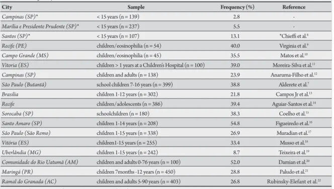
A highly variable prevalence has been reported in Brazil, largely based on descriptive studies using
diferent samples (Table 1 summarizes the studies
reporting the prevalence of anti-Toxocara antibodies
in Brazil published from 1990 to 20098-22).The
only population-based study was published by
Chiei et al8, and this study reported a prevalence
of 2.8-15.5% in children up to 15 years old from four diferent cities in the state of São Paulo.
A prevalence of 33.3-39% was reported in random samples of children at a Children’s Reference
Hospital in Vitória11, 18. In this hospital, a signiicant
association between staphylococcal infection and the
presence of anti-Toxocara antibodies was reported23.
In addition, larva migrans granulomas were observed in 3.2% of livers from routine autopsies at the same
hospital24. herefore, Toxocara infection appears to
be prevalent in children from Brazil.
The Schoolchildren’s Health Program of
the municipality of Vitoria performs clinical
examinations on all children in the first year of public elementary schools. A stool examination, a complete blood cell count and a hemoglobin evaluation are also performed. The aim of the present investigation was to assess the presence
of anti-Toxocara antibodies in a representative
METHODS RESULTS TABLE 1 - Frequency of anti-Toxocara antibodies in children from diferent Brazilian cities.
City Sample Frequency (%) Reference
Campinas (SP)* < 15 years (n = 139) 2.8
-Marília e Presidente Prudente (SP)* < 15 years (n = 237) 5.5
-Santos (SP)* < 15 years (n = 107) 13.1 *Chiei et al.8 Recife (PE) children/eosinophilia (n = 54) 40.0 Virginia et al.9 Campo Grande (MS) children/eosinophilia (n = 45) 35.5 Matos et al.10 Vitoria (ES) children > 1 years at a Children’s Hospital (n = 100) 39.0 Moreira-Silva et al.11 Campinas (SP) children and adults (n = 138) 23.9 Anaruma-Filho et al.12 São Paulo (Butantã) school children 7-16 years (n = 399) 38.8 Alderete et al.7 Brasilia children 1-12 years (n = 302) 21.8 Campos Jr et al.13 Recife children/adolescents (n = 386) 39.4 Aguiar-Santos et al.14 Sorocaba (SP) schoolchildren (n = 180) 38.3 Coelho et al.15 Santo Amaro (SP) children 1-14 years (n = 208) 54.8 Figueiredo et al.16 São Paulo (São Remo) children 1-15 years (n = 338) 26.9 Muradian et al.17 Vitória (ES) children1-15 years (n = 255) 33.4 Musso et al.18 Uberlândia (MG) children 1-15 years (n = 242) 8.7 Teixeira et al.19 Comunidade do Rio Uatumã (AM) children and adults 0-76 years (n = 100) 52.0 Damian et al.20 Maringá (PR) children 7months -12 years (n = 450) 28.8 Paludo et al.21 Ramal do Granada (AC) children and adults 5-90 years (n = 403) 26.8 Rubinsky-Elefant et al.22
SP:São Paulo, PE:Pernambuco, MS:Mato Grosso do Sul, ES:Espírito Santo, MG:Minas Gerais, AM:Amazonas,PR:Paraná, AC:Acre. *his reference includes data from Campinas, Marília and Presidente Prudente
Serum samples were collected from 391 schoolchildren in the irst year at eight elementary schools randomly selected from 39
schools located in low-income neighborhoods of Vitória.
Socio-demographic data including age, gender, family income, parents’ schooling, occurrence of geophagy and onycophagy, use of treated water and sewage facilities, and contact with dogs were collected in interviews with parents that agreed to participate in this investigation.
Stool examinations performed using the sedimentation method, blood cells counts and hemoglobin measurements were performed
in the Central Laboratory of the municipality of Vitória. he blood
cell counts and hemoglobin measurements were performed using automated instruments, with diferential counts checked by Giemsa-stained smears. Aliquots of blood were collected for hematologic studies and were then centrifuged, and the sera were stored at -20ºC
until use for anti-Toxocara antibody detection.
Anti-Toxocara antibodies were detected using a commercial test
(Toxocara IgG ELISA, Cellabs Pty Ltd, Brookvale, Australia) that uses
secretory/excretory antigens of the second stage larvae of Toxocara canis.
he tests were performed according to the manufacturer’s instructions.
Samples with optical densities (OD) ≤ 0.500 were considered negative,
and samples with OD > 0.500 were considered positive.
All children were submited to physical examination and their history of allergy or other disease was determined during an interview with their parents.
Statistical analysis was performed using SPSS 9.0 for Windows. he frequencies of the variables were calculated with 95% conidence intervals. Following statistical tests, p values < 0.05 were considered signiicant.
Ethical considerations
he research was approved by the ethics commitee of Centro de
Ciências da Saúde, Universidade Federal do Espírito Santo.
he detection of anti-Toxocara antibodies was performed on serum
from 391 children. Information pertaining to socio-economic status and sanitary conditions was obtained for approximately 340 of these children. All children were 7 years old, the age for admission to
elementary schools in Vitória.
he results of the ELISA to measure anti-Toxocara antibodies are
shown in Table 2, classiied by gender and OD values in the ELISA.
he overall prevalence of positive results (OD > 0.500) was 51.6%, without signiicant gender diferences.
A fecal examination was performed with 308 children, and 88
(28.5%) children had at least one intestinal helminth. Ascaris
lumbricoides was the most prevalent helminth in this study (53/88, 60.2% of intestinal helminths identiied). Other identiied helminths
were Trichuris trichiura (13/82,14.8%), Enterobius vermicularis
(11/82, 12.5%) and Hymenolepis nana (11/88, 12.5%).
Table 3 shows the ELISA results for anti-Toxocara antibodies in children with negative results upon fecal examination, positive results for at least one intestinal nematode and positive or negative results for
Ascaris lumbricoides. Although the frequencies of positive antibody
tests with diferent OD were higher in children with Ascaris infection,
TABLE 2 - Results of ELISA tests to detect anti-Toxocara antibodies in 391 seven-year-old school children in public elementary schools from the urban periphery of Vitória, State of Espírito Santo, Brazil.
All cases (n = 391) Male (n = 206) Female (n = 185)
ELISA test (OD) n % (95% CI) n % (95% CI) n % (95% CI) Negative tests*
(< 0.500) 189 48.4 (44.8-52.0) 96 46.6 (41.6-51.6) 93 40.3 (36.1-45.4) Positive tests*
(0.501-1.00) 92 23.5 (19.6-27.9) 55 26.6 (20.6-32.6) 37 40.3 (33.3-47.3) (> 1.00) 110 28.1 (23.9-32.7) 55 26.7 (20.6-32.8) 55 29.7 (23.6-36.6)
Total 202 51.6 (46.7-56.5) 110 53.4 (46.6-60.2) 92 49.7 (42.6-56.8) OD: optical density.
*negative or positive tests deined according the manufacturer´s instructions. Chi square test: p = 0.431.
TABLE 3 -Results of ELISA tests for the detection of anti-Toxocara antibodies in 305 seven-year-old schoolchildren atending public elementary schools from the urban periphery of Vitória, State of Espírito Santo, Brazil, separated based on the presence or absence of intestinal helminths.
ELISA test Intestinal helminths Ascaris lunmbricoides
(optical density) negative positive p* negative positive p*
Negative
(< 0.500) 108 36 1.00a 123 21 0.611a
Positive
(0.501 - 1.00) 56 16 60 12
(> 1.00) 60 29 0.200a 69 20 0.477a
All positive cases 116 45 0.651b 129 32 0.286b
acomparison taking in account different optical density levels in negative and
positive cases, bcomparison of all positive and negative cases. ELISA: enzyme-linked
immunosorbent assay. *χ2 test
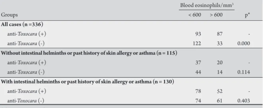
TABLE 4 -Eosinophil counts in seven-year-old children from elementary schools in Vitoria, State of Espírito Santo, Brazil, with or without intestinal helminths or a past history of cutaneous allergy or asthma, according the presence or absence of a positive antibody test for Toxocara canis.
Blood eosinophils/mm3
Groups < 600 > 600 p*
All cases (n =336)
anti-Toxocara (+) 93 87
-anti-Toxocara (-) 122 33 0.000
Without intestinal helminths or past history of skin allergy or asthma (n = 115)
anti-Toxocara (+) 37 20
-anti-Toxocara (-) 44 14 0.114
With intestinal helminths or past history of skin allergy or asthma (n = 130)
anti-Toxocara (+) 78 52
-anti-Toxocara (-) 74 61 0.403 *chi-square test with Yates correction.
As shown in Table 4, eosinophilia was frequent in the individuals
studied (120 of 245 children had an eosinophil count higher than
600cells/mm3)and was detected signiicantly more oten in children
with positive serology for Toxocara. However, the presence of
other intestinal helminths, skin allergy and asthma are important causes of eosinophilia in children. To verify whether a positive test
for anti-Toxocara antibodies is a factor that inluences peripheral
blood eosinophil counts, the proportions of positive and negative antibody results were compared with the frequency of eosinophilia in children with and without intestinal helminths or a past history of skin allergy or asthma. here was no signiicant diference in
eosinophil counts higher than 600cells/mm3 between the children
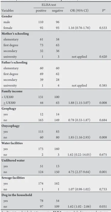
dogs in household and drinking non-iltered water was signiicantly associated with positive antibody tests. A multivariate analysis using a logistic regression demonstrated that drinking non-iltered water and onycophagy were independently associated with positive serology for
Toxocara. Abnormalities upon physical examination were not found in any children in this study.
A past history of asthma, skin allergy and abdominal pain did not correlate with antibodies against Toxocara. Among 84 children with a past history of asthma and 85 children with a past history of skin allergy, anti-Toxocara antibodies were detected in 46 (54.7%) and
42 (49.4%), frequencies similar to those observed in children without
a past history of asthma or skin allergy (86/171, 50.3%; Chi-square test, p=0.475 and 0.825 versus asthma and skin allergy).
with positive or negative anti-Toxocara
antibody tests. Similarly, the presence of positive antibody test results did not inluence the prevalence of eosinophilia in children with intestinal helminths or with a past history of allergy or asthma, thus conirming that the presence of
anti-Toxocara antibodies in the serum
was not associated with increased blood eosinophils in this sample.
The frequencies of positive
anti-Toxocara antibody tests relative to socio-economic status, sanitary conditions, onycophagy, geophagy and contact with
TABLE 5 - Gender, sanitary conditions and familial socio-economic status associated with the presence of a positive result in ELISA test for anti-Toxocara antibodies in serum samples from seven-year-old schoolchildren atending elementary school in of Vitória, State of Espírito Santo, Brazil.
ELISA test
Variables positive negative OR (95% CI) P*
Gender
male 110 96
female 92 93 1.16 (0.76-1.76) 0.533
Mother’s schooling
elementary 61 58 irst degree 73 63
secondary 32 36
university 1 3 not applied 0.420
Father’s schooling
elementary 60 60 irst degree 69 62
secondary 39 28
university 1 4 not applied 0.385
Family income
< U$300 131 100
> U$300 44 63 1.88 (1.15-3.07) 0.008
Geophagy
yes 12 14
no 163 149 0.78 (0.33-1.87) 0.684
Onycophagy
yes 115 83
no 60 80 1.85 (1.16-2.93) 0.008
Water facilities
yes 173 160
no 2 3 1.62 (0.22-14.05) 0.675
Uniltered water
yes 51 13
no 124 150 4.75 (2.37-9.64) 0.001
Sewage facilities
yes 174 162
no 1 1 1.07 (0.98-1.02) 0,733
Dog in the household
yes 78 54
no 97 109 1.62 (1.02 - 2.06) 0.031 *multivariate analysis by logistic regression. ELISA: enzyme-linked immunosorbent assay.
DISCUSSION
hese results demonstrate a high prevalence of anti-Toxocara
antibodies in seven-year-old children living in the urban periphery of Vitória. his prevalence was higher than that reported in other
Brazilian studies but similar to that observed in Santo Amaro, SP16.
Even excluding children with intestinal helminths, the frequency of positive serology was still high. Moreover, 110 children had ELISA-positive results with OD >1.00, considered by the assay manufacturer to be indicative of more recent infection.
Cross-reactivity against other helminth antigens may be responsible for the high frequency of positive ELISA tests for
anti-Toxocara antibodies. his cross-reactivity does exist, mainly against A. lumbricoides25,26, and we did not adsorb the serum samples with
other nematode antigens before performing the ELISA in this study. However, because the frequency of positive ELISA tests in children without worms was 51.7%, similar to the overall frequency of positive test results, we believe that the high frequency of positive serology observed in this study is not due to cross-reactivity. Moreover, no diferences were observed in the frequency of positive results between the individuals with and without intestinal worms or with
and without A. lumbricoides(Table 3). his observation conirms that
although cross-reactivity exists, the frequency of positive serology in children without intestinal worms is high. Although only one fecal sample per child was examined, our conclusion is strengthened by the observation of an increased percentage of positive antibody tests when we considered both children with parasites and children with a past history of worm infection. he cross-reactivity may be responsible for the frequent detection of ELISA-positive samples with high OD in asymptomatic children.
he high prevalence of anti-Toxocara antibodies in seven-year-old
children is consistent with other studies performed in a children’s
hospital in Vitória. hese studies reported prevalences from 30 to 39%
of admited children with positive antibody tests for Toxocara11,18,
and 3.2% of livers obtained from routine autopsies had granulomas
containing Toxocara antigens at the same hospital24.
In contrast to other reports7,11,12,16,21, we did not observe a
signiicant diference for gender regarding the prevalence of positive
tests for anti-Toxocara antibodies. hese results are consistent with
other studies showing no association between gender and risk of Toxocara infection22,27-29. However, positive correlations were
observed between positive ELISA tests for Toxocara and low family
income, the presence of dogs at home and onycophagia. hese results are consistent with the results reported by other authors
using samples from diferent Brazilian cities7,13-15. A multivariate
analysis demonstrated that onycophagia and the use of uniltered
water were associated with positive antibody tests for Toxocara. An
independent association between onycophagia and the presence of
anti-Toxocara antibodies was also reported for children in São Paulo7.
An association between the presence of anti-Toxocara antibodies and
the use of uniltered water has rarely been investigated. However, an investigation in the Amazonia did not reveal a correlation between
positive serology for Toxocara and drinking uniltered water22. Some
authors have suggested the hypothesis that water contaminated with
Toxocara eggs may have a role in Toxocara infection in humans30.
It is possible that eggs transmited by neighborhood dust from neighborhoods may contaminate water within the home. In accordance with this possibility, the presence of dog stool in areas near the dwellings was frequently observed during the visits to the children’s homes.
Eosinophilia was observed in the samples studied and was more
frequent in patientswith positive antibody test results for Toxocara.
However, when we evaluated eosinophil counts in children without other causes of eosinophilia, such as intestinal worms, allergy or asthma, this diference disappeared. In accordance with this observation, there was no signiicant association between positive anti-helminth antibodies and eosinophil counts in children bearing intestinal worms or allergy. Thus, the results presented in this
study demonstrate that the presence of anti-Toxocara antibodies
ACKNOWLEDGMENTS
he authors declare that there is no conlict of interest.
CONFLICT OF INTEREST
FINANCIAL SUPPORT
REFERENCES
We did not ind a correlation between positive antibody tests for
Toxocara and geophagy, as reported in other studies3,5,31-34.
Although controversial, an association between allergic disorders and Toxocara infection has been reported7,34-37. In our sample, no
signiicant association was found between the presence of positive
serology for Toxocara and a past history of asthma or other allergic
manifestations.
Clinical examination of all children did not demonstrate signs
or symptoms that could be correlated with Toxocara infection. he
absence of clinical manifestations in children with anti-Toxocara
antibodies is consistent with the observation that the great majority
of children that acquire Toxocara larvae develop silent infections
with only rare manifestations of visceral or ocular larva migrans38,39.
In conclusion, the results reported here revealed high prevalence
of anti-Toxocara antibodies in seven-year-old children living in the
urban periphery of Vitória as detected by the ELISA using Toxocara
canis secretory/excretory antigens, and this inding suggests that the rate of infection with this nematode in this population is high. Cross-reactivity with other helminth infections may be partially responsible for the observed results, but the prevalence of positive antibody tests was high in children without intestinal worms or a past history of worm
infection. Clinical manifestations related to Toxocara infection were not
observed. he high prevalence of Toxocara antibodies reported here may
be associated with past infections with small number of larvae or with memory responses ater recurrent infections resulting in low larval load.
We thank Rodrigo R. Rodrigues for suggestions and Fabíola Karla Correa Ribeiro for language corrections.
his research was supported by Cia Siderúrgica de
Tubarão-Acelor-Brasil and Fundação de Apoio ao Hospital Universitário Cassiano A Moraes.
1. Hermann N, Glickman LT, Schantz PM, Weston MG, Domanski LM. Seroprevalence of zoonotic toxocariasis in the United States: 1971-1973. Am J Epidemiol 1985; 122:890-896.
2. Magnaval JF, Michault A, Calon N, Charlet JP. Epidemiology of human toxocariasis in La Reunion. Trans R Soc Trop Med Hyg 1994; 88:531-533. 3. Alonso JM, Bojanich MV, Chamorro M, Gorodner JO. Toxocara seroprevalence
in children from a subtropical city in Argentina. Rev Inst Med Trop Sao Paulo 2000; 42:235-237.
4. Despommier D. Toxocariasis: clinical aspects, epidemiology, medical ecology, and molecular aspects. Clin Microbiol Rev 2003; 16:265-272.
5. Glickman LT, Magnaval JF. Zoonotic roundworm infections. Infect Dis Clin North Am 1993; 7:717-732.
6. Holland CV, O’Lorcain P, Taylor MR, Kelly A. Sero-epidemiology of toxocariasis in school children. Parasitology 1995; 110:535-545.
7. Alderete JM, Jacob CM, Pastorino AC, Elefant GR, Castro AP, Fomin AB, et al. Prevalence of Toxocara infection in schoolchildren from the Butanta region, Sao Paulo, Brazil. Mem Inst Oswaldo Cruz 2003; 98:593-597.
8. Chiei PP, Ueda M, Camargo ED, Souza AM, Guedes ML, Gerbi LJ, et al. Visceral larva migrans: a seroepidemiological survey in ive municipalities of Sao Paulo state, Brazil. Rev Inst Med Trop Sao Paulo 1990; 32:204-210.
9. Virginia P, Nagakura K, Ferreira O, Tateno S. Serologic evidence of toxocariasis in northeast Brazil. Jpn J Med Sci Biol 1991; 44:1-6.
10. Matos MF, Militao DN, Brum MA, Omais M, Quiliao ME, Dorval ME, et al. Presence of anti-Toxocara antibodies in children selected at Hospital Universitario, Campo Grande, MS, Brazil. Rev Inst Med Trop Sao Paulo 1997; 39:49-50. 11. Moreira-Silva SF, Leao ME, Mendonca HF, Pereira FE. Prevalence of
anti-Toxocara antibodies in a random sample of inpatients at a children’s hospital in Vitoria, Espirito Santo, Brazil. Rev Inst Med Trop Sao Paulo. 1998; 40:259-261. 12. Anaruma Filho F, Chiei PP, Correa CR, Camargo ED, Silveira EP, Aranha JJ, et al. Human toxocariasis: a seroepidemiological survey in the municipality of Campinas (SP), Brazil. Rev Inst Med Trop Sao Paulo 2002; 44:303-307. 13. Campos Junior D, Elefant GR, Melo e Silva EO, Gandolfi L, Jacob CM,
Tofeti A, et al. Frequency of seropositivity to Toxocara canis in children of diferent socioeconomic strata. Rev Soc Bras Med Trop 2003; 36:509-513. 14. Aguiar-Santos AM, Andrade LD, Medeiros Z, Chiei PP, Lescano SZ, Perez EP.
Human toxocariasis: frequency of anti-Toxocara antibodies in children and adolescents from an outpatient clinic for lymphatic ilariasis in Recife, Northeast Brazil. Rev Inst Med Trop Sao Paulo 2004; 46:81-85.
15. Coelho LM, Silva MV, Dini CY, Giacon Neto AA, Novo NF, Silveira EP. Human toxocariasis: a seroepidemiological survey in schoolchildren of Sorocaba, Brazil. Mem Inst Oswaldo Cruz 2004; 99:553-557.
16. Figueiredo SD, Taddei JA, Menezes JJ, Novo NF, Silva EO, Cristovao HL, et al. Clinical-epidemiological study of toxocariasis in a pediatric population. J Pediatr 2005; 8:126-132.
17. Muradian V, Gennari SM, Glickman LT, Pinheiro SR. Epidemiological aspects of Visceral Larva Migrans in children living at São Remo Community, Sao Paulo (SP), Brazil. Vet Parasitol 2005; 25:93-97.
18. Musso C, Lemos EM, Tsanaclis MC, Pereira EL. Toxocara infection associated with viral or bacterial infections of the central nervous system in children. Neuropediatrics 2006; 37:126-129
19. Teixeira CR, Chiei PP, Lescano SA, Melo Silva EO, Fux B, Cury MC. Frequency and risk factors for toxocariasis in children from a pediatric outpatient center in southeastern Brazil. Rev Inst Med Trop Sao Paulo 2006; 48:251-255. 20. Damian MM, Martins M, Sardinha JF, Souza LO, Chaves A, Tavares AM.
Frequency of the antibody anti-Toxocara canis in a community along the Uatumã river, State of Amazonas. Rev Soc Bras Med Trop 2007; 40:661-664. 21. Paludo ML, Falavigna DL, Elefant GR, Gomes ML, Baggio ML, Amadei LB, et al.
Frequency of Toxocara infection in children atended by the health public service of Maringá, south Brazil. Rev Inst Med Trop São Paulo 2007; 49:343-348. 22. Rubinsky-Elefant G, Silva-Nunes M, Malafronte RS, Muniz PT, Ferreira MU.
Human toxocariasis in rural Brazilian Amazonia: seroprevalence, risk factors, and spatial distribution. Am J Trop Med Hyg 2008; 79:93-98.
23. Moreira-Silva SF, Leite AL, Brito EF, Pereira FE. Nematode infections are risk factors for staphylococcal infection in children. Mem Inst Oswaldo Cruz 2002; 97:395-399.
24. Musso C, Castelo JS, Tsanaclis AM, Pereira FE. Prevalence of Toxocara-induced liver granulomas, detected by immunohistochemistry, in a series of autopsies at a Children’s Reference Hospital in Vitoria, ES, Brazil. Virchows Arch 2007; 450:411-417.
25. Nunes CM, Tundisi RN, Garcia JF, Heinemann MB, Ogassawara S, Richtzenhain LJ. Cross-reactions between Toxocara canis and Ascaris suum in the diagnosis of visceral larva migrans by western bloting technique Rev Inst Med Trop São Paulo 1997; 39:253-256.
26. Romasanta A, Romero JL, Arias M, Sánchez-Andrade R, López C, Suárez JL, et al. Diagnosis of parasitic zoonoses by immunoenzymatic assays-analysis of cross-reactivity among the excretory/secretory antigens of Fasciola hepatica, Toxocara canis, and Ascaris suum. Immunol Invest 2003; 32:131-142
27. Glickman LT, Schantz PM, Cypess RH. Epidemiological characteristics and clinical indings in patients with serologically proven toxocariasis. Trans R Soc Trop Med Hyg 1979; 73:254-258.
29. Jimenez JF, Valladares B, Fernandez-Palacios JM, de Armas F, del Castillo AA. Serologic study of human toxocariasis in the Canary Islands (Spain): environmental inluences. Am J Trop Med Hyg 1997; 56:113-115.
30. Beer SA, Novosil Tsev GI, Mel Nikova LI. he of water factor in the dissemination of Toxocara eggs and the spread of toxocariasis in a megalopolis. Parasitol 1999; 33:129-135.
31. Beaver PC, Snyder CH, Carrera GM, Dent JH, Laferty JW. Chronic eosinophilia due to visceral larva migrans; report of three cases. Pediatrics 1952; 9:7-19. 32. Woodruf AW, DeSavigny D, Jacobs DE. Study of Toxocara infection in dog
breeders. BMJ 1978; 2:1747-1748.
33. Schantz PM, Glickman LT. Toxocaral visceral larva migrans. N Engl J Med 1978; 298:436-439.
34. Chan PW, Anuar AK, Fong MY, Debruyne JA, Ibrahim J. Toxocara seroprevalence and childhood asthma among Malaysian children. Pediatr Int 2001; 43:350-353. 35. Jacob CM, Pastorino AC, Peres BA, Mello EO, Okay Y, Oselka GW. Clinical and laboratorial features of visceral toxocariasis in infancy. Rev Inst Med Trop São Paulo 1994; 36:19-26.
36. Buijs J, Borsboom G, van Gemund JJ, Hazebroek A, van Dongen PA, van Knapen F, Neijens HJ. Toxocara seroprevalence in 5-year-old elementary schoolchildren: relation with allergic asthma. Am J Epidemiol 1994; 140:839-847.
37. Gonzalez-Quintela A, Gude F, Campos J, Garea MT, Romero PA, Rey J, et al.
Toxocara infection seroprevalence and its relationship with atopic features in a general adult population. Int Arch Allergy Immunol 2006; 139:317-324. 38. Agudelo C, Villareal E, Caceres E, Lopez C, Eljach J, Ramirez N, et al. Human
and dogs Toxocara canis infection in a poor neighborhood in Bogota. Mem Inst Oswaldo Cruz 1990; 85:75-78.