Short Communication
Corresponding author: Dr. Marcos L. Moreli. e-mail: [email protected]
Received 30 January 2017 Accepted 17 May 2017
Seropositivity diagnosis for hantavirus
in Jataí, Goiás State, Brazil
Marcos Lázaro Moreli
[1], Daiane Pereira da Silva Novaes
[1], Enia Cristina Flor
[2],
Marielena Vogel Saivish
[1]and Vivaldo Gomes da Costa
[1][1]. Instituto de Ciências da Saúde, Universidade Federal de Goiás, Jataí, GO, Brasil. [2]. Laboratório Elzevir Ferreira Lima, Jataí, GO, Brasil.
Abstract
Introduction: Emerging diseases are of great interest, especially those associated with high mortality rates such as hantaviruses. We aimed to conduct a seroepidemiological survey to determine the levels of hantavirus infection. Methods: In-house enzyme-linked
immunosorbent assay (ELISA) was used to detect speciic antibodies. Results: Of the 429 samples collected, seropositivity of 3.9% to anti-hantavirus immunoglobulin G (IgG) was observed (CI 95%: 2.3-5.7). Moreover, in three cases, immunoglobulin M (IgM)
was detected, of which two were diagnosed as hantavirus cardiopulmonary syndrome (HCPS). Conclusions: Our data indicate the
considerable occurrence of previous hantavirus infections, highlighting occurrences from sub-clinical cases to HCPS.
Keywords: Hantavirus. Zoonosis. Hantavirus cardiopulmonary syndrome.
Hantaviruses are an emerging zoonosis transmitted mainly by the inhalation of aerosols from the excreta of infected wild rodents. Following the inhalation, viral particles may invade
endothelial cells and platelets through tropism by β-integrin
receptors. Subsequently, a heightened immune response can lead to pulmonary edema and cardiogenic shock as observed in hantavirus cardiopulmonary syndrome (HCPS). The evolution
to this clinical picture relects in a high case fatality rate ranging
from 35 to 50% in several American countries1,2.
Hantaviruses are enveloped virus particles with a mean diameter of 112nm, having a spherical morphology and a single-stranded ribonucleic acid (RNA) genome, having negative polarity, and being divided into three segments: small (S), medium (M), and large (L)3. It is reported that more than
21 hantavirus species have already been found, mainly in wild rodents4.
In the Americas, Brazil has the highest number of hantavirus-related cases, with 2,048 disease cases diagnosed by 20165. This fact classiies HCPS as a public health problem in the country. Thus, we aimed to conduct the irst seroepidemiological survey
for hantavirus in the Goiás State to determine the levels of previous hantavirus infections and to evaluate the knowledge of the population about the disease in the Jataí county-Goiás.
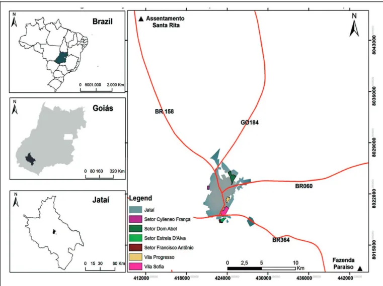
The study site was the municipality of Jataí, located in southwestern part of the state with a distance of 320km from
the capital (Goiânia) and a population of 97,077 individuals (Figure 1). Using a seroepidemiological survey design, the levels of anti-hantavirus immunoglobulin G (IgG) and immunoglobulin M (IgM) were investigated and a questionnaire was administered to the participants. Part of the survey involved epidemiological data related to risk factors such as occupational activity, place of residence and recreational activities such as leisure in the rural area.
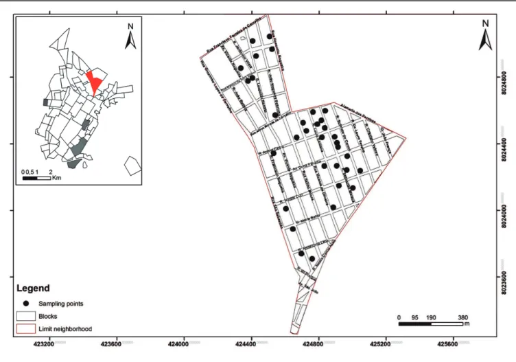
The households drawn to participate in this study, from an urban plan map, were visited from August 2012 to February 2015. Individuals who agreed to participate in the study donated whole blood, collected through a sterile disposable micro lance from the digital pulp and the blood was soaked in paper strips (Nobuto Blood Sampling Paper) of high-quality. After the collection, georeferencing was carried out through the software ArcGis10, for the points where the serological research was carried out (Figure 2).
The ilter paper strips soaked with the biological samples
were stored at a low temperature in the Virology laboratory of
Federal University of Goiás [Universidade Federal de Goiás
(UFG)] in Jataí. Subsequently, they were diluted in phosphate-buffered saline (PBS) buffer and processed by the in-house
enzyme-linked immunosorbent assay (ELISA) test6.
Briefly, a 96-well, flat-bottom ELISA microplate [PolySorp™, Nunc, Chicago, IL, United States of America (USA)] was sensitized at the top (wells A, B, C, and D of rows one to 12) with the recombinant N protein of Araraquara virus (antigen positive). It was also sensitized at the bottom part (wells E, F, G, and H of rows 1 to 12), with Escherichia coli
extract (antigen negative), both diluted in 0.05M carbonate
Moreli ML et al. - Diagnosis for hantavirus in Jataí County
FIGURE 1 - Serological analysis for hantavirus infection in Jataí-GO. The study region is shown with its geographic location in the country. Organization: Martins, Alécio Perini (2016).
and the addition of the antigen (100μl/well), the plates were
incubated in a moist chamber inside a refrigerator at 4°C for 18 hours (overnight). Thereafter, the wash solution (PBS 1X
with tween (T) −20) was used in all phases, except after the addition of the chromogen in the i nal phase. The microplate
was then blocked with skimmed milk powder (SMP, Molico Milk Nestlé®) and remained incubated in humid chambers for 2 hours at 37°C. Subsequently, the test sample, which was diluted 1: 100 in PBS-T and SMP, was added. The next step included adding the conjugate (goat anti-human IgG peroxidase, diluted 1: 2000 in PBS-T and SMP). Finally, the colorimetric substrate used was tetramethylbenzidine (TMB) (Sigma-Aldrich®, St. Louis, MO, USA) and the Stop solution was 1M hydrochloric acid (HCl). The absorbance was measured on an ELISA reader
(Thermoplate) with a primary i lter of 450nm and the secondary i lter of 630 nm. The cut off was determined by the mean optical
densities (OD) of the negative controls plus three standard deviations6.
STATA IC/64 software (version 13.1, College Station,
TX) was used for data analysis. Therefore, for the measure of
association, prevalence ratio (PR) was reported in the statistical
analysis with coni dence interval (CI) of 95%. The study was
approved by the research ethics committee involving human
beings of the Federal University of Goiás (n° 348/2010) and
participants provided written informed consent.
A total of 429 samples were screened by the in-house ELISA method. Of these samples, 52% belonged to males, 84% came from the urban area and the average age of participants was 36.2 years. The seropositivity of anti-hantavirus IgG in the Jataí
population was 3.9% (17/429; 95% CI: 2.3-5.7), with higher seroprevalence for men (5%, 12/238) than for women (2.6%, 5/191) (Table 1). However, no signii cant difference was found
between the seropositivity by gender (PR: 1.9; 95% CI 0.7-5.0), the participants living in rural/urban areas (PR: 1.1; 95% CI 0.3-3.8) and age of the participants (PR: 0.7; 95% CI 0.3-2).
Despite numerous hantavirosis-related mortalities in the county, the participants vaguely remembered symptoms that are suggestive of HCPS, such as intense fever, respiratory problems, chest pain, and headache2,7. Hence, according to Figueiredo
FIGURE 2 - Sector model used in the epidemiological survey. The black points indicate the homes where samples were collected. Organization: Martins, Alécio Perini (2016).
TABLE 1
Information obtained from participants with seropositive hantavirus infection.
Sample Sex Age Residence Contact with
rodent
OD Antibodies Titration
01 M 44 Peri-urban Yes 1,7 IgG 1/3,200
02 M 29 Rural Yes 1,9 IgM/IgG 1/3,200
03 M 32 Rural Yes 1,6 IgM/IgG 1/1,600
04 M 39 Peri-urban Yes 0,69 IgG 1/200
05 M 10 Peri-urban - 0,5 IgG 1/100
06 M 44 Peri-urban Yes 1,0/0,6 IgM/IgG 1/1,800;1/100
07 F 55 Peri-urban Yes 0,45 IgG 1/100
08 M 6 Peri-urban - 0,65 IgG 1/100
09 M 43 Peri-urban Yes 0,54 IgG 1/100
10 F 65 Peri-urban Yes 0,61 IgG 1/100
11 F 12 Peri-urban - 0,75 IgG 1/100
12 M 33 Peri-urban Yes 1,0 IgG 1/200
13 F 34 Peri-urban Yes 0,6 IgG 1/100
14 M 12 Rural - 0,69 IgG 1/100
15 F 25 Peri-urban Yes 0,64 IgG 1/100
16 M 53 Peri-urban Yes 0,99 IgG 1/200
17 M 20 Peri-urban Yes 0,62 IgG 1/100
to only a small part of the totality. Most of the infections could
have been diagnosed as non-speciic acute febrile illnesses such as inluenza A (H1N1), leptospirosis and dengue. Therefore,
owing to the inability of most participants to recall prior clinical presentations and because they also present with antibodies
typical of the chronic phase, it is not possible to conirm the
existence of subclinical infections.
Among the samples included for the serological investigation were samples belonging to two male participants aged 29 and 32 years with IgM anti-hantavirus antibodies. The infection in the two men evolved into the severe form of the disease (HCPS), with the occurrence of death in the younger patient9. The third
IgM reactive case, another man aged 44 years, presented with a disease similar to dengue, but tested negative for the dengue virus. In this case, the diagnosis occurred retrospectively, thereby reinforcing the concern of underreporting of hantavirosis among suspected dengue cases, since the clinical management of the two diseases differs.
The seroprevalence of anti-hantavirus IgG antibodies detected in this study is higher than that reported in the serological survey conducted by Souza et al.10 Despite using
similar techniques, they observed 1.97% seroprevalence in the samples from the urban and rural areas of four municipalities of the Santa Catarina State. In another study conducted in cities near the border between Brazil and Argentina, still in the State
of Santa Catarina, they found a 3.5% seroprevalence11. The
highest reported level of infection with an index of 4.7% was found in the City of Cássia dos Coqueiros, located in the State of São Paulo, in the Southeast region12. It is important to point
out that most of the HCPS cases occurred in the Southeast of the country, along with the South region.
Recently, in the study conducted by Vieira et al.13, high
anti-hantavirus IgG seroprevalence of 13.6% was shown in the City of Sinop in the Mato Grosso State. The study site may justify this in part because the state has the second highest number of HCPS cases nationwide. Vieira et al. also reported higher seropositivity among women from urban areas, differing from our data with a higher seropositivity among men (71%) and from rural areas (4.4%). It is known that hantavirosis is considered primarily a rural disease and, consequently, rural dwellers as well as individuals who are often in contact with the rural area comprise a risk group. Therefore, there are numerous studies from other areas of Brazil demonstrating considerable serological prevalence for hantavirus in men living in rural
areas, a fact that seem to link hantavirus infection and
ield-related activities7,14.
Although the risk group for hantavirus is comprised mainly of men in their productive years (aged 20 to 39 years) residing in the rural areas5, nevertheless, serological surveys for hantavirus
should also be carried out in the urban areas. Residents of urban
areas also have links to the ield, through work or ield-related
activities which might increase the risk of exposure to rodent reservoirs11,13. Thus, when the questionnaire was administered,
a majority of the population expressed having experienced situations considered to enhance the risk for hantavirus infection, including rural tourism (74%) and exposure to house
cleaning in the countryside, sheds, and other similarities (71%). Direct and indirect contact with rodents was also common (81%). Hence, it would be interesting to develop a standardized questionnaire, with a score involving risk activities, since the frequency of performing these activities could better correlate with the risks of viral infection.
In epidemiological studies in Brazil, the Araraquara Hantavirus genotype was detected in the rodent Necromys lasiurus, being found in the Cerrado, a predominant biome in the state4. In this
context, in a study carried out in Ribeirão Preto-São Paulo and Minas Gerais State, near the State of Goiás where sequenced genetic material were collected in patients during the acute phase of infection, showed a high homology with the N gene and partial S segment sequences from the Araraquara virus15,16. Thus, it is
possible that the hantavirus-related morbidities occurring at these sites are caused by this virus, however, it is necessary to carry out
viral identiication in the future owing to the possibility of other
circulating genotypes in this region.
During the course of the study, efforts aimed to raise awareness among the population, through the dissemination of information regarding hantavirosis. Hence, self-explanatory folders on the disease were provided as information regarding symptoms, means of transmission and prevention measures. Our results suggest that most of the participants were unaware of the disease (~ 80%). This reinforces the need for public health measures aimed at raising awareness about the risks of hantavirus. In addition, epidemiological data from the present study provides insights for future studies using molecular
biology tools for the identiication of hantavirus associated with
HCPS and other antigenically related viruses in this region.
Acknowledgments
The authors gratefully acknowledge the medical team and the laboratory staff of Laboratório Elzevir Ferreira Lima, the technical team of Núcleo de Vigilância Epidemiológica e Ambiental em Saúde, Jataí-GO.
Conlict of interest
The authors declare that there is no conlict of interest.
Financial Support
Fundação de Amparo à Pesquisa do Estado de Goiás (FAPEG) research grant
call notice PPSUS n° 004/2010.
REFERENCES
1. Mackow ER, Gavrilovskaya IN. Cellular receptors and hantavirus
pathogenesis. Curr Top Microbiol Immunol. 2001;256:91-115.
2. Hjelle B, Torres-Pérez F. Hantaviruses in the americas and their role
as emerging pathogens. Viruses. 2010;2(12):2559-86.
3. Alfadhli A, Love Z, Arvidson B, Seeds J, Willey J, Barklis E. Hantavirus nucleocapsid protein oligomerization. J Virol.
2001;75(4):2019-23.
4. Suzuki A, Bisordi I, Levis S, Garcia J, Pereira LE, Souza RP, et al. Identifying rodent hantavirus reservoirs, Brazil. Emerg Infect Dis.
2004;10(12):2127-34.
5. Ministério da Saúde. Casos conirmados de Hantavirose: Brasil, Grandes Regiões e Unidades Federadas de 1993-2016 (Internet). Brasília: Secretaria de Vigilância em Saúde. Available from:
http://portalsaude.saude.gov.br/index.php/o-ministerio/principal/ leia-mais-o-ministerio/708-secretaria-svs/vigilancia-de-a-a-z/ hantavirose/11304-situacao-epidemiologica-dados. 2017.
6. Figueiredo LTM, Moreli ML, Borges AA, de Figueiredo GG, Badra SJ, Bisordi I, et al. Evaluation of an enzyme-linked immunosorbent assay based on Araraquara virus recombinant nucleocapsid protein.
Am J Trop Med Hyg. 2009;81(2):273-6.
7. Limongi JE, da Costa FC, Pinto RMC, de Oliveira RC, Bragagnolo C, Lemos ERS, et al. Cross-sectional survey of Hantavirus infection,
Brazil. Emerg Infect Dis. 2009; 15(12):1981-83.
8. Figueiredo LT, Souza WM, Ferrés M, Enria DA. Hantaviruses and cardiopulmonary syndrome in South America. Virus Res.
2014;187:43-54.
9. Moreli ML, da Costa VG, Novaes DPS, Flor EC, Silva JF, Vilela KRG, et al. Hantavirus cardiopulmonary syndrome: a report of two
cases. J Bras Patol Med Lab. 2013;49(5):312-6.
10. Souza WM, Machado AM, Disner GR, Boff E, Machado ARSR, de Padua M, et al. Antibody levels to hantavirus in inhabitants of western Santa Catarina state, Brazil. Rev Inst Med Trop São Paulo.
2012;54(4):193-6.
11. Souza WM, Machado AM, Figueiredo LTM, Boff E. Serosurvey of hantavirus infection in humans in the border region between Brazil
and Argentina. Rev Soc Bras Med Trop. 2011;44(2):131-5.
12. Badra SJ, Maia FGM, Figueiredo GG, Santos Júnior GS, Campos GM, Figueiredo LTM, et al. A retrospective sorologic survey of hantavirus infections in the county of Cássia dos Coqueiros, State
of São Paulo, Brazil. Rev Soc Bras Med Trop. 2012;45(4):468-70.
13. Vieira CJSP, da Silva DJF, Barreto ES, Siqueira CEH, da Costa VG, Lourenço FJ, et al. Serological evidence of hantavirus infection in an urban area in Mato Grosso State, Brazil. Rev Soc Bras Med Trop.
2016;49(3):348-50.
14. Terças ACP, Atanaka dos Santos M, Pignatti MG, Espinosa MM, de Melo Via AVG, Menegatti JA. Hantavirus pulmonary syndrome outbreak, Brazil, December 2009-January 2010. Emerg Infect Dis.
2013;19(11):1824-7.
15. Moreli ML, Sousa RL, Figueiredo LTM. Detection of Brazilian hantavirus by reverse transcription polymerase chain reaction
ampliication of N gene in patients with hantavirus cardiopulmonary syndrome. Mem Inst Oswaldo Cruz. 2004;99(6):633-8.
16. Limongi JE, Oliveira RC, Guterres A, Costa Neto SF, Fernandes J, Vicente LH, et al. Hantavirus pulmonary syndrome and rodent reservoirs in the savanna-like biome of Brazil’s southeastern region.