Clinical Value of High Mobility Group Box 1 and
the Receptor for Advanced Glycation
End-products in Head and Neck Cancer: A Systematic
Review
Austin Nguyen
1Sheila Bhavsar
1Erinn Riley
1Gabriel Caponetti
2Devendra Agrawal
11Department of Clinical and Translational Science, Creighton University School of Medicine, Omaha, Nebraska, United States 2Department of Pathology, Creighton University School of Medicine,
Omaha, Nebraska, United States
Int Arch Otorhinolaryngol 2016;20:382–389.
Address for correspondenceDevendra Agrawal, PhD, Department of Clinical and Translational Science, Creighton University School of Medicine, 2500 California Plaza, Omaha, NE 68172, United States (e-mail: [email protected]).
Introduction
The high mobility group box 1 protein (HMGB1) is a highly conserved and highly versatile nuclear and extracellular protein found in eukaryotic cells. HMGB1 is a non-histone, chromosomal protein that has been implicated in a variety of biologically important processes, including transcription, DNA repair, and extracellular signaling.1 Structurally, HMGB1 consists of 215-amino acid polypeptide organized into two DNA-binding domains (termed A-box and B-box)
and a negatively charged C-terminal tail.2,3 Functionally, HMGB1 appears to have two distinct roles in cellular systems. In the intracellular milieu, HMGB1 localizes to the nucleus and non-specifically binds to the minor groove of DNA, facilitates the assembly of DNA-binding proteins, and is involved in regulating gene transcription.4,5
Following release into the extracellular space, HMGB1 acts as a pro-inflammatory cytokine that is secreted by activated monocytes, macrophages, and natural killer (NK) cells. It is also passively secreted by necrotic cells and is, therefore, Keywords
►
HMGB1 protein
►
advanced
glycosylation end
product-speci
fi
c
receptor
►
head and neck
neoplasms
Abstract
Introduction
High mobility group box 1 is a versatile protein involved in gene
transcription, extracellular signaling, and response to in
fl
ammation. Extracellularly,
high mobility group box 1 binds to several receptors, notably the receptor for advanced
glycation end-products. Expression of high mobility group box 1 and the receptor for
advanced glycation end-products has been described in many cancers.
Objectives
To systematically review the available literature using PubMed and Web of
Science to evaluate the clinical value of high mobility group box 1 and the receptor for
advanced glycation end-products in head and neck squamous cell carcinomas.
Data synthesis
A total of eleven studies were included in this review. High mobility
group box 1 overexpression is associated with poor prognosis and many clinical and
pathological characteristics of head and neck squamous cell carcinomas patients.
Additionally, the receptor for advanced glycation end-products demonstrates potential
value as a clinical indicator of tumor angiogenesis and advanced staging. In diagnosis,
high mobility group box 1 demonstrates low sensitivity.
Conclusion
High mobility group box 1 and the receptor for advanced glycation
end-products are associated with clinical and pathological characteristics of head and neck
squamous cell carcinomas. Further investigation of the prognostic and diagnostic value
of these molecules is warranted.
received
January 20, 2016
accepted
February 28, 2016
published online
April 19, 2016
DOI http://dx.doi.org/ 10.1055/s-0036-1583168.
ISSN 1809-9777.
Copyright © 2016 by Thieme Publicações Ltda, Rio de Janeiro, Brazil
referred as an optimal marker of necrosis.6,7It is involved in the mediation of neurite outgrowth, smooth muscle cell chemotaxis, mesoangioblast migration and proliferation, and tumor growth and metastasis.8–10 The extracellular HMGB1 binds to several cell surface receptors, including the Receptor for Advanced Glycation End-products (RAGE) and the Triggering Receptor Expressed on Myeloid cells 1 (TREM-1).11,12
RAGE is a transmembrane protein that belongs to the immunoglobulin superfamily. It can bind to advanced glycation end-products, the resulting product of non-enzymatic glycation. RAGE is mostly stimulated by cellular stress, such as inflammation and is therefore found to be overexpressed in many diseases, such as different cancers.13In certain cells, HMGB1 and its receptor, RAGE, co-localize on the cell surface. Authors have reported that HMGB1 binding to RAGE may activate signaling pathways, such as Ras/MAKP, PI3K/Akt, NF-kB, which leads to overexpression of genes and the change in the biological
behavior of tumor cells.14Previous studies have found that HMGB1 and RAGE play important roles in the development, growth, and metastasis of multiple tumors.15,16
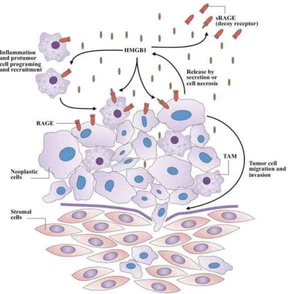
The RAGE-HMGB1 interaction can diminish host anticancer immunity by inducing apoptosis in antigen-presenting dendritic cells and reprogram immune cells by promoting tumor-infiltrating T cell–expressed lympho-toxin a1b2, which leads to the recruitment of CD11bþ
F4/ 80þ
macrophages (tumor-associated macrophages, TAMs) into the tumor for its promotion (►Fig. 1) by providing growth factors and supporting angiogenesis.17 Further-more, HMGB1 released from necrotic cancer cells treated with chemotherapy enhances regrowth and metastasis of remnant cancer cells in a RAGE-dependent manner.18,19 Therefore, blocking the HMGB1–RAGE system may increase the effectiveness of chemotherapy.17 In fact, specialists have used soluble RAGE (sRAGE) to prevent the HMGB1-RAGE signaling from occurring in animal tumor models by acting as a decoy receptor.20
In comparison to RAGE, HMGB1 has been more extensively studied in various cancers. HMGB1 expression appears to be associated with many different tumor types.21–24In addition, the association of HMGB1 overexpression and poor prognosis has been reported in patients with various types of cancers.25–29 To our knowledge, however, a systematic evaluation of the clinical value of HMGB1 and RAGE in head and neck squamous cell carcinoma (HNSCC) has not been performed to date.
HNSCC, which consists of several different subtypes, is the sixth most common malignancy in the world and the most common cause of cancer-related death in South Asia.30Despite aggressive treatment approaches, prognosis of patients with HNSCC is quite poor. Due to the poor outcome, it is of particularly high interest to identify any additional biomarkers that allow early detection or implicate prognosis of HNSCC. In this article, we systematically review the available literature regarding HMGB1 and RAGE in HNSCC and evaluate their potential clinical value.
Review of Literature
We performed a systematic search of the PubMed Database (through July 2015) of the National Library of Medicine using
the following Medical Subject Headings: HMGB1 protein, advanced glycation end-product receptor, neoplasm, and head and neck neoplasms. The articles included discussed original research on the clinical value of HMGB1 or RAGE, including prognostic and diagnostic concordance, in head and neck neoplasms. We cross-referenced the search results with the Web of Science database, using the same terms. We excluded articles based on the following criteria: not written in English, conference abstract, and not performed on pri-mary human subjects/specimens.
An initial search of PubMed and Web of Science retrieved 421 articles (►Fig. 2). After reviewing titles and abstracts, excluding non-English articles and conference abstracts, 16 studies remained as potential candidates for inclusion. Full text review excludedfive studies due to presentation of data irrelevant to the present topics. Ultimately, 11 studies were included in this systematic review, encompassing a total of 2,098 patients (►Table 1). Examination of HMGB1 and/or RAGE was predominantly performed in sectioned tissue, whether frozen (one study) or formalin-fixed and embedded in paraffin (7 studies). Four studies investigated expression levels in serum/blood. Studies in the squamous cell carcinoma (SCC) investigated the following malignancy types: general
head and neck SCC (HNSCC, 2 studies), oral SCC (OSCC, 6 studies, including one study specifically on tongue SCC), nasopharyngeal carcinoma (2 studies), and SCC of the larynx (LSCC, 1 study). Of these,five studies indicated their included patients to be treatment naive. The study populations were between 50.0% and 96.3% male, with a mean/median age of 47.7 to 69.1 years.
The Receptor for Advanced Glycation End-products Four studies evaluated RAGE in head and neck cancer patients31–34(
►Table 2). The earliest study to be included31 analyzed samples from 20 patients with OSCC. The study compared expression of RAGE with angiogenesis and lymphan-giogenesis, evaluated by microvessel density and lymph vessel density. A significant positive correlation was found between RAGE concentration and microvessel density (p¼0.0123), but not between RAGE and lymph vessel density. Additionally, RAGE expression was significantly associated with concentration of vascular endothelial growth factor (VEGF;p¼0.0344). The only clinic pathological factor to be significantly associated with RAGE expression was T-classification, with an elevated level seen in T3-T4 classifications compared with T1-T2 (p¼0.0408).
In a study involving 38 OSCC cases, Landesberg et al32 observed RAGE to be associated with tumor differentiation (p<0.05). RAGE positivity was detected in 100% of well-differentiated OSCC, 75% of well-to-moderately well-differentiated OSCC, 33% of moderately differentiated OSCC, 14% of moder-ate-to-poorly differentiated OSCC, and 0% of poorly differen-tiated OSCC. Additionally, RAGE expression trended toward decreasing staining intensity with less tumor differentiation.
The most recent and largest study34 included 618 OSCC patients. Blood was collected and analyzed for RAGE gene polymorphism. Five polymorphisms of the RAGE gene were assessed: 374T>A (rs1800624), 429T>C (rs1800625), 1704G>T (rs184003), Gly82Ser (rs2070600), and a 63-bp deletion allele ( 407 to 345). The heterozygous 429T>C genotype was found to be significantly associated with an increased incidence of oral cancer [odds ratio (OR), 1.870; 95% confidence interval (CI), 1.323 to 2.644], stage III/IV tumors (OR, 1.736; 95% CI, 1.126 to 2.677), and large tumor size (OR, 1.644; 95% CI, 1.083 to 2.493). None of the other poly-morphisms were found to be significantly associated with incidence or clinical status of OSCC.
Lastly, the clinical value of RAGE was also evaluated in nasopharyngeal carcinoma.33Analysis of 42 nasopharyngeal carcinoma specimens demonstrated expression of RAGE to be associated with tumor microvessel density (p¼0.0049). Additionally, RAGE expression was significantly higher in cases with lymph node metastasis (N1–3 classification), when compared with that of lymph node metastasis-negative (N0) cases (p¼0.0005).
Evaluation of High Mobility Group Box 1
We found seven studies that investigated the clinical role of HMGB1 in head and neck cancers35–41(
►Table 2). Of these, only one study assessed the diagnostic value of HMGB1.39Using the cutoff of 4.80 ng/mL, serum HMGB1 was 42.0% sensitive and 88.3% specific (positive predictive value of 52.9%) for the diag-nosis of laryngeal SCC. However, high serum HMGB1 was significantly associated with T classification (p¼0.005), N Table 1 Study design and population characteristics
Study Malignancy Sample type N (total)
N (cancer)
Age,
mean/median (range)
Sex, M:F
Patient notes
Sasahira et al31 OSCC FFPE tissue 20 20 69.1 (41–83) 10:10
Sasahira et al35 OSCC FFPE tissue 62 62 35:27 Treatment naive
Wu et al36 NPC FFPE tissue 166 166 47.7 (13–74) 133:33 Treatment naive
Landesberg et al32 OSCC FFPE tissue 50 38 A: 69 (28–92). B: 72 (51–88). C: 66 (55–88).
A: 4:7. B: 7:7. C: 6:2
A: Well-differentiated tumor B: Moderately-differentiated
tumor
C: Poorly-differentiated tumor
Tsuji et al33 NPC FFPE tissue 42 42
Liu et al37 HNSCC FFPE tissue 103 103 57.86 (27–80) 99:4
Wild et al38 HNSCC Serum and frozen tissue
52 35 6410
29:6
Treatment naive
Qiu et al39 LSCC Serum 121 71 54.514.0
54:17
Treatment naive
Hanakawa et al40 Tongue SCC FFPE tissue 26 26 69 (29–87) 15:11 Treatment naive Supic et al41 OSCC Tumor tissue
(unspecified) or blood (controls)
246 93 58 (36–80) 69:24 All received primary surgery followed by radiotherapy. 24 received neoadjuvant cisplatin/5-fluorouracil chemotherapy.
Su et al34 OSCC Blood 1210 618 54.2911.28
596:22
Abbreviations: FFPE, formalin-fixed paraffin-embedded; HNSCC, head and neck squamous cell carcinoma; LSCC, laryngeal squamous cell carcinoma; NPC, nasopharyngeal carcinoma; OSCC, oral squamous cell carcinoma; SCC, squamous cell carcinoma.
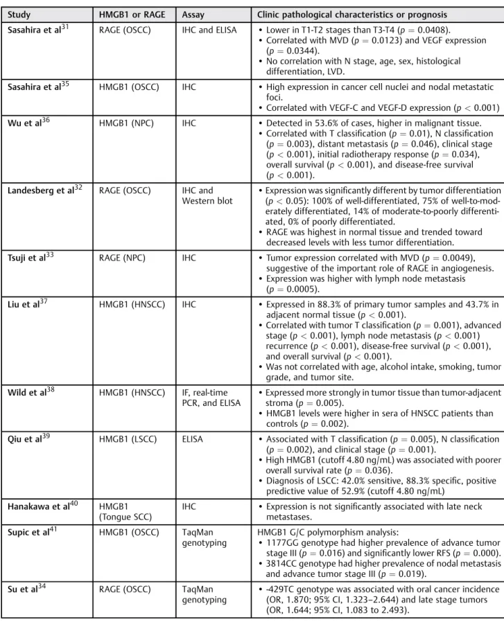
Table 2 Clinical value of HMGB1 and RAGE
Study HMGB1 or RAGE Assay Clinic pathological characteristics or prognosis
Sasahira et al31 RAGE (OSCC) IHC and ELISA •Lower in T1-T2 stages than T3-T4 (p¼0.0408).
•Correlated with MVD (p¼0.0123) and VEGF expression (p¼0.0344).
•No correlation with N stage, age, sex, histological
differentiation, LVD.
Sasahira et al35 HMGB1 (OSCC) IHC •High expression in cancer cell nuclei and nodal metastatic
foci.
•Correlated with VEGF-C and VEGF-D expression (p<0.001)
Wu et al36 HMGB1 (NPC) IHC •Detected in 53.6% of cases, higher in malignant tissue. •Correlated with T classification (p¼0.01), N classification
(p¼0.003), distant metastasis (p¼0.046), clinical stage (p<0.001), initial radiotherapy response (p¼0.034), overall survival (p<0.001), and disease-free survival
(p<0.001).
Landesberg et al32 RAGE (OSCC) IHC and
Western blot •
Expression was significantly different by tumor differentiation (p<0.05): 100% of well-differentiated, 75% of
well-to-mod-erately differentiated, 14% of moderate-to-poorly differenti-ated, 0% of poorly differentiated.
•RAGE was highest in normal tissue and trended toward
decreased levels with less tumor differentiation.
Tsuji et al33 RAGE (NPC) IHC •Tumor expression correlated with MVD (p¼0.0049), suggestive of the important role of RAGE in angiogenesis.
•Expression was higher with lymph node metastasis
(p¼0.0005).
Liu et al37 HMGB1 (HNSCC) IHC •Expressed in 88.3% of primary tumor samples and 43.7% in
adjacent normal tissue (p<0.001).
•Correlated with tumor T classification (p¼0.001), advanced stage (p<0.001), lymph node metastasis (p<0.001)
recurrence (p<0.001), disease-free survival (p<0.001),
and overall survival (p<0.001).
•Was not correlated with age, alcohol intake, smoking, tumor
grade, and tumor site.
Wild et al38 HMGB1 (HNSCC) IF, real-time PCR, and ELISA
•Expressed more strongly in tumor tissue than tumor-adjacent
stroma (p¼0.005).
•HMGB1 levels were higher in sera of HNSCC patients than
controls (p¼0.002).
Qiu et al39 HMGB1 (LSCC) ELISA •Associated with T classification (p¼0.005), N classification (p¼0.002), and clinical stage (p¼0.001).
•High HMGB1 (cutoff 4.80 ng/mL) was associated with poorer
overall survival rate (p¼0.036).
•Diagnosis of LSCC: 42.0% sensitive, 88.3% specific, positive
predictive value of 52.9% (cutoff 4.80 ng/mL)
Hanakawa et al40 HMGB1 (Tongue SCC)
IHC •Expression is not significantly associated with late neck
metastases.
Supic et al41 HMGB1 (OSCC) TaqMan genotyping
HMGB1 G/C polymorphism analysis:
•1177GG genotype had higher prevalence of advance tumor
stage III (p¼0.016) and significantly lower RFS (p¼0.000).
•3814CC genotype had higher prevalence of nodal metastasis
and advance tumor stage III (p¼0.019).
Su et al34 RAGE (OSCC) TaqMan
genotyping
•-429TC genotype was associated with oral cancer incidence
(OR, 1.870; 95% CI, 1.323–2.644) and late stage tumors
(OR, 1.644; 95% CI, 1.083 to 2.493).
classification (p¼0.002), clinical stage (p¼0.001), and poorer overall survival rate (p¼0.036). The study found no association between serum HMB1 level and gender (p¼0.814), age (p¼0.174), tumor site (p¼0.733), and degree of differentiation (p¼0.727). Wild et al38performed the only other evaluation of serum HGMB1 levels. Sera of 35 HNSCC patients contained significantly higher concentrations of HMGB1 compared with that of controls (p¼0.002). HMGB1 levels of patients’sera were also analyzed against clinic pathological characteristics of patients (including cancer location, age, sex, therapy, staging, and grading), but they found no significant associations. Further analysis of frozen sections from HNSCC patients demonstrated stronger HMGB1 expression in tumor islands than in tumor-surrounding stroma (p¼0.005).
Three studies35,40,41 investigated the role of HMGB1 in OSCC. Sasahira et al35 analyzed 62 specimens of primary OSCC. HMGB1 was found to be highly expressed in cancer cell nuclei and nodal metastatic foci. Comparison with expression of VEGF-C and VEGF-D demonstrated a significant correlation (p<0.001). Interestingly, analysis of HMGB1 levels in 26 specimens from patients with tongue SCC40 demonstrated no significant association between high HMGB1 expression and late neck metastases. Supic et al41explored the role of polymorphisms in the HMGB1 gene in OSCC. Using TaqMan genotyping assays, they analyzed four single-nucleotide poly-morphisms within the HMGB1 gene in 246 patients: 2262G
>A (rs1045411), 1177G>C (rs3742305), 3814C>G (rs2249825), and rs4540927. The 1177G>C polymorphism was associated with longer recurrence-free survival (p¼0.000), but not overall survival (though a trend was noted of worse survival with the GG phenotype,p¼0.104). Additionally, the 1177G>C variation was significantly asso-ciated higher histological nucleus grade (wild-type versus combined heterozygote and variant homozygote genotype, p¼0.010), nodal metastasis (p¼0.016), and higher clinical stage (p¼0.030). Additionally, the 3814C>G variant was also significantly associated with nodal metastasis (wild-type versus combined heterozygote and variant homozygote genotype, p¼0.019), and higher clinical stage (wild-type versus combined heterozygote and variant homozygote genotype,p¼0.042).
One study analyzed the role of HMGB1 in 166 nasopharyn-geal carcinoma specimens from treatment-naive patients.36 HMGB1 was detected in 53.6% of the cases, primarily located in the nuclei and cytoplasm of carcinoma cells and a subset of fibroblasts. HMGB1 was positively correlated with T classifi ca-tion (p¼0.01), N classification (p¼0.003), distant metastasis (p¼0.046), and clinical stage (p<0.001). Additionally, HMGB1 expression correlated significantly with initial radiotherapy response in NPC patients (p¼0.034). Prognostically, high HMGB1 expression was associated with a poor overall survival (p<0.001) and disease-free survival (p<0.001). No significant correlation was found between HMGB1 expression and gender, age, pathological classification, or local relapse.
Liu et al37studied 103 specimens from treatment-naive patients with HNSCC. Tumors were located at superglottic (N¼29), glottic (N¼63), subglottic (N¼1), and hypophar-yngeal (N¼10) sites. HMGB1 protein was detected in 88.3%
of cases, but only 43.75% of normal tumor-adjacent tissue controls (p<0.001), with higher expression (staining graded as 6–7 out of 7) in 43.7% of tumor samples. HMGB1 over-expression was significantly associated with advanced clini-cal stage, including larger tumor size (p¼0.001) and lymph node metastasis (p<0.001), and recurrence (p<0.001). No significant relationship was observed between HMGB1 level and age, alcohol history, smoking, tumor grade, and tumor site. Additionally, high HMGB1 expression was associated with shorter disease-free survival (p<0.001) and overall survival (p<0.001). HMGB1 was found to be an independent prognostic factor in HNSCC patients after tumor resection (hazard ratio, 2.133; 95% CI, 1.079–4.218). Further analysis of prognostic value of HMGB1 in patient subgroups demon-strated a high level of HMGB1 expression to be associated with shorter disease-free survival (p¼0.005) and overall survival (p<0.001) in late stage group (stage III / IV) patients.
Discussion
Overall, there was high variation in the included studies, includ-ing sample size (range, 20–1210), presence of controls (range, 0 to 1:1 ratio), and methodology. The majority of studies included treatment-naive patients, with the exception of one study,41in which neoadjuvant therapy was administered to a fraction of the included patients. Additionally, the total number of investiga-tions is a current limiting factor in determining the true clinical values of HGMB1 and RAGE.
We found no studies that investigate the diagnostic poten-tial of RAGE, while only one study39evaluated the diagnostic value of HMGB1. The low sensitivity of HMGB1 in the diagnosis of LSCC potentially makes HMGB1 a suboptimal marker. However, this study was limited in power (N¼121) and by the small sample size of early-stage LSCC patients. Additionally, the multifaceted role of HMB1 and its broad expression likely contribute to its low diagnostic value. Further studies are required to elucidate the diagnostic value of both RAGE and HMGB1, including the possibility of use in conjunction with other markers to improve diagnostic ability. A total of four studies31–34 investigated the prognostic value of RAGE in head and neck cancers (OSCC, N¼3; nasopharyngeal carcinoma,N¼1). The interaction between HMGB1 and RAGE has been shown to stimulate tumor cell migration and invasion.23 Interestingly, Wild et al38 found tumor-infiltrating Tregs to express RAGE as well. Further in vitro analysis demonstrated HMGB1 to induce migration of Treg cells isolated from PBMC of patients with HSNCC. This indicates a possible role of tumor-derived HMGB1 in recruit-ment of Treg cells and, ultimately, suppression of T cell proliferation within the tumor microenvironment.
analysis on polymorphisms in the RAGE gene34may indicate the variants to be involved in tumor cell proliferation but not invasion and differentiation. Perhaps some potential exists for the polymorphisms to be used in assessment of risk. Larger studies are required to determine if RAGE is a clinically useful predictor of survival, and further confirm its prognostic value.
Substantial evidence support the association of HMGB1 with clinic pathological characteristics of patients with head and neck cancers, including tumor angiogenesis and staging. Three of the seven studies investigating HMGB1 found high protein expression to be significantly associated with lower overall survival.36,37,39Interestingly, Wu et al36found high HMGB1 in late-stage nasopharyngeal carcinoma patients (i.e., stages III / IV), to be associated with significantly shorter overall survival when compared with patients with a low level of HMGB1 expression. Conversely, Hanakawa et al40 found HMGB1 to not be significantly associated with late neck metastasis in tongue SCC. This is suggestive of HMGB1 possibly being more valuable in determining the prognosis for late-stage patients.
Furthermore, use of HMGB1 in combination with other clinic pathological indicators could improve the prognostic utility. Liu et al37found the subset of patients with both high HMGB1 expression and positive lymph node metastasis had an even poorer disease-free survival (p<0.001) and overall survival (p<0.001) than that of others.
Final Comments
The diagnostic value of HMGB1 in head and neck cancer, while not exhaustively investigated, may suffer from low sensitivity. No studies were found to evaluate the diagnos-tic value of RAGE. The demonstrated associations of HMGB1 and RAGE with clinic pathological characteristics of head and neck cancer patients offer a strong basis for further investigation into the prognostic value of both of these molecules. Early investigation has demonstrated HMGB1 to have definite potential as a predictor for survival, while RAGE has not been as extensively evaluated. Further studies are needed to confirm the clinical prognostic utility of both RAGE and HMGB1.
Acknowledgment
This work was supported by research grant R01 HL116042 to DK Agrawal from the National Heart, Lung, and Blood Institute, NIH USA.
References
1 Czura CJ, Wang H, Tracey KJ. Dual roles for HMGB1: DNA binding and cytokine. J Endotoxin Res 2001;7(4):315–321
2 Cheng BQ, Jia CQ, Liu CT, et al. Serum high mobility group box chromosomal protein 1 is associated with clinicopathologic features in patients with hepatocellular carcinoma. Dig Liver Dis 2008;40(6):446–452
3 Lee H, Song M, Shin N, et al. Diagnostic significance of serum HMGB1 in colorectal carcinomas. PLoS ONE 2012;7(4):e34318
4 Goodwin GH, Sanders C, Johns EW. A new group of chromatin-associated proteins with a high content of acidic and basic amino acids. Eur J Biochem 1973;38(1):14–19
5 Pallier C, Scaffidi P, Chopineau-Proust S, et al. Association of chromatin proteins high mobility group box (HMGB) 1 and HMGB2 with mitotic chromosomes. Mol Biol Cell 2003;14(8): 3414–3426
6 Lotze MT, Tracey KJ. High-mobility group box 1 protein (HMGB1): nuclear weapon in the immune arsenal. Nat Rev Immunol 2005; 5(4):331–342
7 Zhang Z, Wang M, Zhou L, et al. Increased HMGB1 and cleaved caspase-3 stimulate the proliferation of tumor cells and are correlated with the poor prognosis in colorectal cancer. J Exp Clin Cancer Res 2015;34(1):51–60
8 Evans A, Lennard TW, Davies BR. High-mobility group protein 1 (Y): metastasis-associated or metastasis-inducing? J Surg Oncol 2004;88(2):86–99
9 Palumbo R, Sampaolesi M, De Marchis F, et al. Extracellular HMGB1, a signal of tissue damage, induces mesoangioblast migra-tion and proliferamigra-tion. J Cell Biol 2004;164(3):441–449
10 Wang H, Yang H, Tracey KJ. Extracellular role of HMGB1 in inflammation and sepsis. J Intern Med 2004;255(3):320–331 11 Hori O, Brett J, Slattery T, et al. The receptor for advanced glycation
end products (RAGE) is a cellular binding site for amphoterin. Mediation of neurite outgrowth and co-expression of rage and amphoterin in the developing nervous system. J Biol Chem 1995; 270(43):25752–25761
12 Nguyen AH, Berim IG, Agrawal DK. Chronic inflammation and cancer: emerging roles of triggering receptors expressed on mye-loid cells. Expert Rev Clin Immunol 2015;11(7):849–857 13 Chuah YK, Basir R, Talib H, Tie TH, Nordin N. Receptor for advanced
glycation end products and its involvement in inflammatory diseases. Int J Inflamm 2013;2013:403460
14 Schlueter C, Weber H, Meyer B, et al. Angiogenetic signaling through hypoxia: HMGB1: an angiogenetic switch molecule. Am J Pathol 2005;166(4):1259–1263
15 Srinivasan M, Banerjee S, Palmer A, et al. HMGB1 in hormone-related cancer: a potential therapeutic target. Horm Cancer 2014; 5(3):127–139
16 Diener KR, Al-Dasooqi N, Lousberg EL, Hayball JD. The multifunc-tional alarmin HMGB1 with roles in the pathophysiology of sepsis and cancer. Immunol Cell Biol 2013;91(7):443–450
17 Sasahira T, Sasaki T, Kuniyasu H. Interleukin-15 and transforming growth factor alpha are associated with depletion of tumor-associated macrophages in colon cancer. J Exp Clin Cancer Res 2005;24(1):69–74
18 Luo Y, Chihara Y, Fujimoto K, et al. High mobility group box 1 released from necrotic cells enhances regrowth and metastasis of cancer cells that have survived chemotherapy. Eur J Cancer 2013;49(3):741–751 19 Kang R, Tang D, Schapiro NE, et al. The receptor for advanced glycation end products (RAGE) sustains autophagy and limits apoptosis, promoting pancreatic tumor cell survival. Cell Death Differ 2010;17(4):666–676
20 Taguchi A, Blood DC, del Toro G, et al. Blockade of RAGE-ampho-terin signalling suppresses tumour growth and metastases. Nature 2000;405(6784):354–360
21 Campana L, Bosurgi L, Rovere-Querini P. HMGB1: a two-headed signal regulating tumor progression and immunity. Curr Opin Immunol 2008;20(5):518–523
22 Ellerman JE, Brown CK, de Vera M, et al. Masquerader: high mobility group box-1 and cancer. Clin Cancer Res 2007;13(10): 2836–2848
23 Kang R, Zhang Q, Zeh HJ III, Lotze MT, Tang D. HMGB1 in cancer: good, bad, or both? Clin Cancer Res 2013;19(15):4046–4057 24 Tang D, Kang R, Zeh HJ III, Lotze MT. High-mobility group box 1 and
cancer. Biochim Biophys Acta 2010;1799(1–2):131–140 25 Kuniyasu H, Chihara Y, Kondo H, Ohmori H, Ukai R. Amphoterin
associated with metastatic prostate cancer. Oncol Rep 2003;10(6): 1863–1868
26 Tarbé N, Evtimova V, Burtscher H, Jarsch M, Alves F, Weidle UH. Transcriptional profiling of cell lines derived from an orthotopic pancreatic tumor model reveals metastasis-associated genes. Anticancer Res 2001;21(5):3221–3228
27 Maeda S, Hikiba Y, Shibata W, et al. Essential roles of high-mobility group box 1 in the development of murine colitis and colitis-associated cancer. Biochem Biophys Res Commun 2007;360(2): 394–400
28 Leman ES, Madigan MC, Brünagel G, Takaha N, Coffey DS, Getzen-berg RH. Nuclear matrix localization of high mobility group protein I(Y) in a transgenic mouse model for prostate cancer. J Cell Biochem 2003;88(3):599–608
29 Dolde CE, Mukherjee M, Cho C, Resar LM. HMG-I/Y in human breast cancer cell lines. Breast Cancer Res Treat 2002;71(3): 181–191
30 Siegel RL, Miller KD, Jemal A. Cancer statistics, 2015. CA Cancer J Clin 2015;65(1):5–29
31 Sasahira T, Kirita T, Bhawal UK, et al. The expression of receptor for advanced glycation end products is associated with angiogenesis in human oral squamous cell carcinoma. Virchows Arch 2007; 450(3):287–295
32 Landesberg R, Woo V, Huang L, et al. The expression of the receptor for glycation endproducts (RAGE) in oral squamous cell carcino-mas. Oral Surg Oral Med Oral Pathol Oral Radiol Endod 2008; 105(5):617–624
33 Tsuji A, Wakisaka N, Kondo S, Murono S, Furukawa M, Yoshizaki T. Induction of receptor for advanced glycation end products by EBV
latent membrane protein 1 and its correlation with angiogenesis and cervical lymph node metastasis in nasopharyngeal carcinoma. Clin Cancer Res 2008;14(17):5368–5375
34 Su S, Chien M, Lin C, Chen M, Yang S. RAGE gene polymorphism and environmental factor in the risk of oral cancer. J Dent Res 2015; 94(3):403–411
35 Sasahira T, Kirita T, Oue N, et al. High mobility group box-1-inducible melanoma inhibitory activity is associated with nodal metastasis and lymphangiogenesis in oral squamous cell carcino-ma. Cancer Sci 2008;99(9):1806–1812
36 Wu D, Ding Y, Wang S, Zhang Q, Liu L. Increased expression of high mobility group box 1 (HMGB1) is associated with progression and poor prognosis in human nasopharyngeal carcinoma. J Pathol 2008;216(2):167–175
37 Liu Y, Xie C, Zhang X, et al. Elevated expression of HMGB1 in squamous-cell carcinoma of the head and neck and its clinical significance. Eur J Cancer 2010;46(16):3007–3015
38 Wild CA, Brandau S, LotfiR, et al. HMGB1 is overexpressed in tumor cells and promotes activity of regulatory T cells in patients with head and neck cancer. Oral Oncol 2012;48(5):409–416 39 Qiu G, Li Y, Liu Z, Wang M, Ge J, Bai X. Clinical value of serum
HMGB1 in diagnosis and prognosis of laryngeal squamous cell carcinoma. Med Oncol 2014;31(12):316
40 Hanakawa H, Orita Y, Sato Y, et al. Does HMGB1 predict occult neck lymph node metastasis in early tongue carcinoma? A case-control study of 26 patients. J Laryngol Otol 2014;128(10):926–931 41 Supic G, Kozomara R, Zeljic K, et al. HMGB1 genetic