RESORBABLE EXTRACELLULAR MATRIX GRAFTS IN UROLOGIC
RECONSTRUCTION
RICHARD A. SANTUCCI, THEODORE D. BARBER
Wayne State University School of Medicine, Detroit, Michigan, USA
ABSTRACT
Purpose: There is an increasingly large body of literature concerning tissue-engineering prod-ucts that may be used in urology. Some of these are quite complex (such as multilayer patient-specific cell-seeded implants) yet the most simple and successful products to date are also the most uncompli-cated: resorbable acellular extra-cellular matrices (ECMs) harvested from animals. ECMs have been used in a variety of difficult urologic reconstruction problems, and this review is intended to summa-rize this complex literature for the practicing urologist.
Methods: Medline search of related terms such as “SIS, small intestinal submucosa, ECM, extracellular matrix, acellular matrix and urologic reconstruction”. Manuscripts missed in the initial search were taken from the bibliographies of the primary references.
Results: Full review of potential clinical uses of resorbable extra-cellular matrices in uro-logic reconstruction.
Conclusions: Currently, the “state of the art” in tissue engineering solutions for urologic reconstruction means resorbable acellular xenograft matrices. They show promise when used as a pubovaginal sling or extra bolstering layers in ureteral or urethral repairs, although recent problems with inflammation following 8-ply pubovaginal sling use and failures after 1- and 4-ply SIS repair of Peyronie’s disease underscore the need for research before wide adoption. Preliminary data is mixed concerning the potential for ECM urethral patch graft, and more data is needed before extended uses such as bladder augmentation and ureteral replacement are contemplated. The distant future of ECMs in urology likely will include cell-seeded grafts with the eventual hope of producing “off the shelf” replacement materials. Until that day arrives, ECMs only fulfill some of the requirements for the reconstructive urologist.
Key words: reconstructive surgical procedures; small intestine; intestinal mucosa; extracellular matrix; grafts
Int Braz J Urol. 2005; 31: 192-203
INTRODUCTION
The future of tissue engineering promises nothing less than the manufacture of human replace-ment parts. However, the current reality is that true “replacement part” technology is likely years away, and we must continue to make good with what we have on hand to do urological reconstruction. To this end, resorbable extra-cellular matrix (ECM) graft
of pigs appears to promote the rapid ingrowth of sur-rounding tissue. Other materials have been reported experimentally but not commercially, and still other ECMs are commercially available but are permanent so they differ substantially from ECMs such as SIS (Table-1). The goal of this manuscript is to review the functional characteristics of resorbable graft ma-terials, so that the practicing urologist can understand their potential use in clinical practice.
BENEFITS OF ECM
The modern era of ECM started in the 1980s when SIS was used successfully as a large vessel
re-placement graft in animals (1). The success with these grafts prompted further research and experimental data shows applicability in a variety of animal mod-els: abdominal wall (rodents and dogs), blood ves-sels (dogs), tendon (dogs), skin (rats), dura (rats and dogs) and diaphragm (rats). SIS can support growth of keratinocytes, endothelial cells, smooth muscle cells and bone cells in vitro (2). The potential benefit of ECM is that it recapitulates the tissue it is meant to replace, a process that has been named “smart remod-eling” (2).
Of special note to the urologist is that much animal work using ECM has concentrated on the blad-der, urethra and ureter (see SURGICAL USES, below).
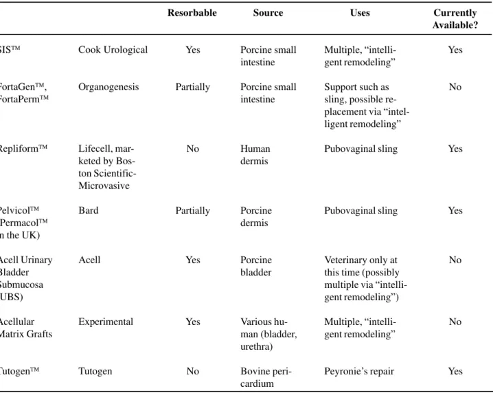
Table 1 – Comparison of various commercial and experimental ECMs.
SIS
FortaGen, FortaPerm
Repliform
Pelvicol
(Permacol
in the UK)
Acell Urinary Bladder Submucosa (UBS)
Acellular Matrix Grafts
Tutogen
Cook Urological
Organogenesis
Lifecell, mar-keted by Bos-ton Scientific-Microvasive
Bard
Acell
Experimental
Tutogen
Resorbable
Yes
Partially
No
Partially
Yes
Yes
No
Source
Porcine small intestine
Porcine small intestine
Human dermis
Porcine dermis
Porcine bladder
Various hu-man (bladder, urethra)
Bovine peri-cardium
Uses
Multiple, “intelli-gent remodeling”
Support such as sling, possible re-placement via “intel-ligent remodeling”
Pubovaginal sling
Pubovaginal sling
Veterinary only at this time (possibly multiple via “intelli-gent remodeling”)
Multiple, “intelli-gent remodeling”
Peyronie’s repair
Currently Available?
Yes
No
Yes
Yes
No
No
In humans, SIS has been used in several settings: as a urethral stricture onlay graft (3); as a pubovaginal sling (4); to repair Peyronie’s disease (5); and to bolster par-tial nephrectomy closures (6). It is perhaps the best-validated graft material in use, although much clinical work must still be done. Pelvicol™ (sold as Permacol™ in Europe; Bard) acellular porcine dermis is also well researched. It has been used in over 60 different surgi-cal procedures, such as abdominal wall defect repair, augmentation cystoplasty (7), pubovaginal slings (8), repair of Peyronie’s disease (9) and augmentation phalloplasty (10).
REPLACEMENT OF ECM GRAFT WITH
TARGET TISSUE
ECM is Completely Absorbed
The first characteristic of most ECMs that are not cross-linked (SIS, experimentally produced AMG, and Acell UBS) is its tendency to become al-most completely absorbed following implantation. The graft is gradually replaced by local tissue in-growth and is eventually no longer needed. It is his-tologically absent by day 28 in studies using both the dog bladder and mouse muscle (11), 90% absent by 60 days when used to replace the dog aorta, and 90% absent from the dog bladder within 3 months using a radiolabelling technique.
ECM Promotes Avid Cell Ingrowth
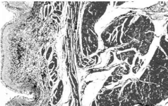
The second benefit of ECM is its tendency to support cell ingrowth, in part by potentiating na-tive cell-cell interactions (12), and in part by provid-ing structural support and the necessary extra-cellu-lar proteins for tissue ingrowth. Over time, the graft is replaced by local tissue; grafts placed into the ure-ter (13) or bladder, for example, have been shown to grow a normal endothelial cell layer, smooth muscle, blood vessels, and nerves (Figure-1) (although the demonstration of nerve functionality in large animals or humans is yet to be seen). In a study of SIS in a rat bladder, the grafts re-grew contractile, functioning bladder with muscarinic, purogenic, and β -adrener-gic receptors (14).
One of the reasons ECM allows cell in-growth is that it is composed mostly of matrix
pro-teins, which have powerful abilities to promote and direct the ingrowth of various cell types. SIS, for example, is made up of 90% Type I and Type IV collagen, which can promote endothelial cell adhe-sion and growth (15,16). Bound into the collagen in SIS are also glyco-proteins such as fibronectin (17); a general adhesion molecule that causes basement membrane assembly and attachment of epidermal cells, fibroblasts, endothelial cells (18,19), and laminin (18); a linker molecule that joins collagen to proteoglycans and promotes endothelial cell ad-hesion and growth (15,16). Glycosaminoglycans (GAGs) in the SIS have functional roles, such as organizing collagen deposition, stimulating angio-genesis and initiating cell differentiation (reviewed in [20]). Examples of GAGs found in SIS are hep-arin (which stimulates angiogenesis, potentiates both epidermal growth factor (EGF) and platelet derived growth factor (PDGF)-induced fibroblast differen-tiation), hylauronic acid (sequesters transforming growth factor beta (TGF-β) into the extracellular matrix), chondroitin sulfate (increases proteoglycan synthesis), and dermatan (interacts with TGF-β1 and may control tissue remodeling).
ECM Growth Factors Favor Cell Ingrowth
ECM appears to maintain functioning growth factors even in sterilized products. These growthtors promote tissue ingrowth and are likely one rea-son robust neovascularization of ECM grafts can be seen within only 3 days of implantation. Growth fac-tors detectible in SIS include basic fibroblast growth factor (bFGF-2) (21), vascular endothelial growth factor (VEGF) (21,22), TGFα, and TGFβ. The role of these compounds varies, but all have functions that might promote active ingrowth.
ECM IS STRONG, AND THEN GETS
STRONGER
In the dry state, ECM in the form of SIS is brittle and sutures will tear through it. However, when reconstituted in saline for 5 minutes, it becomes quite strong and even 0.1 mm single layer SIS is difficult to tear. The breaking point of SIS can be compared to some known materials to give a sense of its strength: SIS 3.4 foot/pounds, polypropylene (Prolene™) su-ture 5 foot/pounds, polyglactin (Vicryl™ or Dexon™) suture 8 foot/pounds, cadaver fascia lata 5.6 foot/ pounds. SIS has an added benefit over these stronger items, however, in that it tends to stretch under force instead of breaking (23).
Although ECM starts out strong, it appears to get even stronger after implantation. A concept known as “Davis’ Law” states that soft tissue heals more strongly along the plane in which it is stressed (24). This principal allows stronger healing of SIS grafts (25): one study of an SIS aorta graft shows it to be 3 times stronger after 3 months than when first implanted (25). A study of SIS replacing the canine abdominal wall is about 5 times stronger than the native abdominal wall (26).
ECM IS IMMUNOCOMPATABLE
ECM causes little detectible experimental im-mune reaction when implanted. Firstly, it is avascular, so hyper-acute rejection cannot occur. It is acellular, so it has a paucity of antigens that might cause hyper-sensitivity. In fact, experimental studies have shown no clinical or histologic evidence of immediate or de-layed rejection to SIS (11). Rabbits implanted with SIS show no signs of antibody production to their major components (27). Even complement activation, which
is a very nonspecific immune response, is absent (11). Some researchers have described a self-limited early acute inflammatory response which is largely resolved by day 10 consisting of polymorphonucleocyte (PMN) infiltration followed by modest monocyte infiltration (11). Interestingly, there is evidence that unknown fac-tors in SIS actually inhibit local immune response by suppressing Helper T cells through interfering with lo-cal interferon-gamma expression (11). This theoreti-cal lack of immunologitheoreti-cal response has not always been found clinically: 8-ply SIS marketed as Stratasis® has induced localized inflammation in about 50% of pa-tients (28).
PREPARATION OF ECM
To prepare SIS, pig jejunum is processed by mechanically removing the mucosa, muscularis externa and serosa. The remaining SIS tissue repre-sents submucosa and basal layers of mucosa. During processing, SIS is defatted, its cells destroyed with paracetic acid,and freeze-dried (18). SIS is not cross-linked, which tends to better preserve its handling characteristics (29). There is some experimental evi-dence that dehydrating the SIS results in less aggres-sive tissue ingrowth experimentally, but the signifi-cance of this is unknown (30), and SIS ingrowth ap-pears to be intact despite this processing step. The final SIS product is 0.2-0.1 mm thick and has the ap-pearance and handling characteristics of cellophane. Because SIS is a natural product, it has an interesting characteristic: Sis is 4 times more permeable to liq-uids in one direction (serosal to mucosal) than the other (31). The clinical implications of this are un-known.
Not all commercially available products are identically processed. Pelvicol™ porcine dermis, for instance, is mildly cross-linked with HMDI, gamma sterilized, and not freeze-dried. FortaPerm™ and FortaGen™ material is mildly cross-linked with EDC, gamma sterilized.
AVAILABLE PRODUCTS
most are not similar to SIS in that they are not de-signed to allow replacement by host tissue. Most other commercially available products are strongly crosslinked, designed to last indefinitely (as apposed to being remodeled by target tissue), and are intended for limited use as pubovaginal slings or abdominal hernia bolsters. Nonetheless, all these products are described here in order to give a complete reckoning of the available armamentarium.
Human Dermis
The predominant acellular matrix grafts made from human cadaver tissue are Repliform™ (Microvasive; Boston, Massachusetts; USA) and Alloderm™ (LifeCell; Branchburg, New Jersey; USA), both of which are processed human dermis. Repliform™ comes in thin and thick forms and is used as a pubovaginal sling material and Alloderm™ as a skin substitute. Repliform™ is freeze-dried. It is unknown if these materials can be used as urologic (bladder, urethral, ureter) substitutes like SIS could be. The company claims preservation of collagen, elastin, and proteoglycans in these materials, but no published data is available.
Porcine Dermis
Pelvicol™ is porcine skin that is gamma ster-ilized and lightly crosslinked. It appears that its per-manence and thickness (it comes in .75 mm and 1.5 mm thickness - much thicker than SIS) might make it useful for pubovaginal slings only.
Small Intestine Submucosa - SIS
SIS is available as a single layer (Surgisis™), a 4 layer product (Surgisis-ES™ and Stratasis™), an 8-layer product for slings (Stratasis®), and as a 10 layer graft for abdominal (ventral) hernia repair. An-other commercially-available abdominal hernia repair product called GraftPatch™ (Organogenesis Inc., Canton Massachusetts) is a 6-layer SIS that is crosslinked for relative permanence (32).
Other ECMs
Acellular matrix grafts that can be made from the bladder, urethra or ureter have been well charac-terized in research studies, but are not yet
commer-cially available. This material is often called “bladder acellular matrix graft” and given the acronym BAMG. However, in many of the research articles the graft is taken from different source tissues, making this moni-ker inaccurate. Acellular matrix grafts seem to have similar characteristics to porcine SIS (33). Specifically, an acellular matrix graft of rabbit ureter placed into a rabbit urethra model allowed regrowth of endothelial, muscle and blood vessels (34), just as SIS does (35). There is clear evidence that the source of the acellular matrix graft affects the quality of subsequent tissue ingrowth (36), but it is too early to determine which is the best donor animal for urologic uses. Finally, a com-mercial ECM product made from porcine bladder is currently available for the veterinary market and may be available for human use in the future (“UBM”; Acell Corporation; Jessup, Maryland).
SURGICAL USES
Pubovaginal Sling
SIS (4 layer Stratasis™, which generally comes in long strips designed for use as a sling) has been used successfully as a pubovaginal sling in 87 humans using a bone-anchoring technique. Gener-ally, results were good: 4/87 (5%) patients did not get relief from their stress incontinence, but 2 of these were due to displaced bone anchors. There were no infections, erosions or graft failures (4). Bovine pericardium has also been used successfully to relieve stress incontinence, with 21/22 patients in one study achieving correction of their urinary incontinence (37). Preliminary studies indicate that Pelvicol™, too, can be used as a pubovaginal sling using a minimally invasive technique in humans (38), but long term data is lacking. 4 layer Surgisis-ES™ SIS comes in sheets and can be cut to fit any size (the largest piece is 7 X 20 cm) and large “T” shaped slings can be fashioned to provide both cys-tocele (posterior) repair and pubovaginal urethral sling (anterior) if desired.
Hypospadias
of patients. All 4 patients had a patent urethra at 22 months and 1 patient with a very long 15 cm defect had a complicating fistula (39).
Urethroplasty
A variety of acellular matrix grafts have been used successfully as experimental urethral onlay grafts. A porcine bladder acellular matrix has been used successfully in an experimental rabbit urethroplasty model, but this was fresh product that may not be the same as commercial-grade product. The urethral caliber was maintained over the study interval, early ingrowth of blood vessels was docu-mented and a healthy transitional cell layer with smooth muscle replaced the graft over time (40). Another interesting study used acellular grafts made from rabbit aorta to bridge long urethral gaps in rabbits, and these formed a patent neourethra (34). Acellular matrix grafts made from rabbit or dog urethra were also used successfully to replace a 1 cm portion of rabbit urethra (41). In this study, there were no fistulae, and the rabbits had both normal urethrograms and urethral pressure profiles. At six months, the urethral grafts had abundant smooth muscle, although they had less muscle than native rabbit urethra. Recently, the successful use of tubularized collagen matrices in rabbit urethral replacement has been reported (42). Finally, SIS was compared to a preputial skin graft in a rabbit urethroplasty model (43). SIS seemed to work bet-ter than the skin, and had the added benefit of re-generating epithelium and smooth muscle into the graft.
Little human data is available about the use of these grafts for urethral reconstruction. Twenty-eight patients were treated in one study, most with long strictures. The success rate was 24/28 (86%) at minimum 36 months follow up, while 1 patient out of 28 (4%) had a fistula which closed spontaneously (44). There is some theoretical evidence from re-search using acellular bladder grafts that protection from urinary extravasation is important for decreas-ing early inflammation (45), so when used in the urethra it may be prudent to provide an extended period of urinary drainage until regrowth of epithe-lium and some underlying muscle is complete.
Bladder Replacement/Augmentation
Bladder augmentation with acellular grafts has the most animal data support of all, yet experts still feel that it will not ultimately be appropriate for human use. A longitudinal study of SIS implanted into a dog bladder, Badylak et al. (46) showed that the augmentation graft re-grew vessels, epithelium and a continuous basement membrane by 4 weeks time. By 8 weeks, there were bundles of smooth muscle and collagen and inflammatory cells were largely absent. By 12 weeks, there was parallel orientation of smooth muscle cells and the complete degradation of SIS, leaving a functioning bladder. Others have confirmed these results and reported that bladder-augmented dogs showed stable preservation of bladder volume over time (47). SIS has also been successful in other animals such as mini-pigs (48). However, the SIS patches seem to contract to 58% of their original size, so augmentations using SIS should be greatly upsized to compensate for this shrinkage (48). SIS in rats cre-ated a bladder that was indistinguishable from a na-tive bladder (49) with the surprising finding that the grafts had become normally innervated. Although these initial positive results in animals have been widely reported, others have described disappoint-ing preliminary results usdisappoint-ing SIS in both dog and porcine models (50,51).
augmenta-tion could be achieved, although the study had small numbers (58).
Human data is largely lacking. Permacol™ (a.k.a. Pelvicol™) has been used in 5 patients as a bladder augmentation but preliminary data has been presented only in abstract form. It appears there were no early complications in this small abstract series (5).
Partial Nephrectomy
Preliminary reports indicate that SIS may be a helpful adjunct in closing the parenchymal defect after partial nephrectomy in humans (6). Theoreti-cally, large sheets of SIS could be used to encase and repair kidneys after partial nephrectomy or even re-nal injury. Severely injured, even shattered kidneys, might be repaired with SIS just as has been done with polyglactin (Vicryl™ or Dexon™) mesh in the past (59). Further data will be needed to further validate this use of ECM.
Peyronie’s Disease
A significant body of literature focusing on the use of acellular matrix grafts in urologic recon-struction of Peyronie’s disease in both humans and animal models has emerged. Both SIS and rabbit tu-nica albuginea derived acellular matrix grafts have been used for successful correction of Peyronie’s in rabbit models (60,61). In humans, both bovine peri-cardium and SIS have been used successfully as a corporal patch after incision of Peyronie’s plaque (5,62) as well as for reconstruction of tunical defi-ciencies following penile prosthesis placement (63). Permacol™ (a.k.a. Pelvicol™) has been used in 5 patients as a Peyronie’s patch with good but prelimi-nary results (9). Finally, a single case report has shown the use of cadaveric tensor fascia lata in cavernosal reconstruction following partial penectomy with sat-isfactory results (64). In our hands, 3 of 4 patients grafted with single or double layer SIS had severe recurrence of penile deformity, and we continue to approach its use with caution.
Abdominal Hernia
Multi-layered SIS patches have proven ex-perimentally effective at repairing ventral hernia
de-fects, although human studies are lacking. GraftPatch™ (Organogenesis Inc., Canton Massachu-setts), a 6-layer SIS graft, repaired experimentally-created abdominal wall defects in rabbits with few adhesions (27). 8-ply SIS was used to repair experi-mentally-created abdominal hernias in dogs and ro-dents, with excellent preservation of strength over a 2-year follow up period (26,65). When compared to repair with polypropylene (Prolene) mesh, the SIS has less inflammation, less foreign-body reaction, and far fewer intestinal adhesions (66).
Ureteral Replacement
Replacement of ureters with acellular grafts is the most controversial because some studies show it works and others show it fails. Rat ureter was suc-cessfully replaced with a ECM graft made of rat ure-ter with excellent recapitulation of ureure-teral tissue: epithelium, blood vessels, smooth muscle and even nerves were all present by 4 months (13). However, a criticism of this study is that the ureters were left stented for the duration of the study and it is unknown if the ureters would have stayed open after the stents were removed. Rabbit ureter was also successfully replaced with SIS grafts, which eventually recapitu-lated the 3 layers of the ureter (epithelium, muscle layer, serosa) and showed robust neovascularity (67). Replacement of swine ureter with SIS resulted in a widely patent re-growth of the missing portion with neovascularization, smooth muscle, epithelium, and scant foreign body reaction (68,69). There is a single human case study where SIS was used to success-fully reconstruct a stenosed uretero-neobladder stric-ture (70). More convincing human data are not yet available.
fas-cial transplants, autologous veins, arterial grafts, skin grafts, freeze-dried ureter, and autologous ureter have also failed in the past (cited in ref 48).
A possible use for SIS in the ureter would be to bolster ureteral repairs after ureteroureterostomy or open ureteral re-implants. Experimentally, this has been shown to be successful when the first genera-tions of implantable collagen membranes were used to bolster ureteral repairs in rabbits (72).
Injectable SIS Bulking Agent
Although this review is not meant to explore the field of injectable bulking agents in urology, it is notable that a paste made from SIS has been devel-oped. Preliminary studies in dogs showed that the injected paste induced smooth muscle regeneration with long-term preservation of 25% of the original injection volume (73).
Urethral Coverage Layer
SIS can be used as an “extra layer” in ure-thral surgery. Rabbit models have shown successful proof of this concept (74) and it may be particularly useful in long urethral suture lines, such as during long hypospadias repair, second stage Johanson ure-throplasty or penile ureure-throplasty in multiply-oper-ated patients.
THE FUTURE
Techniques in tissue engineering also con-tinue to improve and several notable potential im-provements in techniques show promise.
Cell-seeded Acellular Matrix
One potential improvement in existing acel-lular graft material is achieved by pre-seeding the grafts with the patient’s own urothelial cells. These cells can be harvested from the host (usually by blad-der or urethral biopsy), cell cultured and then seeded into acellular matrix (75). Where this approach was attempted using both bladder urothelial cells and bladder smooth muscle cells seeded onto an acellu-lar collagen matrix patch in an animal augmenta-tion cystoplasty model (45), bladder capacity was significantly improved when compared to unseeded
acellular collagen. Subsequent studies using blad-der replacement in dogs with cells seeded onto a biodegradable cell free polymer were equally suc-cessful (76). Clinical success has already been achieved using this approach to engineer skin grafts (77). Recently, two ambitious reports showed that it was possible to seed bladder urothelium and smooth muscle onto an SIS matrix, an approach which im-proved the quantity of smooth muscle in a bladder graft when compared to SIS alone (12). This inno-vation has the potential to be the next “state of the art” in the field.
Modification of Acellular Grafts to Promote
Ingrowth
Special preparation of acellular grafts using gene therapy techniques might also improve the qual-ity of implantable materials in the future. Theoreti-cally, seeded cells as described above could be trans-fected with desirable genes in order to reverse exist-ing bladder or urethral disease or promote healthy tissue ingrowth (75). This remains theoretical today. Acellular grafts might also be modified by adding desirable matrix molecules to improve healthy tissue ingrowth. Glycosaminoglycans such as heparin (78) and chondroitin (79) have been successfully added to collagen matrices to successfully improve neoangiogenesis.
CONCLUSIONS
re-placement are contemplated. The more distant future of ECMs in urology likely will include cell-seeded grafts, with the eventual hope of “off the shelf” re-placement materials. Until that day arrives, ECMs fulfill only some of the requirements for the recon-structive urologist.
REFERENCES
1. Badylak SF, Lantz GC, Coffey A, Geddes LA: Small intestinal submucosa as a large diameter vascular graft in the dog. J Surg Res. 1989; 47: 74-80.
2. Badylak SF: Small intestinal submucosa (SIS): A bio-material conductive to smart tissue remodeling, in Bell E: Tissue Engineering: Current Perspectives. Boston, Birkhauser, 1993, pp. 179-189.
3. Kassaby E, Yoo JJ, Retik AB, Atala A: A novel inert collagen matrix for urethral stricture repair. J Urol. 2000; 163: 70.
4. Rutner AB, Levine SR, Schmaelzle JF: Porcine small intestine submucosa implantation for pubovaginal slings: Results and conclusions after two years in eighty-seven female patients. Society for Urology and Engineering. 16th Annual Meeting. Anaheim, CA, 2001.
5. Knoll LD: Use of porcine small intestinal submucosal graft in the surgical management of Peyronie’s disease. Urology. 2001; 57: 753-7.
6. Corey OCR, Patel RV, Laven BA,Steinberg GD: The use of small intestinal submucosa (SIS) to improve outcomes following partial nephrectomy. North Cen-tral Section American Urologic Association. Chicago, IL, 2002.
7. Speakman MJ: Bladder augmentation cystoplasty with a novel collagen membrane. BJU Int. 2000; 85 (Supl. 5): 2-3.
8. Barrington JW, Edwards G, Arunkalaivanan AS, Swart M: The use of porcine dermal implant in a minimally invasive pubovaginal sling procedure for genuine stress incontinence. BJU Int. 2002; 90: 224-7.
9. Lloyd SN, Hetherington J: Plaque excision and Permacol grafting for Peyronie’s disease. BJU Int. 2000; 85 (Supl. 5): 16.
10. Harper C: Permacol: clinical experience with a new biomaterial. Hosp Med. 2001; 62: 90-5.
11. Allman AJ, McPherson TB, Badylak SF, Merrill LC, Kallakury B, Sheehan C, et al.: Xenogeneic extracel-lular matrix grafts elicit a TH2-restricted immune re-sponse. Transplantation. 2001; 71: 1631-40.
12. Zhang Y, Kropp BP, Moore P, Cowan R, Furness PD 3rd, Kolligian ME, et al.: Coculture of bladder urothelial and smooth muscle cells on small intestinal submucosa: potential applications for tissue engineer-ing technology. J Urol. 2000; 164: 928-34; discussion 934-5.
13. Dahms SE, Piechota HJ, Nunes L, Dahiya R, Lue TF, Tanagho EA: Free ureteral replacement in rats: regen-eration of ureteral wall components in the acellular matrix graft. Urology. 1997; 50: 818-25.
14. Vaught JD, Kropp BP, Sawyer BD, Rippy MK, Badylak SF, Shannon HE, et al.: Detrusor regeneration in the rat using porcine small intestinal submucosal grafts: functional innervation and receptor expression. J Urol. 1996; 155: 374-8.
15. Macarak EJ, Howard PS: Adhesion of endothelial cells to extracellular matrix proteins. J Cell Physiol. 1983; 116: 76-86.
16. Herbst TJ, McCarthy JB, Tsilibary EC, Furcht LT: Differential effects of laminin, intact type IV collagen, and specific domains of type IV collagen on endothe-lial cell adhesion and migration. J Cell Biol. 1988; 106: 1365-73.
17. McPherson TB and Badylak SF: Characterization of fibronectin derived from porcine small intestinal sub-mucosa. Tissue Engineering. 1998; 4: 75-83. 18. Badylak S, Liang A, Record R, Tullius R, Hodde J:
Endothelial cell adherence to small intestinal submu-cosa: an acellular bioscaffold. Biomaterials. 1999; 20: 2257-63.
19. Dejana E, Colella S, Languino LR, Balconi G, Corbascio GC, Marchisio PC: Fibrinogen induces ad-hesion, spreading, and microfilament organization of human endothelial cells in vitro. J Cell Biol. 1987; 104: 1403-11.
20. Hodde J, Badylak S, Brightman A, Voytik-Harbin S: Glycosaminoglycan content of small intestinal submu-cosa: A bioscaffold for tissue replacement. Tissue En-gineering. 1996; 2: 209-17.
21. Voytik-Harbin SL, Brightman AO, Kraine MR, Waisner B, Badylak SF: Identification of extractable growth factors from small intestinal submucosa. J Cell Biochem. 1997; 67: 478-91.
22. Hodde JP, Record RD, Liang HA, Badylak SF: Vascu-lar endothelial growth factor in porcine-derived extra-cellular matrix. Endothelium. 2001; 8: 11-24. 23. Kubricht WS 3rd, Williams BJ, Eastham JA, Venable
24. Tippett SR, Voight ML: Functional Progressions for Sport Rehabilitation. Champlain, IL, Human Kinetics, 1995.
25. Hiles MC, Badylak SF, Lantz GC, Kokini K, Geddes LA, Morff RJ: Mechanical properties of xenogeneic small-intestinal submucosa when used as an aortic graft in the dog. J Biomed Mater Res. 1995; 29: 883-91.
26. Badylak S, Morff R, Tullius R: The use of SIS as a scaffold for abdominal hernia repair: Second SIS Sym-posium. Orlando, FL, 1998, pp. 48-49.
27. Abraham GA, Murray J, Billiar K, Sullivan SJ: Evalu-ation of the porcine intestinal collagen layer as a bio-material. J Biomed Mater Res. 2000; 51: 442-52. 28. Ho KL, Witte MN, Bird ET: 8-ply small intestinal
sub-mucosa tension-free sling: spectrum of postoperative inflammation. J Urol. 2004; 171: 268-71.
29. Khor E: Methods for the treatment of collagenous tissues for bioprostheses. Biomaterials. 1997; 18: 95-105.
30. Lindberg K, Badylak SF: Porcine small intestinal sub-mucosa (SIS): a bioscaffold supporting in vitro pri-mary human epidermal cell differentiation and synthe-sis of basement membrane proteins. Burns. 2001; 27: 254-66.
31. Ferrand BK, Kokini K, Badylak SF, Geddes LA, Hiles MC, Morff RJ: Directional porosity of porcine small-intestinal submucosa. J Biomed Mater Res. 1993; 27: 1235-41.
32. Gloeckner DC, Sacks MS, Billiar KL, Bachrach N: Mechanical evaluation and design of a multilayered collagenous repair biomaterial. J Biomed Mater Res. 2000; 52: 365-73.
33. Sievert KD, Tanagho EA: Organ-specific acellular matrix for reconstruction of the urinary tract. World J Urol. 2000; 18: 19-25.
34. Parnigotto PP, Gamba PG, Conconi MT, Midrio P: Experimental defect in rabbit urethra repaired with acellular aortic matrix. Urol Res. 2000; 28: 46-51. 35. Sievert KD, Bakircioglu ME, Nunes L, Tu R, Dahiya
R, Tanagho EA: Homologous acellular matrix graft for urethral reconstruction in the rabbit: histological and functional evaluation. J Urol. 2000; 163: 1958-65.
36. Wefer J, Sekido N, Sievert KD, Schlote N, Nunes L, Dahiya R, et al.: Homologous acellular matrix graft for vaginal repair in rats: a pilot study for a new recon-structive approach. World J Urol. 2002; 20: 260-3. 37. Pelosi MA 2nd, Pelosi MA 3rd, Pelekanos M: The
YAMA UroPatch sling for treatment of female stress
urinary incontinence: a pilot study. J Laparoendosc Adv Surg Tech A. 2002; 12: 27-33.
38. Edwards G, Barrington J: The use of Pelvicol in a mini-mally invasive pubovaginal sling procedure: 25th An-nual Meeting of the International Urogynecological Association. Rome, Italy, 2000, pp. IDP49.
39. Atala A, Guzman L, Retik AB: A novel inert collagen matrix for hypospadias repair. J Urol. 1999; 162: 1148-51.
40. Chen F, Yoo JJ, Atala A: Acellular collagen matrix as a possible “off the shelf” biomaterial for urethral re-pair. Urology. 1999; 54: 407-10.
41. Sievert KD, Wefer J, Bakircioglu ME, Nunes L, Dahiya R, Tanagho EA: Heterologous acellular matrix graft for reconstruction of the rabbit urethra: histological and functional evaluation. J Urol. 2001; 165: 2096-102.
42. De Filippo RE, Yoo JJ, Atala A: Urethral replacement using cell seeded tubularized collagen matrices. J Urol. 2002; 168: 1789-92; discussion 1792-3.
43. Kropp BP, Ludlow JK, Spicer D, Rippy MK, Badylak SF, Adams MC, et al.: Rabbit urethral regeneration using small intestinal submucosa onlay grafts. Urol-ogy. 1998; 52: 138-42.
44. El-Kassaby AW, Retik AB, Yoo JJ, Atala A: Urethral stricture repair with an off-the-shelf collagen matrix. J Urol. 2003; 169: 170-3; discussion 173.
45. Yoo JJ, Meng J, Oberpenning F, Atala A: Bladder aug-mentation using allogenic bladder submucosa seeded with cells. Urology. 1998; 51: 221-5.
46. Badylak SF, Record R, Lindberg K, Hodde J, Park K: Small intestinal submucosa: a substrate for in vitro cell growth. J Biomater Sci Polym Ed. 1998; 9: 863-78. 47. Kropp BP, Rippy MK, Badylak SF, Adams MC,
Keating MA, Rink RC, et al.: Regenerative urinary bladder augmentation using small intestinal submu-cosa: urodynamic and histopathologic assessment in long-term canine bladder augmentations. J Urol. 1996; 155: 2098-104.
48. Shalhav AL, Elbahnasy AM, Bercowsky E, Kovacs G, Brewer A, Maxwell KL, et al.: Laparoscopic replace-ment of urinary tract segreplace-ments using biodegradable materials in a large-animal model. J Endourol. 1999; 3: 241-4.
49. Kropp BP, Eppley BL, Prevel CD, Rippy MK, Harruff RC, Badylak SF, et al.: Experimental assessment of small intestinal submucosa as a bladder wall substi-tute. Urology. 1995; 46: 396-400.
submu-cosa inferior to autologous bowel for laparoscopic bladder augmentation at one year follow up: North Central Section American Urologic Association. Chi-cago, IL, 2002, pp 109.
51. Pope JC 4th, Davis MM, Smith ER Jr., Walsh MJ, Ellison PK, Rink RC, et al.: The ontogeny of canine small intestinal submucosa regenerated bladder. J Urol. 1997; 158: 1105-10.
52. Wefer J, Sievert KD, Schlote N, Wefer AE, Nunes L, Dahiya R, et al.: Time dependent smooth muscle re-generation and maturation in a bladder acellular ma-trix graft: histological studies and in vivo functional evaluation. J Urol. 2001; 165: 1755-9.
53. Cayan S, Chermansky C, Schlote N, Sekido N, Nunes L, Dahiya R, et al.: The bladder acellular matrix graft in a rat chemical cystitis model: functional and histo-logic evaluation. J Urol. 2002; 168: 798-804. 54. Piechota HJ, Gleason CA, Dahms SE, Dahiya R, Nunes
LS, Lue TF, et al.: Bladder acellular matrix graft: in vivo functional properties of the regenerated rat blad-der. Urol Res. 1999; 27: 206-13.
55. Piechota HJ, Dahms SE, Nunes LS, Dahiya R, Lue TF, Tanagho EA: In vitro functional properties of the rat bladder regenerated by the bladder acellular ma-trix graft. J Urol. 1998; 159: 1717-24.
56. Probst M, Piechota HJ, Dahiya R, Tanagho EA: Ho-mologous bladder augmentation in dog with the blad-der acellular matrix graft. BJU Int. 2000; 85: 362-71. 57. Reddy PP, Barrieras DJ, Wilson G, Bagli DJ, McLorie GA, Khoury AE, et al.: Regeneration of functional bladder substitutes using large segment acellular ma-trix allografts in a porcine model. J Urol. 2000; 164: 936-41.
58. Calvano CJ, Moran ME, Parekh A, Desai PJ, Cisek LJ: Laparoscopic augmentation cystoplasty using the novel biomaterial Surgisis: small-intestinal submucosa. J Endourol. 2000; 14: 213-7.
59. Mounzer AM, McAninch JW, Schmidt RA: Polyglycolic acid mesh in repair of renal injury. Urol-ogy. 1986; 28: 127-30.
60. Monga M, Cosgrove D, Zupkas P, Jain A, Kasyan A, Wilkes N, et al.: Small intestinal submucosa as a tu-nica albuginea graft material. J Urol. 2002; 168: 1215-21.
61. Wefer J, Schlote N, Sekido N, Sievert KD, Wefer AE, Nunes L, et al.: Tunica albuginea acellular matrix graft for penile reconstruction in the rabbit: a model for treat-ing Peyronie’s disease. BJU Int. 2002; 90: 326-31. 62. Egydio PH, Lucon AM, Arap S: Treatment of
Peyronie’s disease by incomplete circumferential
in-cision of the tunica albuginea and plaque with bovine pericardium graft. Urology. 2002; 59: 570-4. 63. Knoll LD: Use of porcine small intestinal submucosal
graft in the surgical management of tunical deficien-cies with penile prosthetic surgery. Urology. 2002; 59: 758-61.
64. Christopher N, Arya M, Brown RS, Payne HA, Woodhouse CR, Ralph DJ: Penile preservation in squa-mous cell carcinoma of the bulbomembranous urethra. BJU Int. 2002; 89: 464-5.
65. Badylak S, Kokini K, Tullius B, Simmons-Byrd A, Morff R: Morphologic study of small intestinal sub-mucosa as a body wall repair device. J Surg Res. 2002; 103: 190-202.
66. Clarke KM, Lantz GC, Salisbury SK, Badylak SF, Hiles MC, Voytik SL: Intestine submucosa and polypropylene mesh for abdominal wall repair in dogs. J Surg Res. 1996; 60: 107-14.
67. Jackson OF: Urolithiasis and experimental urethral obstruction in the cat. J Physiol. 1969; 201: 97-8P. 68. Li BZ: Long-term effect of the pull-through operation
in traumatic posterior urethral stricture. Zhonghua Wai Ke Za Zhi. 1984; 22: 368-9.
69. Smith TG 3rd, Gettman M, Lindberg G, Napper C, Pearle MS, Cadeddu JA: Ureteral replacement using porcine small intestine submucosa in a porcine model. Urology. 2002; 60: 931-4.
70. O’Connor RC, Patel RV, Steinberg GD: Successful repair of a uretero-neobladder stricture using porcine small intestine submucosa. J Urol. 2001; 165: 1995. 71. Xie H, Shaffer BS, Wadia Y, Gregory KW: Use of
re-constructed small intestine submucosa for urinary tract replacement. Asaio J. 2000; 46: 268-72.
72. Scott R, Baraza R, Gorham SD, McGregor I, French DA: Assessment of collagen film for use in urinary tract surgery. Br J Urol. 1986; 58: 203-7.
73. Furness PD 3rd, Kolligian ME, Lang SJ, Kaplan WE, Kropp BP, Cheng EY: Injectable small intestinal sub-mucosa: preliminary evaluation for use in endoscopic urological surgery. J Urol. 2000; 164: 1680-5. 74. Grossklaus DJ, Shappell SB, Adams MC, Brock JW
3rd, Pope JC 4th: Small intestinal submucosa as a ure-thral coverage layer. J Urol. 2001; 166: 636-9. 75. Atala A: Construction of artificial organs and tissues
using autologous cells. AUA Update Series. 2000; 19: 122-8.
77. Casasco A, Casasco M, Zerbinati N, Icaro Cornaglia A, Calligaro A: Cell proliferation and differentiation in a model of human skin equivalent. Anat Rec. 2001; 264: 261-72.
78. Noe HN, Dale GA: Evaluation of children with meatal stenosis. J Urol. 1975; 114: 455-6.
79. Pieper JS, van Wachem PB, van Luyn MJA, Brouwer LA, Hafmans T, Veerkamp JH, et al.: Attachment of glycosaminoglycans to collagenous matrices modulates the tissue response in rats. Biomaterials. 2000; 21: 1689-99.
Received: January 30, 2005 Accepted: March 5, 2005
Correspondence address: Dr. Richard A. Santucci 4160, John R, Suite 1017 Detroit, MI 48167, USA Fax: + 313-745-0464