Síndrome das apnéias-hipopnéias obstrutivas do sono: associação com gênero e obesidade e fatores relacionados à sonolência
Marli Maria Knorst1, Fábio José Fabrício de Barros Souza2, Denis Martinez1
Abstract
Objective: To study the effects that gender and obesity have on excessive daytime sleepiness (EDS) in individuals with obstructive sleep apnea-hypopnea syndrome (OSAHS), as well as to identify factors associated with EDS in such individuals. Methods: A total of 300 consecutive patients who completed the clinical evaluation satisfactorily and whose polysomnography showed an apnea-hypopnea index (AHI) > 10 events/hour of sleep were selected from a sleep clinic population for inclusion in the study. Results: Mean age was 47 ± 11 years, and mean AHI was 52.1 ± 29.2 events/hour of sleep. Females presented higher mean age, lower EDS scores and less time in apnea . Mean EDS score was 14.7 ± 7.2. The EDS score correlated best with body movements (r = 0.43; p < 0.01), respiratory events during sleep (r = 0.40; p < 0.01), duration of apnea (r = 0.40; p < 0.01), peripheral oxygen saturation (SpO2; r = –0.38; p < 0.01) and AHI (r = 0.37;
p < 0.01). Mean body mass index (BMI) was 30.2 ± 5.3 kg/m2. Overweight, obesity and morbid obesity were observed in 41, 44 and 5.3% of
cases, respectively. Disease severity correlated most strongly with BMI (r = 0.51; p < 0.01). Conclusions: Higher mean age, lower EDS scores and less time spent in sleep apnea time in apnea were associated with being female. Fragmented sleep, number/duration of respiratory events during sleep, SpO2 levels and obesity were associated with sleepiness. The BMI had a significant effect on OSAHS severity.
Keywords: Sleep apnea, obstructive; Sleep apnea syndromes; Polysomnography; Sleep stages; Obesity.
Resumo
Objetivo: Estudar os efeitos de gênero e obesidade e identificar fatores relacionados à sonolência diurna excessiva (SDE) em indivíduos com síndrome das apnéias-hipopnéias obstrutivas do sono (SAHOS). Métodos: Foram selecionados para inclusão no estudo 300 pacientes consecutivos, atendidos em clínica do sono, com índice de apnéia/hipopnéia (IAH) > 10 eventos/hora de sono, que completaram adequada-mente a avaliação clínica. Resultados: A média de idade foi de 47 ± 11 anos e o IAH médio foi de 52,1 ± 29,2 eventos/hora de sono. As mulheres apresentaram maior média de idade, menos sonolência e menos tempo em apnéia. O escore médio de SDE foi de 14,7 ± 7,2. O escore de SDE correlacionou-se melhor com movimentos corpóreos (r = 0,43; p < 0,01), eventos respiratórios durante o sono (r = 0,40; p < 0,01), tempo em apnéia (r = 0,40; p < 0,01), valores mínimos da saturação periférica de oxigênio (SpO2; r = –0,38; p < 0,01) e IAH
(r = 0,37; p < 0,01). O índice de massa corpórea (IMC) médio foi de 30,2 ± 5,3 kg/m2. Sobrepeso, obesidade e obesidade mórbida foram
observados em, respectivamente, 41, 44 e 5,3% dos casos. A gravidade da doença correlacionou-se melhor com IMC (r = 0,51; p < 0,01).
Conclusões: Maior média de idade, menor escore de SDE e menor tempo em apnéia foram associados ao gênero feminino. Fragmentação do sono, número e duração de eventos respiratórios durante o sono, níveis de SpO2 e obesidade se associaram à sonolência. O IMC teve efeito significativo na gravidade da SAHOS.
Descritores: Apnéia do sono tipo obstrutiva; Síndromes da Apnéia do Sono; Polissonografia; Fases do sono; Obesidade.
* Study conducted in the Department of Internal Medicine and Postgraduate Program in Respiratory Sciences of the Universidade Federal do Rio Grande do Sul – UFRGS, Federal University of Rio Grande do Sul – School of Medicine, Porto Alegre, Brazil.
1. Associate Professor in the Department of Internal Medicine and Postgraduate Program in Respiratory Sciences of the Universidade Federal do Rio Grande do Sul – UFRGS, Federal University of Rio Grande do Sul – Porto Alegre, Brazil.
2. Masters student in the Postgraduate Program in Respiratory Sciences of the Universidade Federal do Rio Grande do Sul – UFRGS, Federal University of Rio Grande do Sul – Porto Alegre, Brazil.
Correspondence to: Marli Maria Knorst. Serviço de Pneumologia, Hospital de Clínicas de Porto Alegre, Rua Ramiro Barcelos 2350, Sala 2050, 2º andar, Bom Fim, CEP 97035-003, Porto Alegre, RS, Brasil.
Tel 55 51 2101-8241. E-mail: [email protected]
Submitted: 28 March 2007. Accepted, after review: 1 October 2007.
Introduction
Respiration and sleep are two functions that are indis-pensable for the preservation of human life. However, since we sleep an average of eight hours a night, approximately one third of our life is spent in an unconscious state. The alterations that occur during sleep make individuals
vulner-able to various sleep disorders. Therefore, respiration that is normal during waking can become disturbed during sleep.
protocol. The project was approved by the ethics in research committee of the institution.
Clinical data were obtained through anamnesis and physical examination. Upon anamnesis, conducted with the patient and a family member, questions were asked regarding snoring, sleep apneas and daytime sleepiness, as well as regarding the patient history of traffic accidents. We investigated EDS using the criteria proposed by Lavie.(10) The patients were asked how often they fell asleep in different situa-tions, such as reading, watching television, traveling, driving, attending classes, lectures or at work (possible responses: never; hardly ever; sometimes; quite often; and always). A score was attributed to each answer, ranging from 0 to 4, in which 0 corresponded to never and 4 corresponded to always. Therefore, the overall EDS score ranged from 0 to 20.
The body mass index (BMI) for each patient was obtained by dividing weight in kilograms by height in meters squared. Patients were categorized by weight status based on their BMI: normal (between 20 and 24.9 kg/m2), overweight (BMI between 25 and 29.9 kg/m2), obese (BMI between 30 and 39.9 kg/m2) or morbidly obese (BMI ≥40 kg/m2). Blood pressure was measured during the medical appointment, after a 15-min rest, with the patient lying down, and was corrected for arm diameter. Systemic arterial hypertension was defined as a previous medical diagnosis of hypertension, use of antihypertensives or blood pressure equal to or greater than 140/90 mmHg.
Polysomnographic tests were performed using the standard method, with electroencephalo-gram (C3-A2, C4-A1), electro-oculoelectroencephalo-gram (left eye and right eye), electromyogram (chin and anterior tibia) and electrocardiogram. All of the equipment employed was manufactured by Emsa Equipamentos Medicos Ltda. (Rio de Janeiro, Brazil). Airflow was measured using a nasal cannula connected to a pressure transducer, and peripheral oxygen satura-tion (SpO2) was measured using a pulse oximeter (Ohmeda, Boulder, CO, USA).
Sleep stages were classified according to the criteria established by Rechtschafen and Kales. (13) Apnea was defined as a reduction in airflow to ≤10% of the baseline value for 10 s or more, and hypopnea was defined as reductions in airflow of ≥50% accompanied by awakening and a ≥4% drop in SpO2. The AHI was calculated by dividing the total number of apnea-hypopnea episodes by the number airway, within its collapsible segment, during sleep.
Morbidity and mortality rates are elevated among individuals with OSAHS.(1)
The most evident symptom of OSAHS is snoring. (2) Snoring results from the relaxation of the abductor
muscles of the pharynx and is observed in more than 60% of the male population over 60 years of age.(3) Apneas reported by the bed partner is another sign of OSAHS.(4) The incidence of sleep apneas increases with age(5) and in higher among obese individuals,(6) as well as among individuals who use hypnotics or alcohol at bedtime.(7) In general, OSAHS predomi-nantly affects men. Although it is common in elderly individuals of either genders, premenopausal women seem to be protected against apneas.(8) This has been attributed to hormonal influences, particularly to the role of progesterone as a respiratory stimulant.(9)
Sleep-disordered breathing impairs the quality of sleep and can affect the quality of wake time in a variety of ways. Excessive daytime sleepiness (EDS) is the principal symptom seen during waking in patients with OSAHS. It has been shown that 2-3% of industry workers in Israel present EDS caused by sleep disorders.(10)
The combination of OSAHS and EDS is a public health problem. It leads to cognitive impairment and reduced reflexes, thereby increasing the risk of traffic accidents and other mishaps.(11) In addition, EDS can have significant repercussions for work, social life and quality of life.(12) However, the factors associated with EDS are not well known.
The objective of this study was to evaluate the impact of gender and obesity in patients with OSAHS, as well as to identify factors associated with EDS in such individuals.
Methods
of the patients. Clinical and polysomnographic data are shown, by gender, in Table 1.
There was no difference in terms of reported snoring or apnea in relation to gender (chi-square; p > 0.05). Significant differences were observed between males and females in relation to the following: mean age (46.5 ± 10.9 years vs. 52.8 ± 9.5 years; p = 0.002); mean EDS score (15.3 ± 7.0 vs. 9.9 ± 6.9; p = 0.001); mean rapid eye movement (REM) sleep latency (120.5 ± 73.9 min vs. 149.5 ± 73.9 min; p = 0.04); mean number of mixed apneas (12.9 ± 30.9 vs. 1.35 ± 5.9; p = 0.04); and mean time spent in sleep apnea (141.7 ± 95.6 min vs. 105.2 ± 74.0 min; p = 0.04). There were no statistically significant differences between genders in terms of the remaining clinical and polysomno-graphic variables.
The mean EDS score for the sample as a whole was 14.7 ± 7.2. We found that EDS score corre-lated positively and significantly with the following variables: BMI; total sleep time; sleep efficiency; number of body movements; percentage of total sleep time spent in stage 2 sleep; percentage of total sleep time spent in REM sleep; number of obstruc-tive or mixed respiratory events; total number of minutes spent in sleep apnea; and percentage of total sleep time spent in apnea. Statistically signifi-of hours signifi-of sleep. Based on the AHI, OSAHS severity
was classified as mild (<15), moderate (15-30) or severe (>30).(14)
Data were stored and analyzed using the program Statistical Package for Social Sciences, version 14.0 (SPSS Inc, Chicago, IL, USA). Results are expressed as number of cases, percentages, mean and standard deviation or standard error. The Student’s t-test or the Mann-Whitney test was used, together with the chi-square test and analysis of variance for multiple groups, as well as the Kruskal-Wallis test, depending on the distribution of the data. In order to analyze the correlations among variables with normal distri-bution, Pearson’s linear correlation test was used, whereas Spearman’s correlation coefficient was used for variables with non-normal distribution. All of the tests were two-tailed. Values of p < 0.05 were considered statistically significant.
Results
Of the 300 patients with OSAHS, 269 were male and 31 were female. Most (294) of the patients were Caucasian. The mean age was 47.2 ± 10.9 years, ranging from 17 to 82 years. Mode age occurred in the 41 to 50 year age bracket in males, whereas it occurred in the 51 to 60 year age bracket in females. Systemic arterial hypertension was identified in 58%
Table 1 - Clinical and polysomnographic characteristics in 300 patients with obstructive sleep apnea-hypopnea syndrome by gender.a
Characteristics All Males Females p
n = 300 n = 269 n = 31
Age (years) 47.2 ± 10.9 46.5 ± 10.9 52.8 ± 9.5 0.002
Duration of complaints (years) 13.9 ± 12.2 14.0 ± 12.0 12.9 ± 14.0 0.65
Systolic blood pressure (mmHg) 141.1 ± 22.9 141.2 ± 23.0 140.6 ± 22.6 0.89
Diastolic arterial pressure (mmHg) 83.2 ± 14.3 84.5 ± 14.6 81.2 ± 11.4 0.41
Smoking index* (pack-years) 30.8 ± 25.6 31.4 ± 25.9 19.0 ± 14.3 0.16
Sleepiness score 14.7 ± 7.2 15.3 ± 7.0 9.9 ± 6.9 0.0001 Body mass index (kg/m2) 30.2 ± 5.3 30.3 ± 5.2 29.2 ± 6.2 0.30
AHI (events/hour of sleep) 52.1 ± 29.2 52.0 ± 28.5 52.8 ± 35.5 0.88
Time spent in sleep apnea (min) 137.9 ± 94.2 141.7 ± 95.6 105.2 ± 74.0 0.04
Total sleep time (min) 377.3 ± 51.7 378.4 ± 51.6 368.4 ± 51.9 0.30
Sleep latency (min) 21.8 ± 26.7 20.9 ± 26.0 29.3 ± 31.7 0.09
Sleep efficiency (%) 80.9 ± 12.9 81.3 ± 12.7 77.8 ± 14.4 0.16
Slow-wave sleep (%) 5.9 ± 6.6 5.7 ± 6.6 7.9 ± 6.8 0.07
REM sleep (%) 9.5 ± 4.6 9.5 ± 4.6 9.1 ± 4.2 0.63
Minimum SpO2 (%) 71.5 ± 18.7 71.3 ± 18.5 73.4 ± 20.9 0.55
aData presented as mean ± standard deviation. AHI: apnea-hypopnea index; REM: rapid eye movement; and SpO
2: peripheral oxygen
Of the 300 patients studied, 238 drove auto-mobiles. Of those 238 patients, 106 (44.5%) reported falling asleep at the wheel always or quite often. The mean EDS score for these patients was 20.7 ± 3.8, compared with 11.3 ± 6.8 for the group without sleepiness while driving. In comparison with the patients who never fell asleep at the wheel, cant, although negative, correlations were observed
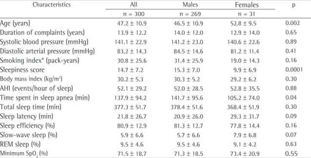
between the EDS score and the following variables: age; sleep latency; percentage of slow-wave sleep; mean SpO2; and minimum SpO2 (Table 2). Figure 1 shows how EDS score correlated with BMI, number of respiratory events, number of body movements and minimum SpO2.
Table 2 - Correlations between the excessive daytime sleepiness score and clinical variables, as well as between the excessive daytime sleepiness score and polysomnographic variables, in 300 patients with obstructive sleep apnea-hypopnea syndrome.
Clinical variables r Polysomnographic variables
Sleep architecture r Respiration r Oximetry r
Age −0.19* TST (%) 0.14** Respiratory events
Duration of complaints
0.03 Sleep efficiency (%) 0.32* Central 0.03 Mean SpO2
−0.27*
Smoking index 0.09 Body movements 0.43* Obstructive 0.35* Min. SpO2
−0.38*
Systolic blood pressure
0.02 Sleep latency −0.26* Mixed 0.15*
Diastolic arterial pressure
0.08 REM latency 0.01 Hypopneas 0.14**
BMI (kg/m2) 0.32* Stage 1 (%) 0.03 Total 0.40*
Stage 2 (%) 0.28* AHI 0.37*
Slow-wave sleep (%) −0.35* Time spent in sleep apnea (total in min)
0.40*
REM sleep (%) 0.17* Time spent in sleep apnea (% of TST)
0.40*
TST: total sleep time; BMI: body mass index; REM: rapid eye movement; AHI: apnea-hypopnea index; SpO2: peripheral oxygen
saturation (oximetry); and Min: minimum. *p < 0.01; and **p < 0.05.
0 5 10 15 20 25
Sleepiness score
Sleepiness score Sleepiness score
Sleepiness score
15 20 25 30 35 40 45 50 55
Body mass index (kg/m2)
r = 0.328; p = 0.0001 a
0 5 10 15 20 25
0 200 400 600 800 1000
Body moviments
r = 0.403; p = 0.0001 b
0 5 10 15 20 25
0 200 400 600 800 1000
Number of respiratory events
r = 0.433; p = 0.0001 c
0 5 10 15 20 25
0 20 40 60 80 100
Minimum SpO2
r = -0.383; p = 0.0001
d
strongly were AHI (r = 0.53; p < 0.0001), number of body movements (r = 0.49; p < 0.0001) and minimum SpO2 (r = −0.49).
The OSAHS was mild (AHI < 15) in 36 patients, moderate (AHI between 15 and 30) in 52 patients and severe (AHI > 30) in 208 patients. The mean EDS score was 11.9 ± 7.3 in the group with mild disease, 9.3 ± 8.2 in the group with moderate disease and 16.7 ± 6.0 in the group with severe disease (p < 0.01). There were no differences among the groups in terms of age (p = 0.80).
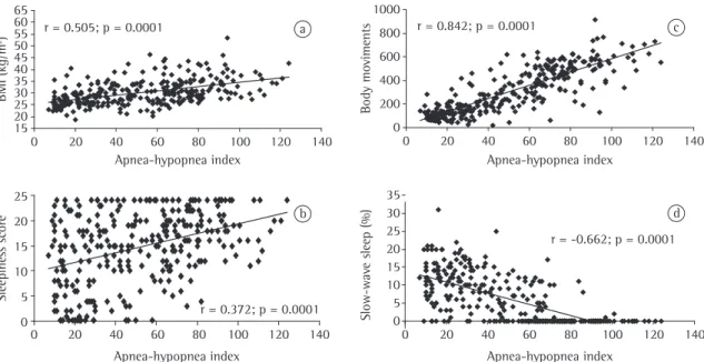
There were significant differences among the three OSAHS severity groups in terms of mean BMI (mild OSAHS: 25.9 ± 2.79 kg/m2; moderate OSAHS: 27.5 ± 3.88 kg/m2; and severe OSAHS: 31.6 ± 5.34 kg/m2). There were also significant differences between the mild OSAHS group and the severe OSAHS group in terms of systolic blood pressure (133.9 ± 36.0 mmHg vs. 144.4 ± 23.8 mmHg; p = 0.001) and in diastolic arterial pressure (77.6 ± 11.0 mmHg vs. 85.5 ± 15.2 mmHg; p < 0.0001). We found AHI to correlate better with BMI and EDS score than with any other clinical variables (Figure 3a and b). As for the poly-somnographic variables, AHI correlated best with number of body movements and slow-wave sleep (Figure 3c and d).
Discussion
We conducted a cross-sectional study involving patients with OSAHS. Gender-related characteristics were compared, factors associated with EDS were evaluated, and the effects of obesity on the disease were analyzed. Our study identified predominance of the disease in males. The severity of the disease was similar in both genders. Females presented higher mean age, lower EDS scores and less time spent in sleep apnea. Various factors were associated with EDS: BMI; sleep latency; percentage of total sleep time spent in slow-wave sleep; fragmented sleep; frequency/duration of respiratory events; and severity of hypoxemia. The degree of obesity, as calculated by BMI, was the clinical parameter that had the greatest impact on the severity of the disease (AHI, percentage of total sleep time spent in apnea and minimum SPO2).
Among the patients in our study, the mean age at diagnosis was similar to that reported in other studies.(15,16) Cases of OSAHS can occur from adoles-cence to old age. The fact that the incidence of the those who often did presented sleep that was
more fragmented, spending a greater percentage of their total sleep time in superficial (stage 2) sleep (58.9 ± 12.5% vs. 54.8 ± 15.2%, p = 0.03), spending a smaller percentage of their total sleep time in slow-wave sleep (4.2 ± 6.1% vs. 7.1 ± 4.2%, p = 0.001) and presenting greater disease severity (AHI, 60.1 ± 27.9 events/hour of sleep vs. 46.2 ± 28.2 events/hour of sleep, p = 0.0001; minimum SpO2, 67.1 ± 19.9% vs. 74.1 ± 14.4%, p = 0.005; and percentage of total sleep time spent in apnea, 47.1 ± 23.2% vs. 35.4 ± 24.2%, p = 0.0001). A total of 19 patients reported having been involved in accidents, and another 27 patients reported "near accidents" due to falling asleep at the wheel. Of those 46 patients, 44 reported falling asleep at the wheel quite often or always.
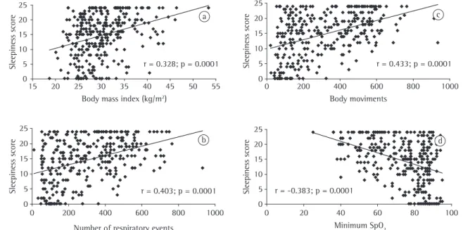
The mean BMI in the sample as a whole was 30.2 ± 5.3 kg/m2. Of the 300 patients evaluated, 271 (91%) presented a BMI ≥25 kg/m2. There were 123 patients (41%) who were classified as over-weight, 132 (44%) who were classified as obese and 16 (5.3%) who were classified as morbidly obese. Figure 2 shows the AHI, the percentage of total sleep time spent in apnea and the minimum SpO2 after the stratification of the patients by BMI. Increases in the degree of obesity were paralleled by increases in AHI and in the percentage of total sleep time spent in apnea, with concomitant drops in the minimum SpO2. We found that BMI correlated more strongly with the EDS score (r = 0.328; p < 0.0001) than with any other clinical variable. The polysom-nographic variables with which BMI correlated most
0 20 40 60 80 100
<25 25-29.9 30-39.9 >40
V
alue
AHI % of time spent in apnea Minimum SpO2 (%) Body mass index (kg/m2)
or near accidents due to falling asleep at the wheel. This is slightly higher than the 9-10% previously reported.(20)
In our study, we also observed that EDS inten-sity and OSAHS severity were significantly greater in the group of patients who reported falling asleep at the wheel. Another study, which investigated 4,002 nonprofessional drivers, demonstrated a significant correlation between traffic accidents and the degree of sleepiness.(11) However, the authors found no association between accidents and AHI. It has been shown that a higher frequency of traffic accidents is also associated with factors other than OSAHS: the use of alcohol, chronic sleep depriva-tion and narcolepsy(21); restless legs syndrome and periodic limb movements(22); and circadian rhythm sleep disorders(23) and the use of medication.(24)
Of the 300 patients, 90.3% were considered overweight or obese. Other authors have described obesity in 66 to 90% of the cases of OSAHS.(25,26) In addition to being associated with OSAHS severity, obesity is one of the risk factors for cardiovascular diseases.(27) Numerous studies have related systemic arterial hypertension to OSAHS. The prevalence of OSAHS is 9 to 20 times greater among patients with hypertension than in the general population.(28,29) It is of note that 58% of our patients suffered from hypertension, a prevalence higher than that found in literature.(30) In addition, in our sample of patients, disease peaks one decade later in females than it
does in males, as found in this and other studies, has been attributed to the effect of hormonal protec-tion of premenopausal females against OSAHS.(9)
A 9:1 male/female ratio was found in our sample of patients, whereas one of the most often cited articles on the prevalence of OSAHS in the litera-ture reported a 2:1 ratio.(17) The lower proportion of females in the present study can be explained by the different presentation of the disease in females and by the fact that diagnosing OSAHS in females continues to be difficult.(8) In addition, the females in our study presented less EDS and remained less time in sleep apnea, although disease severity in females was comparable to that observed in males.
Numerous factors associated with EDS, such as obesity, alterations in sleep architecture and number/ severity of respiratory events during sleep, were iden-tified in our study. However, the correlations between the EDS score and the remaining variables, although significant, ranged from weak to moderate, which suggests that other factors or the summation of factors can be associated with EDS. Factors such as fragmented sleep due to respiratory events or dyspnea have previously been related to EDS.(18,19)
Among the consequences of OSAHS-related EDS is the greater frequency of traffic accidents due to drivers falling asleep at the wheel.(11) Of the patients studied, 15.3% reported having accidents
15 20 25 30 35 40 45 50 55 60 65
BMI (kg/m
2)
0 20 40 60 80 100 120 140
Apnea-hypopnea index Apnea-hypopnea index
Apnea-hypopnea index Apnea-hypopnea index
r = 0.505; p = 0.0001 a
b
0 5 10 15 20 25
Sleepiness score
0 20 40 60 80 100 120 140
r = 0.372; p = 0.0001
0 200 400 600 800 1000
Body mo
viments
0 20 40 60 80 100 120 140
r = 0.842; p = 0.0001
0 5 10 15 20 25 30 35
Slow-wav
e sleep (%)
0 20 40 60 80 100 120 140
r = -0.662; p = 0.0001 d c
11. Masa JF, Rubio M, Findley LJ. Habitually sleepy drivers have a high frequency of automobile crashes associated with respiratory disorders during sleep. Am J Respir Crit Care Med. 2000;162(4 Pt 1):1407-12.
12. Engleman HM, Douglas NJ. Sleep. 4: Sleepiness, cognitive function, and quality of life in obstructive sleep apnoea/ hypopnoea syndrome. Thorax. 2004;59(7):618-22. 13. Rechtschaffen A, Kales A. A manual of standardized
terminology, techniques and scoring system for sleep stages of human subjects. Bethesda, Md: U.S. Dept. of Health, Education, and Welfare; 1968. 62p.
14. Sleep-related breathing disorders in adults: recommendations for syndrome definition and measurement techniques in clinical research. The Report of an American Academy of Sleep Medicine Task Force. Sleep. 1999;22(5):667-89. 15. Smith R, Ronald J, Delaive K, Walld R, Manfreda J, Kryger
MH. What are obstructive sleep apnea patients being treated for prior to this diagnosis? Chest. 2002;121(1):164-72. 16. Scharf SM, Garshick E, Brown R, Tishler PV, Tosteson T,
McCarley R. Screening for subclinical sleep-disordered breathing. Sleep. 1990;13(4):344-53.
17. Young T, Palta M, Dempsey J, Skatrud J, Weber S, Badr S. The occurrence of sleep-disordered breathing among middle-aged adults. N Engl J Med. 1993;328(17):1230-5. 18. Kaneita Y, Ohida T, Uchiyama M, Takemura S, Kawahara K,
Yokoyama E, et al. Excessive daytime sleepiness among the Japanese general population. J Epidemiol. 2005;15(1):1-8. 19. Whitney CW, Enright PL, Newman AB, Bonekat W, Foley
D, Quan SF. Correlates of daytime sleepiness in 4578 elderly persons: the Cardiovascular Health Study. Sleep. 1998;21(1):27-36.
20. Maycock G. Sleepiness and driving: the experience of UK car drivers. J Sleep Res. 1996;5(4):229-37.
21. Olejniczak PW, Fisch BJ. Sleep disorders. Med Clin North Am. 2003;87(4):803-33.
22. Chesson AL Jr, Wise M, Davila D, Johnson S, Littner M, Anderson WM, et al. Practice parameters for the treatment of restless legs syndrome and periodic limb movement disorder. An American Academy of Sleep Medicine Report. Standards of Practice Committee of the American Academy of Sleep Medicine. Sleep. 1999;22(7):961-8.
23. Baker SK, Zee PC. Circadian disorders of the sleep-wake cycle. In: Kryger MH, Roth T, Dement WC, editors. Principles and practice of sleep medicine. Philadelphia: Saunders; 2000. p. 606-14.
24. Qureshi A, Lee-Chiong T Jr. Medications and their effects on sleep. Med Clin North Am. 2004;88(3):751-66, x.
25. Strobel RJ, Rosen RC. Obesity and weight loss in obstructive sleep apnea: a critical review. Sleep. 1996;19(2):104-15. 26. Guilleminault C. Clinical features and evaluation of
obstructive sleep apnea. In: Kryger MH, Roth T, Dement WC, editors. Principles and practice of sleep medicine. Philadelphia: Saunders. 1989. p. 552-8.
27. Young T, Skatrud J, Peppard PE. Risk factors for obstructive sleep apnea in adults. JAMA. 2004;291(16):2013-6. 28. Stradling JR. Sleep apnoea and systemic hypertension.
Thorax. 1989;44(12):984-9.
29. Stradling J, Davies RJ. Sleep apnea and hypertension--what a mess! Sleep. 1997;20(9):789-93.
30. Hedner J, Bengtsson-Boström K, Peker Y, Grote L, Råstam L, Lindblad U. Hypertension prevalence in obstructive sleep apnoea and sex: a population-based case-control study. Eur Respir J. 2006;27(3):564-70.
the level of systemic arterial pressure was associated with obesity and with OSAHS severity.
The major limitation of the present study was the lack of a control group composed of gender- and age-matched individuals. However, due to the number of patients studied in our series, we consider that our sample size was adequate to identify factors related to the intensity of sleepi-ness. Another limitation, in terms of our ability to evaluate differences related to gender and disease severity, was the heterogeneity of the sample; only 31 patients (10.3%) were female, and 208 (69.3%) presented severe OSAHS.
In conclusion, females with OSAHS presented higher mean age at the time of diagnosis, lower EDS score and lesser time spent in sleep apnea. Factors such as fragmented sleep, number/duration of respiratory events, minimum SpO2 levels and degree of obesity were associated with EDS. Obesity has a major effect on the severity of OSAHS.
References
1. Krieger J, McNicholas WT, Levy P, De Backer W, Douglas N, Marrone O, et al. Public health and medicolegal implications of sleep apnoea. Eur Respir J. 2002;20(6):1594-609. Erratum in: Eur Respir J. 2003;21(3):561.
2. Olson LG, King MT, Hensley MJ, Saunders NA. A community study of snoring and sleep-disordered breathing. Symptoms. Am J Respir Crit Care Med. 1995;152(2):707-10.
3. Thomas GN, Jiang CQ, Lao XQ, McGhee SM, Zhang WS, Schooling CM, et al. Snoring and vascular risk factors and disease in a low-risk Chinese population: the Guangzhou Biobank Cohort Study. Sleep. 2006;29(7):896-900. 4. Wiggins CL, Schmidt-Nowara WW, Coultas DB, Samet JM.
Comparison of self- and spouse reports of snoring and other symptoms associated with sleep apnea syndrome. Sleep. 1990;13(3):245-52.
5. Krieger J, Sforza E, Boudewijns A, Zamagni M, Petiau C. Respiratory effort during obstructive sleep apnea: role of age and sleep state. Chest. 1997;112(4):875-84.
6. Arens R, Marcus CL. Pathophysiology of upper airway obstruction: a developmental perspective. Sleep. 2004;27(5):997-1019.
7. Flemons WW. Clinical practice. Obstructive sleep apnea. N Engl J Med. 2002;347(7):498-504.
8. Shepertycky MR, Banno K, Kryger MH. Differences between men and women in the clinical presentation of patients diagnosed with obstructive sleep apnea syndrome. Sleep. 2005;28(3):309-14.
9. Kapsimalis F, Kryger MH. Gender and obstructive sleep apnea syndrome, part 2: mechanisms. Sleep. 2002;25(5):499-506. 10. Lavi P. Sleep habits and sleep disturbances in industrial