Bladder response to acute sacral neuromodulation while
treating rats in different phases of complete spinal cord
injury: a preliminary study
_______________________________________________
Ping Shi
1, Youfang Fang
1, Hongliu Yu
11 Institute of Rehabilitation Engineering and Technology - University of Shanghai for Science and
Technology, Shanghai, China
ABSTRACT
ARTICLE
INFO
______________________________________________________________ ______________________
Background: Compared to conventional therapies, sacral neuromodulation (SNM) may offer an alternative, non-destructive treatment for SCI patients with bladder dysfunc-tion. Understanding bladder response to SNM treatment for SCI in different phases may yield new insights for innovative use of this promising technique.
Materials and Methods: Female Sprague-Dawley rats were used in this study to exami-ne the effects of acute SNM on bladder reflex in complete SCI rats. All rats were aexami-nes- anes-thetized and set up for continuous saline infusion. Acute SNM treatment was imple-mented for about 6 hours for each rat. Cystometric parameters, including time between contractions, contraction duration, bladder peak pressure, and number of uninhibited contractions, were analyzed and compared within rats before and after SNM treatment. Results: For the spinally transected rats during early phase (less than two weeks post spinalization), the time between contractions and contraction duration both increased after SNM treatments, yet the increased amplitude was about or less than 20%. For the spinally transected rats with a longer days survival (about two to four weeks post spinalization), the time between contractions and contraction duration substantially increased after SNM treatment and the changes for their average values were more than 90%. For the spinally transected rats with a much longer days survival (more than five weeks post spinalization), the time between contractions and contraction duration increased after SNM treatments, yet the magnitude of changes were less than 30%. Conclusion: The present study suggested that the significant effectiveness of SNM for complete SCI played its role after the spinal shock phase and prior to the development of detrusor overactivity. It indicated that the time point of SNM treatment is necessary to be paid attention.
Key words:
Spinal Cord Injuries; Urinary Bladder; Urinary Incontinence
Int Braz J Urol. 2015; 41: 1194-1201
_____________________
Submitted for publication: March 20, 2014
_____________________
Accepted after revision: June 08, 2015
INTRODUCTION
Supra-sacral lesions to the spinal cord ne-arly always lead to serious disruption of lower urinary tract function (LUTD). Previous reports showed electrical stimulation has emerged as a valuable minimally invasive treatment option for
an-terior roots (6), or the mixed sacral nerves (7, 8). In practice, only the latter two sites have demons-trated clinical significance. However, stimulation of the sacral anterior roots always combined with posterior sacral rhizotomy can prevent many sui-table patients from accepting this therapy, althou-gh cystometry and clinical examination should show that rhizotomy is effective to suppress reflex incontinence.
Sacral neuromodulation (SNM) may be an alternative solution to sacral deafferentiation, which involves stimulation of sacral afferent pa-thways rather than cutting them to suppress re-flex incontinence (8-10). From early application of SNM until now continuous research is carried out to improve this therapy and to determine the exact mechanism of action. The efficacy of SNM for treatment of LUTD probably relies on spinal and supra-spinal reflex arcs (11). This assump-tion is supported by the observaassump-tion that SNM is not effective in patients with complete or ne-arly complete SCI (12, 13). Recently, Sievert and colleagues’s investigation indicated that early SNM in patients with complete spinal cord in-jury during spinal shock (ie, the bladder arreflexia phase) prevented detrusor overactivity and urina-ry incontinence (14). Prevention of LUTD before irreversible effects occur is a convincing concept and the findings reported by Sievert and collea-gues are exciting. Their research emphasized the significance of the time point of SNM.
Taking into account that SNM is minimally invasive and completely reversible, it is of great interest whether this treatment option is valuable for neurogenic LUTD following complete SCI be-fore resorting to more invasive procedures. Our previous study (15) have substantiated that SNM could offer an alternative, non-destructive treat-ment for complete SCI animal with bladder dys-function about three weeks post-spinalization to resemble the condition of urinary bladder hyper-reflexia. Further, it is necessary to examine the
MATERIAL AND METHODS
Animal Model of SCI
All animal care and experimental procedu-res were reviewed and approved by the Institutio-nal Animal Care and Use Committees of Shanghai University for Science and Technology. Experiments were performed on female Sprague-Dawley (250g--300g) rats. In order to create an urodynamic pattern similar to humans with supra-sacral SCI, T9-T10 le-vel were considered by researchers to be the spinal transection site for the experiment rats (16).
Under general anesthesia with chloral hydra-te (400mg/kg), the rat underwent complehydra-te spinal cord transection by a micro-scissor after laminec-tomy at the T9-T10 level. The rat’s body tempera-ture was maintained at 37ºC during and after the surgery using a heating blanket until it woke up. To ensure complete disconnection of spinal fibers, the open cavity separating the two ends was filled with hemostatic gel foam. The muscle and skin were then sutured. The animal was returned to its cage after full recovery from anesthesia. Upon awakening from anesthesia, animals were given buprenorphine (0.1mL/100g body weight) subcutaneously for pain control. Postoperatively, rats were housed in shallow cages with high absorbent bedding and had access to food and water ad libitum. Penicillin (15-20mg/kg sc) was administered once daily during the feeding. The bladder was manually expressed twice daily (Crede’s maneuver).
Cystometric Studies
The spinally transected rats (7, 12, 15, 18, 20, 27, 36 and 42 days survival, respectively) were anesthetized with chloral hydrate (400mg/kg).
and 1.5mm OD) was inserted into the dome of the bladder. The free end of the implanted catheter was connected via a T-stopcock to a pressure transdu-cer (MLT0380/D, ADInstruments Pty Ltd, Sydney, Australia) for monitoring bladder pressure and an infusion pump for infusion saline. The tube was se-cured with a purse-string suture and the incisions were closed in layers.
During surgery, animal body temperature was maintained at 37ºC using a heating blanket. Room temperature saline solution was infused continuou-sly into the bladder with catheter. The intravesical pressure signal was stored using a biological signal collecting and processing system (PowerLab 4/26, AD Instruments Pty Ltd, Sydney, Australia). Urodynamic characteristics of bladder contractions were investi-gated in this study. Several parameters were calcu-lated based on urodynamic signal, including bladder contraction duration, bladder contraction cycle pe-riod (the time interval between two continuous con-tractions), peak bladder pressure and the number of uninhibited contraction. The cystometric parameters were recorded before and after SNM treatment. The
Figure 1 - Schematic diagram of the experimental set up on the rats for recording bladder activity with SNM treatment. A PE tube was inserted into the bladder dome, which was in turn connected via a three-way stopcock to an infusion pump for filling with saline, and to a pressure transducer for monitoring bladder pressure. The cathode electrode was inserted into the S1 foramina, and the anode was placed under the skin of the back.
number of uninhibited contractions was also recor-ded. The experimental setup is shown in Figure-1.
SNM Treatment
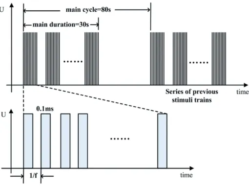
Unilateral sacral foramen electrode has been the gold standard for SNM (17). Indeed, there is no evidence that bilateral simultaneous tion has any added benefits to unilateral stimula-tion (18). Unilateral sacral segmental stimulastimula-tion with an electrode at the level of the sacral foramen S1 was accepted and performed by most resear-chers in the rat with SNM experiment (19, 20). In the experiment with acute SNM, the time of SNM treatment was not the same between experiments, however, a short or a long treatment time was con-sidered to be inefficient or lead to increased mor-tality in experimental animals. Therefore, in this study the unilateral S1 roots of rat were electrically stimulated for 6 hours using stainless steel electro-des inserted into the S1 foramina.
and train period of 80 sec (Figure-2). The stimulation amplitude was adjusted to 80% of the value that in-duced a visible tail tremor (about 1.5-4.0 V, which is variable for individual rat). Before and after stimu-lation, the rats underwent continuous urodynamic recording with saline infusion at the rate of 0.1mL/ min. During the period of experiment study, analge-sic depth was assessed continuously by the eyelash reflex and the paw retraction on moderate pinching. Anesthesia was maintained with low dose of chloral hydrate (100mg/kg).
Data Analysis
In the study, 20 values of the cystometric parameters were applied to analysis. Therefore, 20 cycles of bladder response for each rat before and after SNM treatment were recorded. The intravesical pressure was recorded and analyzed by two different persons, and the latter was blinded to the rat’s con-ditions. The results are expressed as mean and stan-dard deviation (±SD). The significance of differences
before and after SNM treatment was compared. A P<0.05 was considered statistically significant. The statistical analyses were run in MATLAB software (Math Works Inc., MA, USA).
RESULTS
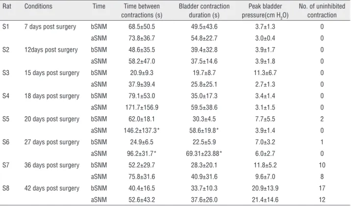
SCI rats in different phases were treated with SNM. Cystometric parameters before and after SNM treatment, including contraction cycle period, con-traction duration, bladder peak pressure, and number of uninhibited contractions, are presented in Table-1. The SCI rats were analyzed individually with bladder infusion because the condition of rats and the para-meters used in electrical stimulation are not identical, and it is also necessary to evaluate the effect of the therapeutic neuromodulation for each rat. For the spinally transected rats during early phase less than two weeks post spinalization, i.e. within 7 and 12 days survival in the present study, the time between contractions and contraction duration both
Figure 3 - Changes of average values of parameters after SNM treatment. The parameters, i.e. time between contractions and bladder contraction duration, for the SCI rat in the late phase of spinal shock or shortly after the spinal shock phase present a substantial increase.
Table 1 - Measurement from the SCI rat 7, 12, 15, 18, 20, 27, 36 and 42 days post surgery before and after SNM treatment under saline infusion with 0.1mL/min. bSNM: before Sacral Neuromodulation; aSNM: after Sacral Neuromodulation.
Rat Conditions Time Time between contractions (s)
Bladder contraction duration (s)
Peak bladder pressure(cm H2O)
No. of uninhibited contraction
S1 7 days post surgery bSNM 68.5±50.5 49.5±43.6 3.7±1.3 0
aSNM 73.8±36.7 54.8±22.7 3.0±0.4 0
S2 12days post surgery bSNM 48.6±35.5 39.4±32.8 3.9±1.7 0
aSNM 58.2±47.0 37.5±14.6 3.9±1.8 0
S3 15 days post surgery bSNM 20.9±9.3 19.7±8.7 11.3±6.7 0
aSNM 37.9±39.4 25.8±25.1 2.7±1.3 0
S4 18 days post surgery bSNM 79.1±53.0 35.0±17.3 3.4±1.4 0
aSNM 171.7±156.9 59.5±38.6 3.1±1.5 0
S5 20 days post surgery bSNM 62.0±18.1 30.3±4.5 7.7±5.5 2
aSNM 146.2±137.3* 58.6±19.8* 3.9±1.4 0
S6 27 days post surgery bSNM 24.9±6.5 22.5±5.9 7.0±3.2 1
aSNM 96.2±31.7* 69.31±23.88* 6.0±2.7 0
S7 36 days post surgery bSNM 52.2±29.7 28.3±20.1 11.8±5.2 10
aSNM 75.8±31.6 40.9±31.6 9.6±7.0 8
S8 42 days post surgery bSNM 40.4±16.5 33.7±10.3 20.9±13.9 17
aSNM 52.6±43.2 37.6±26.0 21.4±14.6 12
*P<0.05 vs bSNM
sed after SNM treatments (Table-1), yet the increased amplitude was about or less than 20% (Figure-3). The peak bladder pressure decreased or had little changes. For the spinally transected rats with longer survival days (about two to four weeks post spinalization, i.e. with 15, 18, 20 and 27 days survival in the present study), the time between contractions and contrac-tion duracontrac-tion showed substantial increases after SNM treatment (Figure-3). Especially, the parameters of time between contractions and contraction duration for SCI rats with 20 and 27 days survival dramati-cally increased (P<0.05, Table-1) and the changes for their average value were more than 90% (Figure-3). For the spinally transected rats with more than five weeks post spinalization, i.e. 36 and 42 days survival in the present study, the time between contractions and contraction duration increased after SNM treat-ments (Table-1), yet the magnitude of changes were about or less than 45% (Figure-3). The parameter of peak bladder pressure decreased or changed little af-ter SNM treatment (Figure-3). The uninhibited
urinary tract complications and decubitus ulcers in-fection, the details of the parameters for SCI rats with a very long survival days have not been included.
DISCUSSION
The control of the lower urinary tract is a complex, multilevel process that involves both the peripheral and central nervous systems (21). The le-sion of spinal cord produces LUTD by eliminating the brain mechanisms. SNM has become a well--established and widely accepted treatment moda-lity in recent years for LUTD (22), whereas SNM has been attempted without success in complete SCI pa-tients (13). However, a recent study (14) in which bi-lateral SNM was initiated early during the recovery period from complete thoracic spinal cord injury prevented the development of neurogenic detrusor overactivity and urinary incontinence. It is assumed that the effectiveness of the SNM treatment to some extent was determined by the dynamic neurologic process and reorganization or neuroplasticity that might occur after a spinal lesion, emphasizing the significance of the time point of SNM treatment. In the present study, SNM was treated on completely spinalized rats to understand its effectiveness via urodynamic parameters during different SCI phases.
With SCI the normal connections between the sacral cord and the supraspinal circuits that con-trol urine storage and release are disrupted. Patients or animals in the early phase of SCI, e.g. spinal sho-ck phase, will typically present with overflow in-continence due to detrusor failure or severe bladder outlet obstruction (15). Experimental studies in rats have shown that, soon after SCI, bulbospinal pa-thways are damaged and this disrupts the control of sympathetic preganglionic neurons (23). In the pre-sent study, SNM treated during spinal shock phase (rats S1, S2, S3 and S4) didn’t achieve a significant result. However, it is worth mentioning that during the late phase of spinal shock phase (rat S4) the treatment of SNM improved the bladder function
(24). During late spinal shock phase, electrical sti-mulation influences spinal cord plasticity and per-formance including promoting synaptic maturation and refinement of neural circuits (24).
storage and voiding (30), suggesting the SNM works through the remaining intact sympathetic trunk ex-tending to the brain (bypassing the SCI).
In the results from the present study, the efficacy of SNM substantially weakened about five weeks post spinalization although the number of uninhibited contraction decreased due to SNM pro-bably. Also in clinical practice, the biggest obstacle to the acceptance of SNM is its potential to fail over time. The reasons for these failures are not clear, but the natural plasticity of the nervous system, leading to reactivation of pathological reflex arcs (31), was considered one of possible explanations.
CONCLUSION
Electrical stimulation has been investigated for many years for the purpose of restoring function to the neurogenic bladder. SNM for treating func-tional voiding dysfunction has become established in urology. SNM was originally not considered an option for neurogenic LUTD, and SNM has been attempted without success in complete chronic SCI patients. The present study indicated that the time point of SNM treatment is necessary to be paid at-tention. Although SNM seems to be a promising therapy for neurogenic disease, further studies and long-term results with an extended cohort of com-plete SCI patients are yet to be obtained.
ACKNOWLEDGEMENT
This work is sponsored by Natio-nal Natural Science Foundation of Chi-na (Grant No. 81201174). The authors thank Miss Xueyan Zhao for the technical assistance.
CONFLICT OF INTEREST
None declared.
REFERENCES
1. Magasi P, Simon Z. Electrical stimulation of the bladder and gravidity. Urol Int. 1986;41:241-5.
2. Opisso E, Borau A, Rijkhoff NJ. Urethral sphincter EMG-controlled dorsal penile/clitoral nerve stimulation to treat neurogenic detrusor overactivity. J Neural Eng. 2011;8:036001. 3. Horvath EE, Yoo PB, Amundsen CL, Webster GD, Grill WM.
Conditional and continuous electrical stimulation increase cystometric capacity in persons with spinal cord injury. Neurourol Urodyn. 2010;29:401-7.
4. Nashold BS, Friedman H, Grimes J. Electrical stimulation of the conus medullaris to control bladder emptying in paraplegia: a ten-year review. Appl Neurophysiol. 1982;45:40-3.
5. van Balken MR, Vandoninck V, Gisolf KW, Vergunst H, Kiemeney LA, Debruyne FM, et al. Posterior tibial nerve stimulation as neuromodulative treatment of lower urinary tract dysfunction. J Urol. 2001;166:914-8.
6. Kutzenberger J, Domurath B, Sauerwein D. Spastic bladder and spinal cord injury: seventeen years of experience with sacral deafferentation and implantation of an anterior root stimulator. Artif Organs. 2005;29:239-41.
7. Schmidt RA, Jonas U, Oleson KA, Janknegt RA, Hassouna MM, Siegel SW, et al. Sacral nerve stimulation for treatment of refractory urinary urge incontinence. Sacral Nerve Stimulation Study Group. J Urol. 1999;162:352-7.
8. Kirkham AP, Knight SL, Craggs MD, Casey AT, Shah PJ. Neuromodulation through sacral nerve roots 2 to 4 with a Finetech-Brindley sacral posterior and anterior root stimulator. Spinal Cord. 2002;40:272-81.
9. Van Kerrebroeck PE, Marcelissen TA. Sacral neuromodulation for lower urinary tract dysfunction. World J Urol. 2012;30:445-50. 10. Al-zahrani AA, Elzayat EA, Gajewski JB. Long-term outcome and
surgical interventions after sacral neuromodulation implant for lower urinary tract symptoms: 14-year experience at 1 center. J Urol. 2011;185:981-6.
11. Schmidt RA, Doggweiler R. Neurostimulation and neuromodulation: a guide to selecting the right urologic patient. Eur Urol. 1998;34:23-6.
12. Hohenfellner M, Schultz-Lampel D, Dahms S, Lampel A, Matzel K, Wienhold D, et al. Functional Rehabilitation of the Neurogenic Bladder by Chronic Sacral Neuromodulation. Aktuel Urol. 1996; 27:89-91.
13. Schurch B, Reilly I, Reitz A, Curt A. Electrophysiological recordings during the peripheral nerve evaluation (PNE) test in complete spinal cord injury patients. World J Urol. 2003;20:319-22.
14. Sievert KD, Amend B, Gakis G, Toomey P, Badke A, Kaps HP, et al. Early sacral neuromodulation prevents urinary incontinence after complete spinal cord injury. Ann Neurol. 2010;67:74-84. 15. Shi P, Zhao X, Wang J, Lan N. Effects of acute sacral
neuromodulation on bladder reflex in complete spinal cord injury rats. Neuromodulation. 2013;16:583-9; discussion 589. 16. Inskip JA, Ramer LM, Ramer MS, Krassioukov AV. Autonomic
17. Weil EH, Ruiz-Cerdá JL, Eerdmans PH, Janknegt RA, Van Kerrebroeck PE. Clinical results of sacral neuromodulation for chronic voiding dysfunction using unilateral sacral foramen electrodes. World J Urol. 1998;16:313-21.
18. Scheepens WA, de Bie RA, Weil EH, van Kerrebroeck PE. Unilateral versus bilateral sacral neuromodulation in patients with chronic voiding dysfunction. J Urol. 2002;168:2046-50. 19. Ghiselli R, Lucarini G, Filosa A, Minardi D, Pelliccioni G, Orlando
F, et al. Nitric oxide synthase expression in rat anorectal tissue after sacral neuromodulation. J Surg Res. 2012;176:29-33. 20. Minardi D, Ghiselli R, Lucarini G, Mocchegiani F, Filosa A, Zizzi
A, et al. Activity and expression of nitric oxide synthase in rat bladder after sacral neuromodulation. Int J Immunopathol Pharmacol. 2008;21:129-35.
21. Fowler CJ, Griffiths D, de Groat WC. The neural control of micturition. Nat Rev Neurosci. 2008;9:453-66.
22. Van Kerrebroeck PE, Marcelissen TA. Sacral neuromodulation for lower urinary tract dysfunction. World J Urol. 2012;30:445-50.
23. Krassioukov AV, Weaver LC. Morphological changes in sympathetic preganglionic neurons after spinal cord injury in rats. Neuroscience. 1996;70:211-25.
24. Ditunno JF, Little JW, Tessler A, Burns AS. Spinal shock revisited: a four-phase model. Spinal Cord. 2004;42:383-95.
25. Sekhon LH, Fehlings MG. Epidemiology, demographics, and pathophysiology of acute spinal cord injury. Spine (Phila Pa 1976). 20015;26:S2-12.
26. de Groat WC. Mechanisms underlying the recovery of lower urinary tract function following spinal cord injury. Paraplegia. 1995;33:493-505.
27. Van Kerrebroeck PE. The role of electrical stimulation in voiding dysfunction. Eur Urol. 1998;34:27-30.
28. Blok BF, Groen J, Bosch JL, Veltman DJ, Lammertsma AA. Different brain effects during chronic and acute sacral neuromodulation in urge incontinent patients with implanted neurostimulators. BJU Int. 2006;98:1238-43.
29. van der Pal F, Heesakkers JP, Bemelmans BL. Current opinion on the working mechanisms of neuromodulation in the treatment of lower urinary tract dysfunction. Curr Opin Urol. 2006;16:261-7. 30. Blok BF, Holstege G. The pontine micturition center in rat receives
direct lumbosacral input. An ultrastructural study. Neurosci Lett. 2000;282:29-32.
31. Zvara P, Sahi S, Hassouna MM. An animal model for the neuromodulation of neurogenic bladder dysfunction. Br J Urol. 1998;82:267-71.
_______________________ Correspondence address:
Ping Shi, MD Institute of Rehabilitation Engineering and Technology University of Shanghai for Scienceand Technology,