Laparoscopic Sacral Uteropexy with Cravat
Technique--Experience and Results
_______________________________________________
Murat Api
1, Semra Kayatas
1, Aysen Boza
1, Hakan Nazik
2, Hakan Aytan
21Zeynep Kamil Maternity and Children’s Training and Research Hospital, Department of Obstetrics and
Gynecology, Istanbul and 2Adana Numune Training and Research Hospital, Department of Obstetrics and
Gynecology, Adana, Turkey
ABSTRACT
ARTICLE
INFO
______________________________________________________________ ______________________
Objective: The aim of the present study was to evaluate the safety and efficacy of a “Cravat’’ technique for the management of uterine prolapse in patients who want to preserve uterus, involving suspension of the uterus from the sacral promontory by using polypropylene mesh.
Materials and Methods: A prospective observational study between January 2011 and September 2013 was conducted. Prior to surgery, prolapse assessment was undertaken with Baden-Walker halfway system to grade the degree of prolapse at all sites. Patients with severe uterine prolapse (stage II-IV) who want to preserve uterus, were operated with Cravat technique. All patients were evaluated at 2 weeks and 6 weeks after surgery and followed for 6 months. Outcomes were evaluated objectively by vaginal examination using Baden-Walker halfway classification, and subjectively classifying patients as ‘very satisfied’, ‘satisfied’ and ‘not satisfied’ at the 6th month postoperatively.
Results: Sacral uteropexy was successfully performed by laparoscopy in 32/33 pa-tients (one needed to be converted to laparotomy). Nine papa-tients also had a concur-rent procedure as colporaphy anterior, colporaphy posterior or transobturator tape. Postoperative recovery has been uneventful with subjective and objective cure rates were 96.9% and 93.9%, respectively at six month. One recurrence of total prolapse needed to be reoperated and two patients with sacrouteropexy still remained at stage 2 prolapse. There have been no cases of graft exposure, rejection or infection with a median follow-up of 23.9 months.
Conclusions: Laparoscopic sacral uteropexy with “Cravat technique” was found to be safe and simple procedure.
Key words:
Laparoscopy; Pelvic Organ Prolapse; Suburethral Slings; Abdominal operations
Int Braz J Urol. 2014; 40: 526-32
_____________________
Submitted for publication: December 01, 2013
_____________________
Accepted after revision: March 22, 2014
INTRODUCTION
Although pelvic organ prolapse (POP) is a common and disabling condition, the exact pre-valance is difficult to ascertain due to different classification systems. Furthermore, many women do not seek medical attention despite of sympto-matic POP. Population based studies report a 11
to 19 percent lifetime risk in women undergoing surgery for prolapse or incontinence (1,2).
correct anatomical defects maintaining the uterus in normal anatomic position. Preservation of the uterus not only supports the pelvic floor, but also preserves fertility, improves sexual function and wellbeing. The increasing desire for uterine pre-servation provoked the development of new te-chniques with less morbidity and more patients’ satisfaction. Several conservative (pessary) or sur-gical treatment options were defined to preserve uterus in patients with POP (1,2). Surgical appro-ach could be either vaginal or abdominal route.
The advancement of the minimal invasive surgery with respect to equipment and skills has provided the added option of endoscopic pelvic reconstructive surgery (1). Conventional laparos-copic and robot-assisted routes result in a shorter hospital stay, faster time to recovery, and lesser postoperative pain than laparotomy, with compa-rable short-term efficacy (5-8). The United King-dom multicenter ranKing-domized equivalence trial found that after one year follow-up, there were no differences in anatomic or subjective pelvic floor outcomes compared to open and laparoscopic te-chniques; however, the blood loss, postoperative hemoglobin values, and hospital stay were better in the laparoscopic arm (9).
The objective of this study was to describe and evaluate the safety and efficacy of the sur-gical technique, laparoscopic sacrohysteropexy, a uterus preserving procedure for correction of ute-rine prolapse, involving suspension of the uterus with a mesh surrounding the isthmic portion of the uterus like cravat surrounding the neck.
MATERIAL AND METHODS
The investigation was designed as a pros-pective observational study from January 2011 through September 2013. The study protocol was conducted according to the revised Declaration of Helsinki and was approved by the Local Research and Ethics Committee of our hospital. All subjects provided written informed consent. Thirty-six wo-men with symptomatic uterine prolapse (stage II--IV), who wanted to retain their uteri, underwent a laparoscopic sacro-uteropexy. Three of them were evaluated at 2 weeks and 6 weeks but could not be followed to 6 months so that 33 of them were
included in the final analyses. Women with pre-vious abnormal cervical cytological examination, abnormal uterine bleeding, significant uterine en-largement (e.g. uterine fibroids) and concomitant medical problems precluding general anaesthesia were excluded. The assessment of the prolapse was performed by the principal author (M.A) using the Baden-Walker halfway system (10). Urodynamic studies were performed in women with urinary incontinence complaint. Urodynamic results were evaluated in accordance with criteria established by the International Continence Society (11).
Surgical Technique
least two, then 2/0 polpropylene suture and mesh arms were adjusted according to the need of ute-rine suspension by lifting the cervix at least 10cm above the level of introitus from the vaginal way (Figure-1G, Figure-1H). The rest of the mesh arms were trimmed over the sacral promontory after the fixation (Figure-1I). Some patients also underwent concomitant additional surgery as anterior and/or posterior colporrhaphy or transobturator tape (TOT).
All women were prospectively evaluated at 2 weeks and 6 weeks after surgery and followed up for at least 6 months. Operation time was defi-ned as time from skin incision to final closure
wi-thout including the concomitant surgery; duration of stay in hospital, complications, objective and subjective success rates were evaluated.
Postoperatively pelvic examination was performed to assess objective success rate defined as Baden-Walker grade 1 or 0 uterine prolapse. Subjective satisfaction of the patient was classi-fied as ‘very satisclassi-fied’, ‘satisclassi-fied’and ‘not satisclassi-fied’. Qualitative data are expressed in percen-tages (%) and quantitative data are expressed as the means ± standard deviation. Differences between the means in normally distributed va-riables were performed by using Student’s t-test.
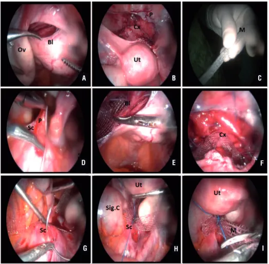
Figure 1 - The surgical procedure of Cravat technique. A) Opening of left broad ligament. B) Incision of vesicouterine peritoneum. C) 25x1.5 cm2 prolene mesh. D) Opening of peritoneum over the sacral promontorium. E) Mesh inserted to the windows in broad ligaments. F) Suturing of the mesh over the anterior cervix. G,H) Suturing of the mesh over the sacral promontory. I) Trimming of the rest of the mesh.
A
D
G
B
E
H
C
F
I
Chi-square test was done on categoric variables. Baden-Walker grades was analyzed by the Wil-coxon signed-rank test for pre and postoperati-ve grades of related samples. Analyses was un-dertaken using Statistics Package for the Social Sciences 15 (SPSS Inc. Chicago, IL). A p value of < 0.05 was accepted as statistically significant.
RESULTS
A total of 33 patients underwent a sacro--uteropexy, a uterus sparing procedure for the management of uterovaginal prolapsus. The de-mographic characteristics of the patients are sho-wn in Table-1 and operative data are given in Ta-ble-2. The mean age was 42.5 ± 7.4 years, mean parity was 3.2 ± 0.96 and mean body mass index was 25.3 ± 3.26 kg/m2. Eight (24.2%) of them have a desire for children in the future. Preoperatively, all women had significant uterine descent of grea-ter than or equal to grade 2, as measured using the Baden-Walker halfway system (Table-1). Thirteen women (39.3%) had grade 2 uterine prolapse, 18 women (54.5%) had grade 3 uterine prolapse, and the remaining 2 (6%) had grade 4 uterine prolapse. In addition, 3(9%) of the women had grade 1-3 anterior vaginal wall prolapse, 2 (6%) had poste-rior vaginal wall prolapse and 2 (6%) of women had both of them.
Nine patients (27.2%) had additional sur-gery performed at the same time. Five patients (15.1%) with urodynamically confirmed urinary incontinence had TOT, 4 patients had colpography anterior, 6 of them had colpography posterior at the time of sacro-uteropexy.
Sacral uteropexy was performed successfully by laparoscopy in 32/33 patients; one of them was converted to laparotomy because of massive hemor-rhage, but bleeding was succesfully controlled and the same procedure was proceeded. Mean operation time was 46.39 ± 5.8 minute. The maximum and mean operation time decreased with increasing the surgeons’ experience on Cravat technique. The me-dian hospital stay was 1.3 ± 0.4 day.
Major intraoperative vascular, genitouri-nary or gastrointestinal complications were not recorded. Only one patient developed middle sa-cral bleeding requiring laparotomy conversion.
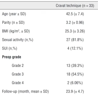
Table 1 - The demographic characteristics of patients.
Cravat technique (n = 33)
Age (year ± SD) 42.5 (± 7.4)
Parity (n ± SD) 3.2 (± 0.96)
BMI (kg/m2, ± SD) 25.3 (± 3.26)
Sexual activity (n,%) 27 (81.8%)
SUI (n,%) 4 (12.1%)
Preop grade
Grade 2 13 (39.3%)
Grade 3 18 (54.5%)
Grade 4 2 (6.06%)
Follow-up (month, mean ± SD) 23.9 (± 4.7)
SUI = stress urinary incontinence; BMI = body mass index; SD = standard deviation
Table 2 - The operation characteristics of Cravat technique.
Cravat technique (n = 33)
Operation time (minutes, mean, range) 46.39 (40-65)
Duration of stay (day, mean ± SD ) 1.3 (± 0.4)
Concomitant procedure (n,%) 9 (27.2%)
CA 0
CP 3
CA+CP 2
TOT 1
CA+TOT 2
CP+TOT 1
Complications (n)
Transfusion 1
Bowel injury 0
Ureteric injury 0
Postoperative recovery was uneventful. Among all participants, 96.9% (32/33) were either very satisfied 84.8% (28/33) or satisfied 12.1% (4/33). One (1.8%) patient reported not satisfied because of laparotomy conversion, blood transfusion and long hospital stay.
The mean postoperative Baden-Walker grades of the patients were statistically signifi-cantly lower compared to preoperative grades (p < 0.001). Subjective and objective cure rates were 96.3% and 93.9%, respectively at six months. The-re was no cases of graft exposuThe-re, The-rejection or in-fection with a median follow-up of 23.9 months and no case of recurrence of total prolapse.
DISCUSSION
The demand for uterine preservation du-ring surgical management of uterovaginal pro-lapse is increasing and it is difficult to select the ideal uterus-sparing procedure for a given patient. Several alternative operations for prolapse repair with uterine preservation, using either a vaginal or an abdominal approach, have been proposed (1,2,12-20).
Vaginal operations are well defined in the literature; as in 2001, posterior intravaginal slin-goplasty was first described with a reported mesh complications as infection and erosion (12). Other technique is the Manchester operation, that in-cludes the vaginal shortening of the uterosacral and cardinal ligaments with cervical amputation which has a deleterious effect on fertility and also complicates with dyspareunia, dysmenorrhea, re-current uterine prolapse and enterocele formation (3,4). Transvaginal sacrospinous fixation is the other vaginal operation that includes the fixation of cervix to the sacrospinous ligament by dissec-ting pararectal space down to the sacrospinous ligament. The proximity of the sacrospinous liga-ment to the sciatic nerve and pudendal vessels and nerves may cause significant buttock and leg pain and haemorrhage (13).
Abdominal operations performed for pre-serving the uterus have also been defined. The suspension of uterus from round ligaments (ven-trosuspension) is associated with high recurrence rate. In a case series of nine women, recurrence
of prolapse in eight women was reported within 3 months of surgery (2,14). This technique causes significant change of normal vaginal axis, hence it results in transmission of abdominal pressure to the cul-de-sac increasing the formation of entero-cele (13,14).
Uterosacral plication is the other descri-bed technique in literature as placing three purse--string sutures from the uterosacral ligaments to the posterior cervix (15). Wu et al. reported a case series of seven women with no recurrence of pro-lapse at 9-17 months follow-up and also, Maher et al. reported an objective success rate of 79% in 43 women after a mean follow-up of 12 months (1,16). Lantzsch et al. described complications in-cluding massive haemorrhage, buttock pain and recurrent cystocele, and also Stepp and Paraiso described ureteral injury after uterosacral plica-tion (17,18).
Suspension of prolapsed uterus from sacral promontorium by using polypropylene mesh was called sacrohysteropexy. This technique yields a satisfactory anatomic and functional results with normal vaginal axis (19). It was first described by Cutner et al. by passing Mersilene tape through uterosacral ligaments to resuspend the uterus to the sacral promontory bilaterally (20). Then this procedure was changed and non-absorbable mesh was started to be used by suturing the cervix pos-teriorly to the sacral promontorium with or wi-thout pelvic peritoneum closure via abdominal or laparoscopic way. The disadvantage of these te-chniques is the possible extrusion of mesh from cervix where it has been sutured before (21). Also, in sacrohysteropexy pulling the uterus from only one location seems to be less secure than wrap-ping the mesh around the uterus as described in our Cravat technique.
medially to the right ureter, and also the peritoneum at the level of the insertion of uterosacral ligaments was mobilised to ease complete peritonisation of the mesh. Bifurcated polypropylene mesh was sutured anteriorly to the anterior cervical wall, posteriorly to the anterior longitudinal ligament over the sacral promontorium and then, complete peritonisation of mesh was performed (2). The difference from the bi-furcated mesh described by Price et al. and our mesh was the 25x1.5cm2 tape. The advantage of our mesh was that it can be either available in every operation theather or produced by slicing the 25x25cm2 squa-re mesh into squa-rectangular 25x1.5 cm2 long pieces.
Price et al. connected the bifurcated tips of the mesh anterior to the uterine cervix and fixed these tips by 5-6 non-absorbable suture material (2). In our technique, the mesh was wrapped around the uterus anteriorly and the tips were connected on the sacral promontory. For the anterior fixation we used one absorbable polyglactin 910 suture. We believe that mesh located between the vesica and cervix may potentially irritate the bladder, if mesh tips and 5-6 non-absorbable suture material are left in-situ in this area which may theoretically cause de-novo detrusor instability or bladder fistula afterwards. Absorbable suture material might not cause perma-nent irritation on the bladder but it is premature to say that absorbable material is superior to the non--absorbable one. Although we could not prove this fact by urodynamic examination, none of our sub-jects complained of overactive bladder symptoms in the postoperative follow-up period.
In Price’s technique, all of the operations were performed with a standardized mesh length placed under standardized tension. On the other hand, we adjusted the mesh length described as follows: we pulled the mesh between the prolap-sed uterus and sacral promontory with a moderate tension in order to suspend the cervix at least abo-ve the interspinous leabo-vel of the bony pelvis. Uterus was pushed with a ZUMI vaginally. The sacral pro-montory mesh was fixed to the anterior longitudi-nal ligament by non-absorbable suture and then the extra mesh was incised and taken out. We thought that every patient needs a different length of mesh because of the variation in lenght of the prolapsed portion, so putting the uterus in normal anatomic position by raising the cervix and adjusting the
len-ght of the mesh milen-ght be the key components of the our technique which increased the success rate.
In our technique, we did not make a perito-neal relaxing incision down into the pelvis, we only incised the peritoneum over the sacral promontory and avoided the bruise of mesh which might con-tribute to complications (22,23). We believe that suturing the peritoneum may cause direct/ indirect ureteric injury (kinking) or inadvertent bowel injury (22). In addition, the insertion of sutures into the pe-ritoneum may cause bleeding and the formation of haematomas (24). As a result of non-peritonisation of mesh in abdominal vault suspension operations done by Elneil et al. none of the patients developed problems with adhesions, only 3 out of 128 patients with a rate of 2.3% had vaginal mesh erosion (22).
Cravat technique had shorter mean opera-tion time than other techniques (1,2). Surgical steps that likely contributed most to improve the ope-ration time were eliminating the need to open the peritoneum from the sacral promontory to the cul--de-sac, non-peritonisation of mesh and since mesh was wrapped around the uterus, there was only one area that we should secure to mesh instead of two or more. The number and localisation of the sutures may also decrease the operation time; one suture rather than 6 and using anterior cervical wall rather than posterior one may fasten the operation.
Although this follow up period cannot pro-vide conclusive epro-vidence we are most encouraged that in long term follow-up more successful results might be expected from hysteropexy with Cravat technique with lower complication rate and shorter operation time.
CONCLUSIONS
for larger randomized controlled trial evaluating its efficacy, safety and long term outcome measures still remains.
CONFLICT OF INTEREST
None declared.
REFERENCES
1. Maher CF, Carey MP, Murray CJ: Laparoscopic suture hysteropexy for uterine prolapse. Obstet Gynecol. 2001; 97: 1010-4.
2. Price N, Slack A, Jackson SR: Laparoscopic hysteropexy: the initial results of a uterine suspension procedure for uterovaginal prolapse. BJOG. 2010; 117: 62-8.
3. O’Leary JA, O’Leary JL: The extended Manchester operation. A review of 289 cases. Am J Obstet Gynecol. 1970; 107: 546-50.
4. Tipton RH, Atkin PF: Uterine disease after the Manchester repair operation. J Obstet Gynaecol Br Commonw. 1970; 77: 852-3.
5. Paraiso MF, Walters MD, Rackley RR, Melek S, Hugney C: Laparoscopic and abdominal sacral colpopexies: a comparative cohort study. Am J Obstet Gynecol. 2005; 192: 1752-8.
6. Higgs PJ, Chua HL, Smith AR: Long term review of laparoscopic sacrocolpopexy. BJOG. 2005; 112: 1134-8. 7. Geller EJ, Siddiqui NY, Wu JM, Visco AG: Short-term outcomes
of robotic sacrocolpopexy compared with abdominal sacrocolpopexy. Obstet Gynecol. 2008; 112: 1201-6.
8. Klauschie JL, Suozzi BA, O’Brien MM, McBride AW: A comparison of laparoscopic and abdominal sacral colpopexy: objective outcome and perioperative differences. Int Urogynecol J Pelvic Floor Dysfunct. 2009; 20: 273-9. 9. Freeman RM, Pantazis K, Thomson A, Frappell J, Bombieri
L, Moran P, et al.: A randomised controlled trial of abdominal versus laparoscopic sacrocolpopexy for the treatment of post-hysterectomy vaginal vault prolapse: LAS study. Int Urogynecol J. 2013; 24: 377-84.
10. Baden WF, Walker TA: Genesis of the vaginal profile: a correlated classification of vaginal relaxation. Clin Obstet Gynecol. 1972; 15: 1048-54.
11. Abrams P, Cardozo L, Fall M, Griffiths D, Rosier P, Ulmsten U, et al.: The standardisation of terminology of lower urinary tract function: report from the Standardisation Sub-committee of the International Continence Society. Am J Obstet Gynecol. 2002; 187: 116-26.
12. Baessler K, Hewson AD, Tunn R, Schuessler B, Maher CF: Severe mesh complications following intravaginal slingplasty. Obstet Gynecol. 2005; 106: 713-6.
13. Richardson DA, Scotti RJ, Ostergard DR: Surgical management of uterine prolapse in young women. J Reprod Med. 1989; 34: 388-92.
14. O’Brien PM, Ibrahim J: Failure of laparoscopic uterine suspension to provide a lasting cure for uterovaginal prolapse. Br J Obstet Gynaecol. 1994; 101: 707-8.
15. Diwan A, Rardin CR, Strohsnitter WC, Weld A, Rosenblatt P, Kohli N: Laparoscopic uterosacral ligament uterine suspension compared with vaginal hysterectomy with vaginal vault suspension for uterovaginal prolapse. Int Urogynecol J Pelvic Floor Dysfunct. 2006; 17: 79-83. 16. Wu MP: Laparoscopic uterine suspension for the treatment
of uterovaginal prolapse. Int J Gynaecol Obstet. 1997; 59: 259-60.
17. Lantzsch T, Goepel C, Wolters M, Koelbl H, Methfessel HD: Sacrospinous ligament fixation for vaginal vault prolapse. Arch Gynecol Obstet. 2001; 265: 21-5.
18. Stepp KJ, Paraiso MF: Laparoscopic management of ureteric obstruction after uterosacral vaginal vault suspension. J Minim Invasive Gynecol. 2005; 12: 70-2.
19. Leron E, Stanton SL: Sacrohysteropexy with synthetic mesh for the management of uterovaginal prolapse. BJOG. 2001; 108: 629-33.
20. Cutner A, Kearney R, Vashisht A: Laparoscopic uterine sling suspension: a new technique of uterine suspension in women desiring surgical management of uterine prolapse with uterine conservation. BJOG. 2007; 114: 1159-62. 21. Moulder JK, Cohen SL, Morse AN, Einarsson JI: Mesh
extrusion through the internal cervical os: an unusual complication following laparoscopic sacrocervicopexy. Female Pelvic Med Reconstr Surg. 2013; 19: 309-11. 22. Elneil S, Cutner AS, Remy M, Leather AT, Toozs-Hobson
P, Wise B: Abdominal sacrocolpopexy for vault prolapse without burial of mesh: a case series. BJOG. 2005; 112: 486-9.
23. Lindeque BG, Nel WS: Sacrocolpopexy--a report on 262 consecutive operations. S Afr Med J. 2002; 92: 982-5. 24. Ong AW, Cohn SM, Cohn KA, Jaramillo DH, Parbhu R,
McKenney MG, et al.: Unexpected findings in trauma patients dying in the intensive care unit: results of 153 consecutive autopsies. J Am Coll Surg. 2002; 194: 401-6.
_______________________ Correspondence address: Aysen Boza, MD Zeynep Kamil Maternity and Children’s Training and