Evaluation of the use of captopril on peritoneal fibrosis
induced in rats by the use of glucose solution 4.25%
Authors
Adriana Fátima Menegat Schuinski1
Gilberto Baroni1 Roberto Flávio Pecoits Filho2
Fernando Meyer3 Marina Lindstrom Wittica Cerqueira2
Maria Angélica Alexandre da Silva2
Viviane de Carvalho2
1 State University of Ponta
Grossa (UEPG).
2 Pontifical Catholic University
of Paraná (PUCPR), Curitiba - PR, Brazil.
3 Department of Urology
- Catholic University of Paraná (PUCPR). Cajuru University Hospital, PUCPR, Curitiba-PR, Brazil.
Submitted on: 08/16/2012. Approved on: 09/05/2013.
Correspondence to: Fernando Meyer.
Pontifical Catholic University of Paraná (PUCPR). Department of Urology. Rua Portugal, nº 307. Curitiba, PR, Brazil. CEP: 80510-280. Tel: (55 41)3074-7478. Fax: (55 41)3015-0303. E-mail: [email protected]
Introduction: Chronic renal failure has alarming incidence all over the world in this century. Among the modalities of dia-lytic treatment, peritoneal dialysis has a major spot. This method of dialytictreat-ment may present complications, and among those is peritoneal fibrosis. It oc-curs in patients submitted to peritoneal dialysis along years. It's most dangerous form is sclerosing encapsulant perito-nitis, wich leads to a need of change in modality and many times lead to death.
Objective: Study the influence of using captopril on the peritoneal fibrosis in-duced in rats using solution with glucoses 4.25%. Methods: Prospective controlled study in 20 non-uremic Wistar rats. The animals received a peritoneal infu-sion of 10 ml/100g of peritoneal dialysis solution glucose 4.25% on a daily basis. The animals were divided in two groups: experimental and control. The experimen-tal group received captopril 30 mg/kg/d, by a gastric tube. The control group did not receive any drug. The follow-up was 21 and 49 days. At the end, one surgi-cal procedure was performed to get his-tological samples of visceral and parietal peritoneum. The samples were analyzed using Hematoxylin Eosin and Sirius Red, to evaluate the severity of the fibrosis.
Results: The analysis showed that the intensity of the fibrosis, the peritoneal thickness and the cell number in experi-mental and control groups were not statis-tically significant different in experimen-tal and control groups. Conclusion: Our findings indicate that captopril do not decrease the intensity of fibrosis on the peritoneal membrane that happens on rats on peritoneal dialysis.
A
BSTRACTKeywords: captopril, peritoneal dialysis, peritoneal fibrosis.
I
NTRODUCTIONChronic renal failure (CRF) has had an alarming increase in incidence, and it is considered a world health problem.1
In Brazil, there has been an increase of 8.8% per annum in the dialysis population since the year 2001.1
CRF can be defined as the progressive and irreversible loss of kidney function for at least three months, having a kidney structural or functional lesion; or glomerular filtration rate less than 60 mL/min/1.73 m2.1 In its more advanced
stages, the so-called renal replacement therapy (RRT) is necessary, which can be performed by hemodialysis (HD), peritoneal dialysis (PD) - which may be divided into continuous ambulatory peritoneal dialysis (CAPD), automated peritoneal dialysis (APD), intermittent peritoneal dialysis (IPD) and renal transplantation (TX).
PD was popularized after the advent of CAPD, because it enables the patient to undergo treatment at home, favoring better adaptation to social and work activities. However, this treatment mode bears complications, such as peritonitis and peritoneal fibrosis - which may progress to encapsulating peritoneal fibrosis, greatly limiting its use. Peritoneal fibrosis is the main morphological change that occurs in CAPD. The pathophysiology is complex, in which there may be peritonitis, PD solutions acidic pH, uremia per se.2
In the early 1970s, scientists discovered polypeptides that inhibit the
formation of angiotensin II or block their receptors. Several experimental studies have shown that the renin-angiotensin system (RAS) has important physiological and pathophysiological functions. Subsequent to these findings, there was a great interest in developing a new class of agents that acted in this system by inhibiting its deleterious actions, thus creating the ACE inhibitors. Later studies revealed that ACE inhibitors acted on the pathophysiological mechanism of hypertension, kidney and heart failure.3
Studies are being carried out to evaluate its antifibrotic action because a study showed the existence of an independent renin-angiotensin system in various organs,4 including the peritoneum.
ACE inhibitors are currently inexpensive drugs available in the public healthcare network, and besides its antihypertensive efficacy, they have effects on the cardiovascular system by inhibiting the deleterious actions of angiotensin and its kidney protective effect.
Considering the need to reduce the occurrence of these complications, we developed an experimental study in an animal model using peritoneal dialysis solution with 4.25% hypertonic glucose. This model was based on the model developed by Ishii et al.5
Authors such as Bui et al.6 and Baroni et al.,7
performing daily infusion of hypertonic peritoneal dialysis solution, used the N-acetylcysteine and statins, respectively, to assess the ability to protect the peritoneum by the peritoneal irritant agent.
Angiotensin-converting enzyme inhibitors (ACE inhibitors) have the ability to modify the course of disease in various organs such as kidneys, heart and lungs.
Due to the increasing number of patients on PD as RRT, our study evaluated the inhibitory effects of captopril - an ACE inhibitor in peritoneal fibrosis, with the intent to create the possibility of chronic kidney patients to have a preventive therapy for peritoneal fibrosis in PD, which often mandates a change in dialysis treatment.
M
ETHODSThis study was approved by the Ethics Committee for use in Animals (CEUA) of the Pontifical
Catholic University of Paraná (PUC-PR). The experiments were performed according to the Brazilian Committee for animal studies (COBEA).
This was a prospective controlled study in experimental animal model. Twenty male Wistar rats (Rattus norvegicus albinus, Rodentia mammalia) at 90 days of age and weighing between 250-300g we included in the study. They were divided into two groups: experimental and control, with ten animals in each. They were kept in proper cages, with a maximum of five animals in each cage, at 21°C of room temperature with 12h light-dark cycle. The animals were tagged and placed in cages with free access to food and water. They were assessed and weighed daily.
All rats were subjected to daily abdominal puncture in the left lower quadrant, using a 25 x 7 needle. A commercially available peritoneal dialysis solution was infused with 4.25% glucose, once a day. The infused dose was 10 ml/100g/body weight. The animals were divided into control and experimental groups with ten animals each and followed for 21 and 49 days, when they were euthanized. The experimental group received 30 mg/kg/wt solution of captopril orally by gavage once a day, through an adequate enteral tube used for studies in rats. This solution contained 1 mg/ml prepared by the pharmacy at PUC-PR. The control group did not receive captopril.
counting mononuclear cells per field and measured peritoneal thickness, according to the methodology used by the laboratory.
The results obtained in the study were expressed as mean, standard deviation, median, minimum and maximum values. To compare the groups we used the nonparametric Mann-Whitney test. P values < 0.05 were con-sidered statistically significant. The data was analyzed using the Statistica v.8.0 software.
R
ESULTSDuring this study, four animals died and at the end of the experiment and the sample was: control group 21 days: three animals; control group 49 days five animals; experimental group 21 days: five animals in the control group and at 49 days we had three animals. The deaths in the control group had no defined cause and in the experimental group it was related to trauma during gavage.
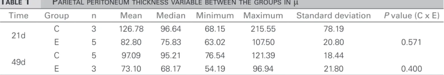
Table 1 shows the parietal peritoneum thickness. We found a reduction in thickness of the visceral peritoneum in both experimental groups at 21 and 49 days, without statistical significance when compared with the control group at the same time frames.
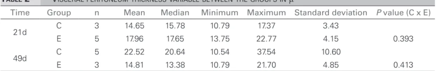
We also found thickness of the visceral peritoneum, though to a lesser degree, which can be explained by the quadruped position of the animal, causing the peritoneal solution to be in gre-ater contact with the parietal peritoneum surface. The experimental group also had a trend towards lower thickness compared to the control group in the same periods, as seen on Table 2.
The analysis of the mature visceral collagen variable showed greater deposition in the control group both at 21 and 49 days, with a trend toward lower deposition in the experimental groups at the same time frames. This analysis can be seen on Figure 1A.
Upon assessing the young visceral collagen variable, we found a statistically significant
reduction, p-value of 0.016 in the 49 days
experimental group, compared with the control group during the same period. At 21 days, there was a tendency for lower deposition in the experimental group. The results are shown on Figure 1B.
The mature parietal collagen variable tended to higher deposition in the experimental group at both time frames 21 and 49 days; although without statistical significance. This finding is shown on Figure 1C.
The young parietal collagen variable had the same behavior of the young collagen variable, being higher in the experimental group. This result is shown on Figure 1D.
The visceral peritoneum cellular reaction is summarized on Table 3.
The parietal peritoneum cellular reaction is summarized on Table 4.
The micrographs below, seen on Figure 2, showed thickening of the visceral and parietal peritoneum and the measurement was carried out in microns (μ).
D
ISCUSSIONThis study was carried out in order to evaluate the use of captopril in peritoneal fibrosis, which was caused by the use of dialysis solution with 4.25% hypertonic glucose.
This solution was used for a minimum period of 21 days and a maximum of 49 days of exposure, and we measured the changes caused to the abdominal wall parietal peritoneum and the spleen visceral peritoneum. The choice of these organs aimed to avoid choice bias in the area to be examined.
Peritoneal fibrosis is the major complication vis-à-vis the morphological changes that occur in CAPD. The mesothelial cells undergo a transition to mesenchymal cells, and fibroblasts derived from
TABLE 1 PARIETALPERITONEUMTHICKNESSVARIABLEBETWEENTHEGROUPSINμ
Time Group n Mean Median Minimum Maximum Standard deviation P value (C x E)
21d C 3 126.78 96.64 68.15 215.55 78.19
E 5 82.80 75.83 63.02 107.50 20.80 0.571
49d C 5 97.09 95.21 76.54 121.39 18.44
E 3 73.10 68.17 54.19 96.94 21.80 0.400
TABLE 2 VISCERALPERITONEUMTHICKNESSVARIABLEBETWEENTHEGROUPSINμ
Time Group n Mean Median Minimum Maximum Standard deviation P value (C x E)
21d C 3 14.65 15.78 10.79 17.37 3.43
E 5 17.96 17.65 13.75 22.77 4.15 0.393
49d C 5 22.52 20.64 10.54 37.54 10.60
E 3 14.81 13.38 10.79 21.70 4.85 0.413
C: Control group; E: Experimental group; µ: Microns.
Figure 1. A: Visceral peritoneum HE; B: Parietal peritoneum HE; C: Visceral peritoneum SR; D: Parietal peritoneum HE.
Figure 2. Microphotography of the visceral and parietal peritonea. Upper figure: HE dye; Lower figure: Sirius red stain.
mesothelium are primarily responsible for the fibrinogenic process.8,9
Cellular changes in our study revealed an increase in the number of cells, higher in the
experimental group at times 21 and 49 days, in the visceral peritoneum. The parietal peritoneum showed a decrease in the number of cells in the experimental group - it can be inferred that captopril was able to reduce the inflammatory reaction in the parietal peritoneum, although without statistical significance.
Parietal peritoneum thickness was lower in the experimental group at both periods 21 and 49 days, although without statistical significance.
The visceral peritoneum mature collagen variable was lower in the experimental group on days 21 and 49 of the experiment compared to the control group. The young collagen also appeared at a lesser extent in the experimental groups, compared to what was expected.
The results revealed a tendency towards the beneficial effects of captopril, but the data was not statistically significant and it may be related to the small sample size.
The models of experimental peritoneal sclerosis may use several peritoneal cell irritants, such as asbestos, iron dextran, sodium hypochlorite and silica, causing hydrogen peroxide production by mesothelial cells, which in contact with the PD glucose solution increased substantially.10
Several hypotheses have been considered to explain the peritoneal changes that occur during the PD, and the main one is that PD solutions are biocompatible, leading researchers to develop new solutions which could be less irritating to the peritoneum, e.g. icodextrine.11
Sawada et al.12 used an experimental model in
TABLE 3 VISCERALPERITONEUMCELLULARREACTION
Time Group n Mean Median Minimum Maximum Standard deviation P value (C x E)
21d C 3 18.60 23.10 6.60 26.10 10.50
E 5 21.58 22.10 4.00 33.20 11.28 1
49d C 5 31.72 27.70 22.10 45.40 9.10
E 3 13.93 9.65 3.00 33.40 14.05 0.111
C: Control group; E: Experimental group.
TABLE 4 PARIETALPERITONEUMCELLULARREACTION
Time Group n Mean Median Minimum Maximum Standard deviation P value (C x E)
21d C 3 23.13 19.80 12.30 37.30 12.83
E 5 20.46 21.00 11.70 32.80 8.02 1 49d C 5 24.16 21.70 15.10 34.60 7.58
E 3 21.10 18.80 16.20 28.30 6.37 0.571
C: Control group; E: Experimental group; µ: Microns.
noting that quinalapril was able to reduce the fibrosis effects. Still with this model, Bozkurt et al.13 studied
N-acetylcysteine as an anti-fibrotic agent, which did not prove effective, but showed promising anti-inflammatory effects.
Ultrafiltration failure is a common complication of CAPD, with greater prevalence along the years of treatment in this modality. Enalapril, an ACE inhibitor, widely used in medical practice as an antihypertensive agent, significantly reduced the production of TGF-B1 (B1 growth factor), but the peritoneal thickening was partially reduced. They concluded that TGF-B1 inhibition by enalapril can preserve peritoneal function.14
Renin angiotensin system (RAS) block by lisinopril, ACE inhibitor or valsartam - an angiotensin receptor blocker (ARB) had a protective effect on the peritoneal membrane against the effects of hypertonic glucose.15
ACE inhibitors have been studied by authors as antioxidants in the prevention of fibrosis in various organs, i.e. kidneys, lungs, heart and pancreas, with very pleasing results. Chronic kidney patients usually have hypertension and they frequently need to use antihypertensive medication. ACE inhibitors are among the most prescribed drugs. Captopril was the drug of choice in this study because it is commonly used, with well-established side effects, it is commercially available at low cost and it is also available in the public healthcare system, facilitating its acquisition.
C
ONCLUSIONCaptopril did not prove to be statistically significant in reducing peritoneal fibrosis induced in rats by hypertonic glucose solution.
This finding in this study may be related to the small sample size, and new studies should be carried out using a sample with a larger number of animals.
R
EFERENCES1. Sesso Rde C, Lopes AA, Thomé FS, Lugon JR, Burdmann Ede A. Brazilian dialysis census, 2009. J Bras Nefrol 2010;32:374-8. 2. Lugon JR. Doença Renal Crônica no Brasil: um problema de
saúde pública. J Bras Nefrol 2009;31:2-5.
3. Okada M, Kikuzuki R, Harada T, Hori Y, Yamawaki H, Hara Y. Captopril attenuates matrix metalloproteinase-2 and -9 in monocrotaline-induced right ventricular hypertrophy in rats. J Pharmacol Sci 2008;108:487-94. DOI: http://dx.doi. org/10.1254/jphs.08174FP
4. Nakamoto H, Imai H, Fukushima R, Ishida Y, Yamanouchi Y, Suzuki H. Role of the renin-angiotensin system in the pathoge-nesis of peritoneal fibrosis. Perit Dial Int 2008;28:S83-7. 5. Ishii Y, Sawada T, Shimizu A, Tojimbara T, Nakajima I,
Fu-chinoue S. An experimental sclerosing encapsulating peritonitis model in mice. Nephrol Dial Transplant 2001;16:1262-6. DOI: http://dx.doi.org/10.1093/ndt/16.6.1262
6. Bui DS, Seguro AC, Shimitzu MH, Schliemann I, Martini D, Romão JE Jr, et al. N-Acetylcysteine protects the peritoneum from the injury induced by hypertonic dialysis solution. J Ne-phrol 2012;25:90-5.
7. Baroni G, Schuinski AF, Berticelli PT, Silva MA, Gouveia DS, Pecoits Filho R, et al. The influence of simvastatin in induced peritoneal fibrosis in rats by peritoneal dialysis solution with glucosis 4.25%. Acta Cir Bras 2012;27:350-6. DOI: http:// dx.doi.org/10.1590/S0102-86502012000400012
9. Aroeira LS, Aguilera A, Selgas R, Ramírez-Huesca M, Pérez-Lozano ML, Cirugeda A, et al. Mesenchymal conversion of mesothelial cells as a mechanism responsible for high solute transport rate in peritoneal dialysis: role of vascular endothelial growth factor. Am J Kidney Dis 2005;46:938-48. DOI: http:// dx.doi.org/10.1053/j.ajkd.2005.08.011
10. De Vriese AS, Tilton RG, Mortier S, Lameire NH. Myofi-broblast transdifferentiation of mesothelial cells is mediated by RAGE and contributes to peritoneal fibrosis in uraemia. Nephrol Dial Transplant 2006;21:2549-55. DOI: http://dx.doi. org/10.1093/ndt/gfl271
11. Nakao A, Nakao K, Takatori Y, Kojo S, Inoue J, Akagi S, et al. Effects of icodextrin peritoneal dialysis solution on the perito-neal membrane in the STZ-induced diabetic rat model with par-tial nephrectomy. Nephrol Dial Transplant 2010;25:1479-88. DOI: http://dx.doi.org/10.1093/ndt/gfp479
12. Sawada T, Ishii Y, Tojimbara T, Nakajima I, Fuchinoue S, Teraoka S. The ACE inhibitor, quinapril, ameliorates perito-neal fibrosis in an encapsulating peritoperito-neal sclerosis model in mice. Pharmacol Res 2002;46:505-10. DOI: http://dx.doi. org/10.1016/S1043661802002281
13. Bozkurt D, Hur E, Ulkuden B, Sezak M, Nar H, Purclutepe O, et al. Can N-acetylcysteine preserve peritoneal function and morphology in encapsulating peritoneal sclerosis? Perit Dial Int 2009;29:S202-5.
14. Jearnsujitwimol V, Eiam-Ong S, Kanjanabuch T, Wathanavaha A, Pansin P. The effect of angiotensin II receptor blocker on peritoneal membrane transports in continuous ambulatory pe-ritoneal dialysis patients. J Med Assoc Thai 2006;89:S188-95. PMID: 17044472
15. Duman S, Sen S, Duman C, Oreopoulos DG. Effect of valsartan