INTRODUCTION
The endodontic infection in primary teeth with pulp necrosis and apical periodontitis is of polymicrobial nature with predominance of anaerobic bacteria (1), which are disseminated within the root canal system
Antibacterial Activity of Root Canal Filling
Materials for Primary Teeth: Zinc Oxide and
Eugenol Cement, Calen Paste Thickened with
Zinc Oxide, Sealapex and EndoREZ
Alexandra Mussolino de QUEIROZ1
Paulo NELSON-FILHO1
Léa Assed Bezerra da SILVA1 Sada Assed1
Raquel Assed Bezerra da SILVA1 Izabel Yoko ITO2
1Department of Pediatric Clinic, Preventive and Community Dentistry, Ribeirão Preto Dental School,
University of São Paulo, Ribeirão Preto, SP, Brazil
2Department of Clinical Analysis, Toxicology and Bromatology, School of Pharmaceutical Sciences of Ribeirão Preto,
University of São Paulo, Ribeirão Preto, SP, Brazil
This study evaluated in vitro the antibacterial activity of 4 root canal filling materials for primary teeth - zinc oxide and eugenol cement (ZOE), Calen paste thickened with zinc oxide (Calen/ZO), Sealapex sealer and EndoREZ sealer - against 5 bacterial strains commonly found in endodontic infections (Kocuria rhizophila, Enterococcus faecalis, Streptococcus mutans, Escherichia coli and Staphylococ-cus aureus) using theagar diffusion test (agar-well technique). Calen paste, 1% chlorhexidine digluconate (CHX) and distilled water served as controls. Seven wells per dish were made at equidistant points and immediately filled with the test and control materials. After incubation of the plates at 37oC for 24 h, the diameter of the zones of bacterial growth inhibition produced around the wells was measured (in mm) with a digital caliper under reflected light. Data were analyzed statistically by analysis of variance and Tukey’s
post-hoc test (α=0.05). There were statistically significant differences (p<0.0001) among the zones of bacterial growth inhibition
produced by the different materials against all target microorganisms. K. rhizophila wasinhibited more effectively (p<0.05) by ZOE, while Calen/ZO had its highest antibacterial activity against E. faecalis (p<0.05). S. mutans was inhibited by Calen/ZO, Sealapex and ZOE in the same intensity (p>0.05). E. coli wasinhibited more effectively (p<0.05) by ZOE, followed by Calen/ZO and Sealapex. Calen/ZO and ZOEwere equally effective (p>0.05) against S. aureus, while Sealapex had the lowest antibacterial efficacy (p<0.05)
against this microorganism. EndoREZ presented antibacterial activity only against K. rhizophila and S. aureus. The Calen paste and Calen/ZO produced larger zones of inhibition than 1% CHX when the marker microorganism was E faecalis. In conclusion, the in vitro antibacterial activity of the 4 root canal filling materials for primary teeth against bacterial strains commonly found in endodontic infections can be presented in a decreasing order of efficacy as follows: ZOE>Calen/ZO>Sealapex>EndoREZ.
Key Words: Calen paste, Sealapex, EndoREZ, zinc oxide and eugenol cement, antibacterial activity.
(i.e., the dentinal tubules, lateral canals, accessory canals, secondary canals, apical delta ramifications, apical foramen and apical root cementum surface) and physiological resorptive areas on the external apical root surface (extraradicular infection) (2), pulpectomies being necessary to treat these cases (3). However, as
these microorganisms may persist even after biome-chanical preparation and use of intracanal dressing (4), root canal filling materials should be able to eliminate residual pathogens, neutralize their toxic products and prevent canal reinfection to in order to create a favorable environment for the healing process to proceed (5). It is therefore important that root canal filling materials used in primary teeth have antimicrobial activity (6-11) and the antimicrobial spectrum of action of these materials should be investigated. The agar diffusion test has been widely used for such purpose (5-10,12-14).
This study evaluated the in vitro antibacterial activity of 4 root canal filling materials for primary teeth - zinc oxide and eugenol cement (ZOE), Calen paste thickened with zinc oxide (Calen/ZO), Sealapex sealer and EndoREZ sealer - against 5 bacterial strains commonly found in endodontic infections.
MATERIAL AND METHODS
The in vitro antibacterial activity of the follow-ing materials against 5 reference strains of bacteria was evaluated by the agar diffusion test (agar-well technique): zinc oxide and eugenol cement (ZOE) (S.S.White Arti-gos Dentários Ltda., Rio de Janeiro, RJ, Brazil); Calen paste (S.S.White Artigos Dentários Ltda.) thickened with zinc oxide (ZO) (Calen/ZO), Sealapex sealer (Sybron Endo, Glendora, CA, USA); EndoREZ sealer (Ultradent Products Inc., South Jordan, UT, USA). Calen paste, 1% chlorhexidine digluconate (CHX) (Farmácia Doce Erva, Ribeirão Preto, São Paulo, SP, Brazil) and distilled water served as controls. Table 1 presents the composition and mixing ratio of each material.
The following bacterial strains obtained from the American Type Culture Collection (ATCC) were used as indicator microorganisms in the study: Kocuria rhizophila
(ATCC 9341; gram-positive coccus), Enterococcus faeca-lis (ATCC 10541; gram-positive coccus); Streptococcus mutans (ATCC 25175; gram-positive coccus), Escherichia coli (ATCC 10538; gram-negative bacillus),
Staphylococ-cus aureus (ATCC 6538; gram-positive coccus).
The inocula for the bacterial strains were prepared as follows: saline suspension of the strains cultivated in MHb (Mueller Hinton Broth; Difco, Detroit, MI, USA) was prepared and adjusted to a density equivalent to the 2-3 standard of the McFarland scale for K. rhizophila
(formerly known as Micrococcus luteus) and the 0.5 standard for E. coli and S. aureus; dilution in saline of
the strains cultivated in TSb (Tryptic Soy Broth; Difco) was done and adjusted to a density equivalent to the 1 standard of the McFarland scale for E. faecalis and the 1-2 standard for S. mutans.
For the agar diffusion test (double layer agar-well technique), the MH culture medium (Mueller-Hinton-Medium, Difco) was used for K. rhizophila, E. coli and
S. aureus, and the TSA culture medium
(Tryptic-Soy-Agar; Difco) was used for E. faecalis and S. mutans. In a laminar flow chamber, a base layer composed of 12 mL of MH or TSA at 50oC was poured into 20 x 10 mm
sterile Petri dishes. After solidification of the culture medium, a seed layer composed of 8 mL of MH or TSA at 50oC with addition of 106 colony forming units (cfu)
per milliliter of original inoculum was poured onto the base layer. After solidification of the seed layer, seven 5-mm-diameter wells were made in each dish by removal of the agar at equidistant points using a sterile straw, and were immediately filled with the test and control materials (one well for each substance). The commercial materials were mixed according to the manufacturers’ instructions. The Calen/ZO paste was prepared by mix-ing 1 g of Calen paste with 1 g of ZO on a sterile glass plate. All materials were used immediately after mixing. Six repetitions of the test were done, that is, 6 plates were used for each test microorganism. The plates were maintained at room temperature during 2 h for pre-diffusion of the materials, and then the MH plates were incubated in aerobiosis and the TSA plates were incubated in microaerophilia (candle jar system) at 37ºC during 24 h. After incubation, the diameter of the zones of bacterial growth inhibition formed around the wells was measured in millimeters with a digital caliper (Mitutoyo, Tokyo, Japan) under reflected light. Data of antibacterial activity of the root canal materials were analyzed statistically by ANOVA and Tukey’s post-hoc test at a significance level of 5% using the Graph Pad Prism 4 software (Graph Pad Inc., San Diego, CA, USA).
RESULTS
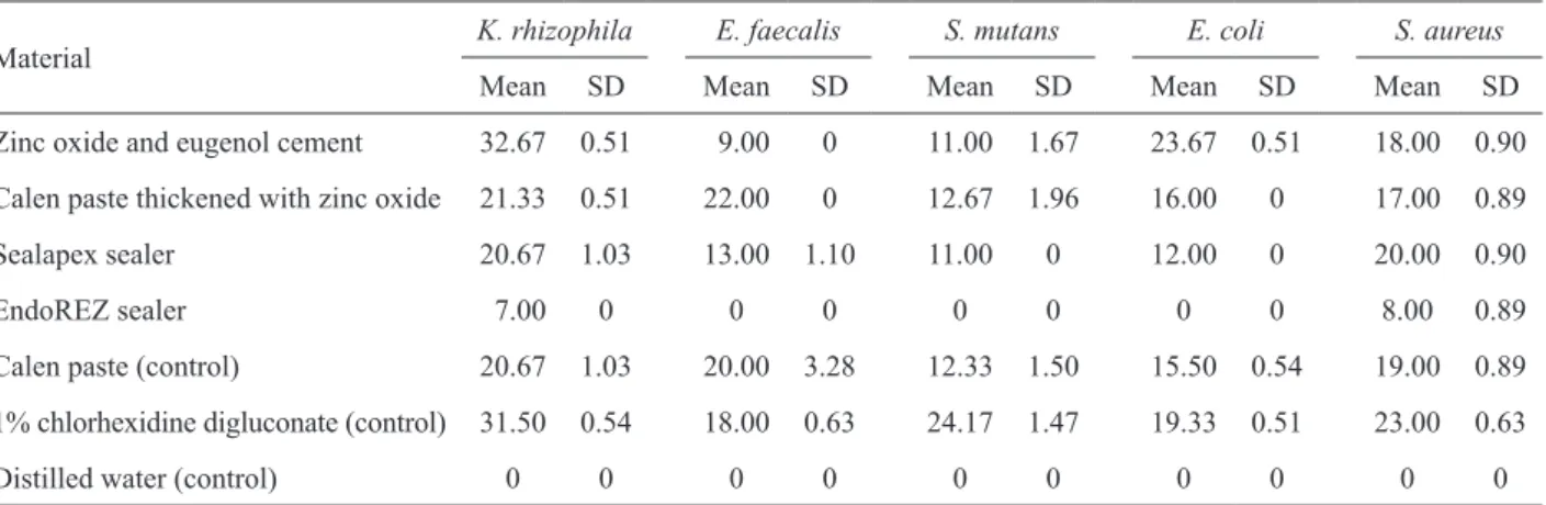
Table 2 shows the diameters (means and standard deviations) of the zones of bacterial growth inhibition zones (in mm) obtained for the tested materials. There
were statistically significant differences (p<0.0001)
microorganisms (K. rhizophila, E. faecalis, S. mutans, E. coli and S. aureus) produced by the root canal filling materials for primary teeth (ZOE, Calen/ZO, Sealapex and EndoREZ) and the control materials (Calen paste, 1% CHX and distilled water), by the agar diffusion test.
Regarding K. rhizophila, there was no statisti-cally significant difference (p>0.05) among Calen paste, Calen/ZO and Sealapex, which presented zones of bacterial growth inhibition with mean diameters ranging from 20.67 to 21.33, and neither between ZOE
Table 2. Diameters (in mm) of the zones of bacterial growth inhibition against of the bacterial strains.
Material
K. rhizophila E. faecalis S. mutans E. coli S. aureus
Mean SD Mean SD Mean SD Mean SD Mean SD
Zinc oxide and eugenol cement 32.67 0.51 9.00 0 11.00 1.67 23.67 0.51 18.00 0.90
Calen paste thickened with zinc oxide 21.33 0.51 22.00 0 12.67 1.96 16.00 0 17.00 0.89
Sealapex sealer 20.67 1.03 13.00 1.10 11.00 0 12.00 0 20.00 0.90
EndoREZ sealer 7.00 0 0 0 0 0 0 0 8.00 0.89
Calen paste (control) 20.67 1.03 20.00 3.28 12.33 1.50 15.50 0.54 19.00 0.89
1% chlorhexidine digluconate (control) 31.50 0.54 18.00 0.63 24.17 1.47 19.33 0.51 23.00 0.63
Distilled water (control) 0 0 0 0 0 0 0 0 0 0
The values are expressed as means of 6 repetitions and standard deviations (SD).
Table 1. Chemical composition of the experimental and control materials and mixing ratios.
Material Composition Mixing ratio
Zinc oxide and eugenol
cement Zinc oxide and eugenol
1.4 g zinc oxide to 0.4 mL eugenol
Calen paste thickened with zinc oxide
Calen paste: 2.5 g calcium hydroxide, 0.5 g zinc oxide, 0.05 g colophony and 1.75 mL polyethylene glycol 400 (vehicle); Zinc oxide
1 g Calen paste to 1 g zinc oxide
Sealapex sealer
20% calcium oxide, 29% bismuth trioxide, 2.5% zinc oxide, 3.0% submicron silica, 2.0% titanium dioxide, 1.0% zinc stearate,
3.0% tricalcium phosphate, *39% mixture
Equal amounts of the base and catalyst pastes
EndoREZ sealer Zinc oxide, barium sulfate, pigments, urethane dimethacrylate resin matrix
Equal amounts of the base and catalyst paste
Calen paste (control) 2.5 g calcium hydroxide, 0.5 g zinc oxide, 0.05 g colophony
and 1.75 mL polyethylene glycol 400 (vehicle)
-1% chlorhexidine digluconate (control)
1% chlorhexidine digluconate
Vehicle qsp 100 mL
-Distilled water (control) -
Figure 1. Zones of bacterial growth inhibition formed around the wells filled with the root canal filling materials for primary teeth and control substances in the agar diffusion test (agar-well technique): a: distilled water, b: Calen paste, c: Sealapex, d: zinc oxide and eugenol, e: Calen paste thickened with zinc oxide, f: EndoREZ, g: 1% digluconate chlorhexidine.
A: Kocuria rhizophila
B: Enterococcus faecalis
C: Streptococcus mutans
D: Escherichia coli
and CHX (p>0.05), which presented zones of bacterial growth inhibition with diameters of 32.67 and 31.5 mm, respectively. EndoREZ produced inhibition zones with mean diameter of 7 mm.
The largest mean zones of bacterial growth inhi-bition against E. faecalis were produced by Calen/ZO, followed by Calen paste and 1% CHX (22, 20 and 18 mm diameter, respectively). There was no statistically significant difference (p>0.05) between Calen paste and Calen/ZO, and neither between Calen paste and 1% CHX, as for their antibacterial activity against this
mi-croorganism. Statistically significant difference (p<0.05)
was found between Calen/ZO (22 mm diameter) and 1% CHX (18 mm diameter). EndoREZ and distilled water did not show antibacterial activity. Sealapex and ZOE presented intermediate antibacterial activity, with mean zones of bacterial growth inhibition of 13.00 and 9.00 mm diameter, respectively, statistically different
(p<0.05) from each other.
In the dishes seeded with S.mutans, no significant difference (p>0.05) was observed between Calen paste, Calen/ZO, Sealapex and ZOE, which produced mean zones of bacterial growth inhibition of 12.33, 12.67, 11.00 and 11.00 mm diameter, respectively. The largest
mean zone of bacterial growth inhibition (p<0.05) was
produced by 1% CHX (24.17 mm diameter), which dif-fered significantly from the other materials. EndoREZ and distilled water did not present antimicrobial activity.
Regarding E. coli, no statistically significant difference (p>0.05) was found between Calen paste and Calen/ZO, which produced mean zones of bacte-rial growth inhibition of 15.50 and 16.00 mm diameter, respectively, and neither between EndoREZ and distilled water, which did not present antibacterial activity. ZOE and 1% CHX presented the largest mean zones of bacte-rial growth inhibition (23.67 and 19.33 mm diameter, respectively), with statistically significant difference
between them (p<0.05).
The diameters of the zones of bacterial growth inhibition produced by the root canal filling materi-als against S. aureus were the following: 23 mm (1% CHX), 20 mm (Sealapex), 19 mm (Calen paste), 18 mm (ZOE), 17 mm (Calen/ZO) and 8 mm (EndoREZ). There were no statistically significant differences (p>0.05) between the following pairs of materials: Calen paste x Sealapex, Calen paste x ZOE and Calen/ZO x ZOE.
Statistically significant difference (p<0.05) was found
between Sealapex (20 mm diameter) and ZOE (18 mm
diameter), and between Calen paste (19 mm diameter) and Calen/ZO (17 mm diameter).
The antibacterial activity of the root canal filling materials for primary teeth against the 5 bacterial strains can be summarized as follows: K. rhizophila: ZOE > Calen/ZO = Sealapex > EndoREZ; E. faecalis: Calen/ZO > Sealapex > ZOE > EndoREZ; S. mutans: Calen/ZO = Sealapex = ZOE > EndoREZ; E. coli: ZOE > Calen/ZO > Sealapex > EndoREZ; S. aureus: Sealapex > ZOE = Calen/ZO > EndoREZ. The signal “=” indicates absence
of statistically significant difference. Regardless of the microorganism, ZOE showed the highest antibacterial activity, followed by Calen/ZO, Sealapex and EndoREZ.
DISCUSSION
In the present study, ZOE showed antibacterial activity against all test microorganisms. This material produced the largest zones of bacterial growth inhibi-tion against K. rhizophila and E. coli, and the smallest zones of bacterial growth inhibition against E. faecalis. Similar results have been reported (14).
Apart from K. rhizophila, which is a microorgan-ism with low intrinsic resistance, the largest zones of bacterial growth inhibition were formed around E. coli
(gram-negative bacillus). These results agree with those of Tchaou et al. (6), who observed that ZOE inhibited more effectively gram-negative than gram-positive bacteria.
Savioli et al. (15) evaluated the antimicrobial activity of a ZOE-based root canal filling material for permanent teeth (Grossman’s sealer) and its components against different microbial strains using the double layer well-diffusion method, and found that the eugenol com-ponent inhibited K. rhizophila, E. faecalis, S. mutans, E. coli and S. aureus, which were also evaluated in the present study. ZO alone inhibited only the growth of S. sobrinus and E. coli. Perhaps the fact that E. coli was inhibited by both eugenol and ZO may explain why in the present study ZOE produced the largest bacterial growth inhibition zones against E. coli (23.67 mm di-ameter), even larger than those produced by 1% CHX (19.33 mm diameter). Several authors (5,10,16) have attributed the antimicrobial effects of ZOE to eugenol. The pre-incubation, which consisted of maintain-ing the culture media innoculated for about 2 h at room temperature, was an important factor to evince the anti-microbial activity of the calcium hydroxide pastes (5,14).
extensively demonstrated (4,5,14,17). In the present study, the Calen paste presented antibacterial activity against all test microorganisms with the diameters of the zones of bacterial growth inhibition ranging from 12.33 to 20.67 mm. In order to be used as a root canal filling material for primary teeth, the Calen paste must be thickened with ZO, and the antibacterial activity of this material had not yet been evaluated. Our findings showed that Calen/ZO had antibacterial activity against all test microorganisms with the diameters of inhibition zones ranging from 12.67 to 22.00 mm.
Comparing the diameters of the zones of bacte-rial growth inhibition produced by Calen paste alone or thickened with ZO, it was observed that slightly larger inhibition zones were formed around Calen/ZO, except for S. aureus. Therefore, it may be inferred that the ad-dition of ZO to thicken the Calen paste did not interfere in its antimicrobial activity, with a mild increase of this property. It might be explained by the fact that ZO also presents antibacterial activity (18). Leonardo et al. (5) has shown that ZO associated with water inhibited the growth of K. rhizophila, E. faecalis, S. mutans, E. coli, S. aureus, S. epidermidis and Pseudomonas aeruginosa.
E. faecalis is a facultative anaerobic gram-posi-tive coccus that is considered as one of the most resistant microorganisms to calcium hydroxide-based intracanal medications (19). However, in the present study, Calen paste and Calen/ZO produced the largest zones of bacte-rial growth inhibition against this pathogen.
Sealapex has been indicated as a root canal filling material for primary teeth (11). In the present investiga-tion, Sealapex showed antibacterial activity against K. rhizophila, E. faecalis, S. mutans, E. coli and S. aureus, as reported elsewhere (5). These findings are in accor-dance with those of Mickel et al. (16) and Bodrumlu and Semiz (12), which evaluated the antibacterial activity of endodontic sealers against E. faecalis, using the us-ing the agar diffusion test. In all experiments, Sealapex showed antibacterial activity against this microorganism. On the other hand, Miyagak et al. (13) did not observe antimicrobial activity of Sealapex against E. faecalis,
S. aureus and Candida albicans, using the agar diffu-sion test. This result is probably due to the fact no pre-diffusion was done in their study, which is a step of the methodology that permits the dissociation and diffusion of the calcium hydroxide contained in the pastes and sealers based on this substance (5).
The use of methacrylate-based sealers as root
canal filling materials for primary teeth was first pro-posed by Woods et al. (20) in the 1980s’ after observing biological compatibility and phagocytosis at the same rate of the physiological root resorption of the primary teeth. EndoREZ was evaluated in the present study for this reason.
Few studies have evaluated the antimicrobial efficacy of EndoREZ. A previous study using the agar diffusion test found that EndoREZ did not present an-timicrobial activity against E. faecalis, E. coli, Micro-coccus luteus, S. aureus, S. epidermidis, P. aeruginosa
and Candida albicans (7). Using the agar diffusion test as well, Eldeniz et al. (9) did not observe antibacterial activity of EndoREZ against E. faecalis, S. aureus and
P. aeruginosa, and mild antibacterial activity against these microorganisms was observed only when the di-rect contact test was used. In the present investigation, EndoREZ exhibited antibacterial activity, though small, against K. rizhophila (7 mm diameter) and S. aureus (8 mm diameter).
Based on the statistically significant differences among the root canal filling materials for primary teeth evaluated in this study, it may be inferred that overall, ZOE presented the highest antibacterial activity against the test microorganisms, followed by Calen/ZO and Sealapex. EndoREZ presented the lowest antibacte-rial activity among the tested mateantibacte-rials. However, it is important to mention this alleged superior antibacterial activity of ZOE compared to Calen/ZO was partially due to the fact that K. rizhophila was more effectively inhibited by ZOE than by Calen/ZO, with diameters of the zones of bacterial growth inhibition of 32.67 mm and 21.33 mm, respectively. However, it is known that the intrinsic resistance of K. rizhophila is considerably low, which means that an intense antibacterial activity is not necessary to inactivate this microorganism. E. coli
ZOE, with the formation of zones of bacterial growth inhibition of 22 and 9 mm diameter, respectively. In this case, an intense antibacterial activity is essential to inactivate this microorganism. There was no statistically significant difference between the antibacterial activity of ZOE and Calen/ZO against S. mutans and S. aureus.
Based on the obtained results and within the limitations of the methodology, it may be concluded that the in vitro antibacterial activity against the tested bacterial strains in a decreasing order was: ZOE, Calen/ ZO, Sealapex and EndoREZ.
RESUMO
Este estudo avaliou in vitro a atividade antibacteriana de 4 materi-ais obturadores de canmateri-ais radiculares de dentes decíduos - cimento de óxido de zinco e eugenol (OZE), pasta Calen espessada com óxido de zinco (Calen/OZ), cimento Sealapex e cimento EndoREZ - sobre 5 cepas bacterianas comumente encontradas em infecções endodônticas: Kocuria rhizophila, Enterococcus faecalis, Strepto-coccus mutans, Escherichia coli e StaphyloStrepto-coccus aureus, usando o teste de difusão em ágar (técnica do poço). A pasta Calen, digluconato de clorexidina a 1% (CHX) e água destilada foram usados como controle. Sete poços por placa foram preparados em pontos eqüidistantes e imediatamente preenchidos com os materiais experimentais e controle. Após incubação das placas a 37oC por 24 h, o diâmetro dos halos de inibição do crescimento bacteriano formados ao redor dos poços foi medido (em mm) com um paquímetro digital sob luz refletida. Os dados obtidos foram submetidos à análise de variância e ao pós-teste de Tukey
(α=0,05). Com relação à atividade antibacteriana, evidenciaram-se diferenças estatisticamente significantes (p<0,0001) entre os
halos de inibição formados pelos diferentes materiais, para todos os microrganismos avaliados. A K. rhizophila foi inibida mais
eficazmente pelo OZE (p<0,05), enquanto que o E. faecalis foi
inibido mais eficazmente pela Calen/OZ (p<0,05). O S. mutans
foi inibido pela Calen/OZ, cimento Sealapex e OZE na mesma intensidade (p>0,05). A E. coli foi inibida mais eficazmente pelo
OZE, seguido pela Calen/OZ e pelo cimento Sealapex (p<0,05). O
S. aureus foi inibido pela Calen/OZ e OZEna mesma intensidade
(p>0,05), e menos intensamente pelo cimento Sealapex (p<0,05).
O cimento EndoREZ apresentou atividade antibacteriana apenas frente a K. rhizophila e ao S. aureus. A pasta Calen e a Calen/ OZ ocasionaram halos de inibição maiores que a CHX quando o microrganismo indicador foi o E. faecalis. Pode-se concluir que a atividade antibacteriana, in vitro, dos 4 materiais obturadores de canais radiculares de dentes decíduos sobre cepas bacterianas comumente encontradas em infecções endodônticas pode ser apresentada numa ordem decrescente de eficácia da seguinte maneira: OZE>Calen/OZ>Sealapex>EndoREZ.
REFERENCES
1. Ruviére DB, Leonardo MR, da Silva LA, Ito IY, Nelson-Filho P. Assessment of the microbiota in root canals of human primary teeth by checkerboard DNA-DNA hybridization. J Dent Child
2007;74:118-123.
2. Rocha CT, Rossi MA, Leonardo MR, Rocha LB, Nelson-Filho P, Silva LA. Biofilm on the apical region of roots in primary teeth with vital and necrotic pulps with or without radiographically evident apical pathosis. Int Endod J 2008;41:664-669.
3. Carrotte PV, Waterhouse PJ. A clinical guide to endodontics-update part 2. Br Dent J 2009;206:133-139.
4. Faria G, Nelson-Filho P, Freitas AC, Assed S, Ito IY. Antibacte-rial effect of root canal preparation and calcium hydroxide paste (Calen) on intracanal dressing in primary teeth with apical peri-odontitis. J Appl Oral Sci 2005;13:351-355.
5. Leonardo MR, Silva LAB, Tanomaru Filho M, Bonifácio KC, Ito IY. In vitro evaluation of antimicrobial activity of sealers and pastes used in endodontics. J Endod 2000;26:391-394.
6. Tchaou WS, Turng BF, Minah GE, Coll JA. Inhibition of pure cultures of oral bacteria by root canal filling materials. Pediatr Dent 1996;18:444-449.
7. Sipert CR, Hussne RP, Nishiyama CK, Torres SA. In vitro antimi-crobial activity of Fill Canal, Sealapex, Mineral Trioxide Aggre-gate, Portland cement and EndoREZ. Int Endod J 2005;38:539-543. 8. Amorim LF, Toledo OA, Estrela CR, Decurcio DA, Estrela C. Antimicrobial analysis of different root canal filling pastes used in pediatric dentistry by two experimental methods. Braz Dent J 2006;17:317-322.
9. Eldeniz AU, Erdemir A, Hadimli HH, Belli S, Erganis O. Assess-ment of antibacterial activity of EndoREZ. Oral Surg Oral Med Oral Pathol Oral Radiol Endod 2006;102:119-126.
10. Reddy S, Ramakrishna Y. Evaluation of antimicrobial efficacy of various root canal filling materials used in primary teeth: a micro-biological study. J Clin Pediatr Dent 2007;31:193-198.
11. Sari S, Okte Z. Success rate of Sealapex in root canal treatment for primary teeth: 3-year follow-up. Oral Surg Oral Med Oral Pathol Oral Radiol Endod 2008;105:e93-e96.
12. Bodrumlu E, Semiz M. Antibacterial activity of a new end-odontic sealer against Enterococcus faecalis. J Can Dent Assoc 2006;72:637.
13. Miyagak DC, de Carvalho EM, Robazza CR, Chavasco JK, Levorato GL. In vitro evaluation of the antimicrobial activity of endodontic sealers. Braz Oral Res 2006;20:303-306.
14. Tanomaru-Filho M, Tanomaru JM, Barros DB, Watanabe E, Ito IY. In vitro antimicrobial activity of endodontic sealers, MTA-based cements and Portland cement. J Oral Sci 2007;49:41-45. 15. Savioli RN, Pecora JD, Mian H, Ito IY. Evaluation of the
antimi-crobial activity of each component in Grossman’s sealer. Braz Oral Res 2006;20:127-131.
16. Mickel AK, Nguyen TH, Chogle S. Antimicrobial activity of end-odontic sealers on Enterococcus faecalis. J Endod 2003;29:257-258. 17. Soares JA, Leonardo MR, Tanomaru Filho M, Silva LA, Ito IY. Residual antibacterial activity of chlorhexidine digluconate and camphorated p-monochlorophenol in calcium hydroxide-based root canal dressings. Braz Dent J 2007;18:8-15.
18. Jones N, Ray B, Ranjit KT, Manna AC. Antibacterial activity of ZnO nanoparticle suspensions on a broad spectrum of microorgan-isms. FEMS Microbiol Lett 2008;279:71-76.
19. Onçag O, Cogulu D, Uzel A. Efficacy of various intracanal medi-caments against Enterococcus faecalis in primary teeth: an in vivo study. J Clin Pediatr Dent 2006;30:233-237.
20. Woods RL, Kildea PM, Gabriel SA, Freilich LS. A histologic com-parison of Hydron and zinc oxide-eugenol as endodontic filling materials in the primary teeth of dogs. Oral Surg Oral Med Oral Pathol 1984;58:82-93.