• Nenhum resultado encontrado

Braz. j. . vol.77 número3

N/A
N/A
Protected

Academic year: 2018

Share "Braz. j. . vol.77 número3"

Copied!
6
0
0

Texto

(1)

Complications in blepharoplasty: how to avoid and manage them

Abstract

Tomas Gomes Patrocinio1, Bruno Alvarenga Silva Loredo2, Carlos Eduardo Arnez Arevalo3, Lucas Gomes

Patrocinio4, José Antonio Patrocinio5

1 Otorhinolaryngologist; Head of the Ocular Plasic Surgery Division - Otorhinolaryngology Department - Medical School of the Federal University of Uberlândia. 2 Resident physician - Otorhinolaryngology Department - Medical School of the Federal University of Uberlândia.

3 Resident physician - Otorhinolaryngology Department - Medical School of the Federal University of Uberlândia.

4 PhD - Medical School of São José do Rio Preto, Head of the Maxillo-Facial Surgery Division - Otorhinolaryngology Department - Medical School of the Federal University of

Uberlândia.

5 Full Professor - Otorhinolaryngology Department - Medical School of the Federal University of Uberlândia.

Divisão de Plásica Ocular, Serviço de Otorrinolaringologia, Universidade Federal de Uberlândia, Uberlândia, Minas Gerais, Brasil. Send correspondence to: Tomas Gomes Patrocínio - Rua Arthur Bernardes 555 1º andar Uberlândia MG 38.400-368.

Tel/Fax: 34-3215-1143 - E-mail: tomaspatrocinio@hotmail.com

Paper submited to the BJORL-SGP (Publishing Management System – Brazilian Journal of Otorhinolaryngology) on July 08, 2010;

and accepted on August 02, 2010. cod. 7193

C

omplications in blepharoplasty are uncommon and, when they occur, they are usually mild and transient, such as hematomas and chemosis. However, sometimes they can be severe, such as blindness, or they might require surgical correction, such as ectropion.

Objective: To evaluate the results and complications of transcutaneous blepharoplasty performed in the same procedure, discussing how to treat and how to avoid them.

Methods: A retrospective study of 200 medical records of consecutive patients that underwent transcutaneous blepharoplasty from January 2007 to January 2009. The variables analyzed were age, gender, complications, clinical outcome, patient satisfaction, and photographic documentation.

Results: The incidence of complications was 9.5% (19 patients). The complications were 1 hematoma, 12 cases of chemosis and 13 patients who underwent canthoplasty, 6 patients with malposition of the lower eyelid (5 retractions and 1 ectropion). Medical treatment was performed in 12 patients and revision surgery in 7 cases of all the patients who had complications.

Conclusions: We demonstrated that blepharoplasty is a procedure with a high satisfaction and a low complication rate, and it is an excellent surgical procedure, when properly indicated.

ORIGINAL ARTICLE Braz J Otorhinolaryngol.

2011;77(3):322-7.

BJORL

Keywords: blepharoplasty, postoperative complications, eyelid diseases, eyelids.

(2)

Brazilian Journalof otorhinolaryngology 77 (3) May/June 2011

INTRODUCTION

Blepharoplasty is among the most performed cos-metic surgeries in the world today, and its main goal is to promote rejuvenation, functional and cosmetic enhance-ment of the periorbital region1.

Complications are uncommon and when they happen they are usually mild and transitional, such as hematomas and chemosis. Nonetheless, sometimes they can be definitive, such as blindness, or they require new surgical approaches for correction, such as ectropion and eyelid ptosis2.

Complication prevention or even their forecast starts with a careful preoperative assessment. It must include a detailed clinical history (comorbidities, use of medication, ophthalmic past, personal and family members), and care-ful physical exam. The assessment of the patient’s psycho-logical aspects and the patient’s expectations regarding surgery are also very important. The surgical technique to be used is based on the anatomical changes found and the patient’s complaints, always taking into account patient expectation and the real surgical possibilities for cosmetic improvement3.

The treatment of complications include from a cli-nical approach all the way to new surgical interventions, which must be properly employed by face surgeons pro-perly trained to do a blepharoplasty.

The goal of the present paper is to assess the results and complications of transcutaneous blepharoplasties done by the Ocular Plastic Surgery service from the Otorhino-laryngology Department from a University Hospital, from January of 2007 through January of 2009, showing how to treat them and, especially, how to avoid them.

MATERIALS AND METHODS

Series

We reviewed the charts from 200 consecutive patients submitted to transcutaneous blepharoplasty sur-gery between January of 2007 and January of 2009 in an otorhinolaryngology department in a university hospital. The variables analyzed were: patient age and gender, the surgical procedure (duration, anesthesia and surgical technique), complications (hematoma, chemosis, lower eyelid mal-positioning, lagophthalmos, epicanthus, eyelid ptosis, eyebrow ptosis, senile orbit, diplopia) and periodic clinical evolution carried out in the outpatient ward during one year after the procedure, emphasizing the degree of patient satisfaction and pre and post-operative pictures.

All the surgeries were carried out under supervision of the same surgeon. The procedures were done under local anesthesia with local injection of lidocaine with epinephrine at 1:100.000 concentration in the place of in-cision and for a regional block. After heart and peripheral

oximetry were monitored, the patient was sedated with the slow intravenous injection of 2ml of fentanyl chlorhydrate and 3 mg of midazolam.

Surgical Technique

The upper blepharoplasty technique used was:

• The eyelid incision was marked with the patient seating down and looking straight ahead to a fixed point, highlighting the excess tissue to be resected;

• Upper eyelid incision with the 15 blade scalpel in the highlighted place;

• Removal of a strip of orbicular muscle from the eye, its pre-septal portion and the fat pouches are dis-sected;

• The excess fat in the pouches are excised and cauterized with the bipolar cautery;

• Intradermal continuous skin suture with 6-0 nylon wire.

The lower blepharoplasty surgical technique was:

• Creation of the myocutaneous flap by a subciliary incision;

• Careful removal of the excess fat, muscle and skin;

• Canthopexy or canthoplasty, depending on the degree of lower eyelid laxity and that of the canthal tendon;

• Orbit oculoplastics;

• Skin suture with simple 6-0 nylon suturing wire.

RESULTS

Of the 200 patients included in the study, 81% (162) were females and 19% (38) were males, with mean age of 44.5 years (35 to 60).

The incidence of complications was 9.5% (19 pa-tients). The complications found were: 1 patient (0.5%) with hematoma; 12 cases (6%) of chemosis, and this ha-ppened to 61.5% of the 13 patients submitted to cantho-plasty; 6 patients (3%) with lower eyelid malpositioning and from these, 83% corresponded to retraction (2.5% from the total, 5 patients) and 17% to ectropion (0.5% of the total, 1 patient) (Graph 1).

All the patients who had lower eyelid malpositio-ning were not submitted to lateral canthal anchoring in the primary surgery. We did not find some of the complications reported in the literature, such as: eyelid closure changes, lagophthalmos, epicanthus, eyelid ptosis, eyebrow drop, senile orbit and diplopia.

As far as the management of complications is con-cerned, a clinical treatment was carried out with outpatient follow up in 12 patients (63%) and revision surgery in 7 patients (37%) of all the patients with complications. The revision surgeries happened because of a hematoma in one patient and 6 patients were submitted to a second look because of the malpositioning of the lower eyelid border.

(3)

be-cause of the lower eyelid mal-positioning and consequent apparent sclera. They were submitted to revision surgery and were pleased with the outcomes.

DISCUSSION

Blepharoplasty is a surgery with rare complications; nonetheless, when they happen they may cause important functional and/or cosmetic damage. Nonetheless, besides being rare, most of the times these complications are sim-ple to manage when treated by an experienced surgeon with anatomical knowledge of the eyelid and orbit regions4.

Among possible complications we stress hematoma, chemosis, lower eyelid mal-positioning, eye closure deficit, epicanthus, lagophthalmos, eyelid ptosis, eyebrow ptosis, senile orbit and diplopia1,5.

Hematoma

Hematoma is a complication that, depending on its magnitude and evolution, may cause amaurosis. Its preven-tion starts with preoperative care, a detailed clinical history involving coagulopathies, comorbidities which may pre-dispose to bleeding and the use of anticoagulant drugs6,7.

They may be classified as:

(1) Pre-septal: confined to the eyelid alone, not causing any risk to vision and resolving with ice packs.

(2) Post-septal: it happens because of intraorbital hemorrhage, causing a pressure increase in the orbit and compromising the optical nerve.

Demere and Wood reported 40 cases of blindness caused by retrobulbar hematoma in a series of 98,514 blepharoplasties (1 for each 2,500 procedures = 0.04%)4.

Drugs which predispose to hemorrhage are antipla-telet adhesion drugs and anti-inflammatory agents - which must be suspended at least 10 days before the procedure. Chronic disorders such as systemic arterial hypertension, must be controlled before surgery.

During surgery, hemorrhage control must be rigo-rous, with the cauterization of the fat pouches resected in order to avoid retrobulbar hematoma, as well as the cauterization of bleeding points in the orbicular muscle and

adjacent tissues. There are controversies about the type of coagulation that must be used during surgery1,8. Most of

the authors prefer to use the bipolar cautery to avoid the risk of damaging posterior eye structures.

In cases of small hematomas, without visual or pupillary changes, one must be very conservative, using systemic steroids and strict follow up.

In cases of pupillary and visual changes, with the tense orbit, intervention must be immediate, with surgical exploration for hemostatic purposes. Depending on the hematoma severity, one must do a canthotomy, cantholysis or orbit decompression.

Post-operative strong pain is extremely rare, and when it happens one must consider the possibility of a hematoma formation. Routinely, one must avoid compres-sive dressing, and ice packs are used.

In our investigation, there was one case of hemato-ma (0.5% of all the patients), in which the patient had a fast evolution, with a large bulging which drew our attention for a surgical approach before there were pupillary or visual changes. This case shows the importance of also assessing the time it took the hematoma to form, its mag-nitude and evolution. Results are satisfactory if treatment is instituted early on.

Chemosis

Chemosis is an infiltrative edema of the eye con-junctive. It results from liquid collection arising from in-flammatory reaction, lymphatic drainage obstruction and deficient eye closure, originating a bulged and reddish border around the cornea. In our series, chemosis was the most common complication (6% of all the patients) (Figure 1). It happened more frequently after surgery of the lower eyelid, especially when canthoplasty was carried out, because of the intense handling of the conjunctiva. Spontaneous resolution happened within 3 to 4 weeks, and a vasoconstrictor and steroid eye drops may be used in order to speed up the process1,5.

Graph 1. Graph showing the distribution of the main complications happened to patients submitted to blepharoplasty.

(4)

Lower eyelid mal-positioning

The ideal position of the lower eyelid is at 1mm or above the lower limbus. The lower eyelid malpositioning was the second most common complication found in our patients. The main causes of lower eyelid malpositioning are excessive skin resection (anterior lamella); inadvertent scarring of the orbital septum (middle lamella)9; and the

failure in cantus anchoring10-14.

In order to prevent lower eyelid malpositioning, it is important to be careful in removing the excess skin on the lower eyelid; to suture the orbital septum, reducing the possibilities of an inadvertent scarring; and to anchor the lateral cantus, either with a canthopexy or cantoplasty1,5,15,16

.

In order to correct the lower eyelid malpositioning, one must anchor the eyelid cantus associated with the orbit oculoplasty with a suspension of the myocutaneous flap, free skin graft or upper eyelid flap in some cases, and even use a posterior lamella expander.9,16

Some anatomical characteristics are true traps, which increase the risk of malpositioning and the sur-geon should treat them more carefully. They are: elderly patients with laxity and deep eye sockets; and patients with bulging eyes and negative vector (anterior portion of the eye bulb anterior to the lower eyelid and to the malar promontory)16,17,18.

In the present study, the malpositioning cases ha-ppened because of retraction and ectropion (Figures 2, 3 and 4). In such cases we did not do any type of lateral canthal anchoring in the primary surgery. The retraction correction was carried out by means of the canthoplasty using the McCord technique (full thickness), and the ec-tropion was corrected using a tarsal strip associated to a skin graft, achieving good post-operative results11,12.

Lagophthalmos

The eyelid slit had 10mm of distance between the upper eyelid and the lower one, and 30mm of distance between the medial and the lateral canthi. Lagophthalmos is an alteration in which the eye cannot be fully closed, usually because of an excessive retraction of the upper eyelid tissue, orbicular muscle paralysis or lower eyelid retraction.

An inadvertent lesion of the upper eyelid elevator muscle aponeurosis may cause eyelid ptosis, also changing inter-eyelid distance. Lateral canthoplasty may reduce the distance between the medial and lateral canthi.

It is the most common cause of dry eye after ble-pharoplasty and in some cases it is necessary to operate the patient in order to correct the situation, doing a tar-sorrhaphy, skin grafts and middle third suspension. Dry eye may be treated with massage and artificial tear drops, and most of the times it resolves spontaneously. We did not have this complication in our sample1,5,19,20.

Eyelid ptosis

Eyelid ptosis is a condition in which the upper eyelid falls below its normal level and the upper eyelid elevator muscle is not able to raise it. It is classified in two major groups: congenital or acquired21,22. Preoperative

assessment is very important, one should always look for ptosis, because it may become even more apparent after blepharoplasty is done, frustrating patient’s expectations both in the cosmetic and in the functional aspects. The ptosis mechanism can be: myogenic, neurogenic, mecha-nical or aponeurotic.

The main cause of postoperative ptosis is eyelid edema, which causes a mechanical restriction to the eyelid elevator muscle action, with spontaneous resolution in most of the cases. Hematomas may also cause restrictions to the eyelid elevator muscle movement, and when deep and persistent it may cause fibrosis, making the situation last longer or even become permanent, although most of the times it resolves spontaneously. Ptosis may happen if the orbital septum is inadvertently joined to the elevator muscle during blepharoplasty suture, as well as forming adherences between the septum and the eyelid elevator, in cases of careless removal of the post-septal fat pouches. Damage to the eyelid elevator aponeurosis with the cautery may also cause ptosis.

Figure 2. Patient with left lower eyelid malpositioning after blepharo-plasty caused by retraction.

Figure 4. Patient with lower eyelid malpositioning after blepharoplasty caused by ectropion. Below: postoperative picture after correction by means of canthoplasty with the tarsal strip and skin grafting.

(5)

Most of the ptoses cases resolve spontaneously wi-thin 3 months. When it does not happen, the patient must be submitted to surgery. The correction of post-surgery eyelid ptosis, usually the result of injury to the eyelid ele-vator muscle aponeurosis, involves the exploration of the upper eyelid and aponeurosis repair3,22-24.

Eyebrow ptosis

Eyebrow ptosis must be assessed in the preoperative visit. The patient must be educated as to the limitations of blepharoplasty in cosmetic improvement and possible complications, such as a reduction in the distance betwe-en the eyelid border and the eyebrow. Surgical options to reposition the eyelid must be offered to the patient by means of an endoscopic or transpalpebral surgery.

When the surgeon disregards this complication, the patient will be dissatisfied with the blepharoplasty’s cosmetic results, and the eyebrow ptosis can be misjud-ged by the patient as being a complication arising from inadequate surgical technique25,26,27.

Senile orbit

As time goes by, there is a loss of support and also a reduction in facial soft tissue volume. This is extremely important because as one performs blepharoplasty, the excessive removal of fat pouches can increase upper and lower eyelid grooves, giving the patient the appearance of aging. This situation may not be perceived in the immedia-te post-op, becoming more evident in the long run, when the patient develops the so called “senile orbit”. Even in a younger patient, one must avoid the excessive removal of fat pouches, because the loss of soft tissue is part of natural aging and the excess fat removed will be missed in the future. The correction of such complication can be done by the surgical technique described by Maniglia et al28. Diplopia

Diplopia, or double vision is a rare alteration which can happen in cases involving the muscles responsible for eye movement, or scars around the eyeball. The most vulnerable muscle is the inferior oblique. It may be tran-sitional or permanent, requiring surgical correction when it does not evolve properly1,2.

Unhappy patient

Patient satisfaction with the blepharoplasty result depends on the following points: 1) detailed preoperative assessment; 2) contraindication of the cases which patient expectation is unreal; 3) indication of the most adequate surgical technique, including possible associations such as eyebrow elevation, ptosis correction, canthoplasty, and others; 4) carefully performed surgical technique, espe-cially in hemostasis and the resection of skin and fat; 5) frequent postoperative follow up3,18.

In the present study, there was a high patient satis-faction rate, especially because the surgeons followed the points listed above. The complications found are within the regular rates published in the world literature and could be corrected by clinical or surgical management.

CONCLUSION

Blepharoplasty is a surgery not only with a cosmetic component, making the person’s look younger and less tense, but it also has a functional component, providing the patient with a better visual field without the need to stress eyelid muscles. In order to achieve a good result, it is necessary to do a careful pre-operative evaluation, educating the patient as to the benefits and limitations of the procedure and also to customize the approach accor-ding to the characteristics of each patient. When surgery is carefully done, it entails few complications, thus gua-ranteeing its success.

The present study showed that blepharoplasty is a procedure with a high satisfaction rate and low number of complications, yielding excellent results when properly indicated.

REFERENCES

1. Glavas IP. The diagnosis and management of blepharoplasty complica-tions. Otolaryngol Clin North Am.2005;38(5):1009-21.

2. Morax S, Touitou V. Complications of blepharoplasty. Or-bit.2006;25(4):303-18.

3. Jelks GW, Jelks EB. Preoperative evaluation of the blepharoplasty patient. Bypassing the pitfalls. Clin Plast Surg.1993;20(2):213-23. 4. DeMere M, Wood T, Austin W. Eye complications with

blepharo-plasty or other eyelid surgery. A national survey. Plast Reconstr Surg.1974;53(6):634-7.

5. Hartstein ME, Kikkawa D. How to avoid blepharoplasty complica-tions. Oral Maxillofac Surg Clin North Am.2009;21(1):31-41. 6. Wolfort FG, Vaughan TE, Wolfort SF, Nevarre DR. Retrobulbar hema-Wolfort FG, Vaughan TE, Wolfort SF, Nevarre DR. Retrobulbar hema-Retrobulbar

hema-toma and blepharoplasty. Plast Reconstr Surg.1999;104(7):2154-62. 7. Hass AN, Penne RB, Stefanyszyn MA, Flanagan JC. Incidence of

postblepharoplasty orbital hemorrhage and associated visual loss. Ophthal Plast Reconstr Surg.2004;20(6):426-32.

8. Wiggs EO. Blepharoplasty complications. Trans Am Acad Ophthalmol Otolaryngol.1976;81(4 Pt 1):603-6.

9. Patipa M. The evaluation and management of lower eyelid retraction following cosmetic surgery. Plast Reconstr Surg.2000;106(2):438-53. 10. Flowers RS. Canthopexy as a routine blepharoplasty component. Clin

Plast Surg.1993;20(2):351-65.

11. Anderson RL, Gordy DD. The lateral tarsal strip procedure. Arch Ophthalmol.1979;97(11):2192-6.

12. McCord CD, Hester TR, Codner MA. The lateral canthoplasty. Op Tech Plast Reconstr Surg.1998;5(2):90-8.

13. McCord CD, Shore JW. Avoidance of complications in lower lid blepharoplasty. Ophthalmology.1983;90(9):1039-46.

14. McCord CD, Boswell CB, Hester TR. Lateral canthal anchoring. Plast Reconstr Surg.2003;112(1):222-37.

15. Jordan DR, Anderson RL. The lateral tarsal strip revisited. The enhan-ced tarsal strip. Arch Ophthalmol.1989;107(4):604-6.

(6)

17. Murakami CS, Orcutt JC. Treatment of lower eyelid laxity. Facial Plast Surg.1994;10(1):42-52.

18. Patrocinio LG, Dolci JEL, Patrocinio JA, Scalia RA. Blefaroplastia e Otoplastia. In: Melo Jr JF, Martins RHG, Imamura R, Caldas Neto S (editores). Manuais de Otorrinolaringologia. Bucofaringologia e Cirurgia Cérvico-Facial. São Paulo: ABORL-CCF; 2008. p. 164-73. 19. Lima CGMG, Siqueira GB, Cardoso IH, Sant’Anna AEB, Osaki MH.

Avaliação do olho seco no pré e pós-operatório da blefaroplastia. Arq Bras Oftalmol.2006;69(2):227-32.

20. Lessa SF, Lena EH, Araújo MRC, Pitanguy I. Modificações anatômicas da fenda palpebral após blefaroplastia. Rev Bras Cir.1997;87(4):179-88.

21. Knize DM. An anatomically based study of the mechanism of eyebrow ptosis. Plast Reconstr Surg.1996;97(7):1321-33.

22. Hornblass A. Ptosis and pseudoptosis and blepharoplasty. Clin Plast Surg.1981;8(4):811-30.

23. Baylis HI, Sutclife T, Fett Dr. Levator injury during blefaroplasty. Arch Ophthalmol.1984;102(4):570-1.

24. Edmonson BC, Wulc AE. Ptosis evaluation and management. Oto-laryngol Clin North Am.2005;38(5):921-46.

25. Horibe EK, Horibe K, Lodovici O. Lifting of the eyebrows in blepha-roplasties. Aesthetic Plast Surg.1989;13(1):79-82.

26. Patrocinio LG, Patrocinio JA. Forehead-lift: 10-year review. Arch Facial Plast Surg.2008;10(6):391-4.

27. Patrocinio LG, Reinhart RJY, Patrocinio TG, Patrocinio JA. Fronto-plastia endoscópica: três anos de experiência. Braz J Otorhinolaryn-gol.2006;72(5):624-30.

Imagem

Figure  1.  Patient  with  bilateral  chemosis  after  blepharoplasty  with  canthoplasty.
Figure 2. Patient with left lower eyelid malpositioning after blepharo- blepharo-plasty caused by retraction.

Referências

Documentos relacionados

Aside from changes in IgA secretion, a few authors have suggested that increased deposition of collagen in AIDS patients may also deplete CD4 T cells. Increased

The microscopic analysis tried to assess the morpho- logy of the region where the defect was created, observing the abundant bone neogenesis in 68.53% of the defects cre- ated on

The present study assessed the prevalence of bronchopulmonary and otorhinolaryngological symptoms in children below 12 years of age submitted to 24 hour pH probe for

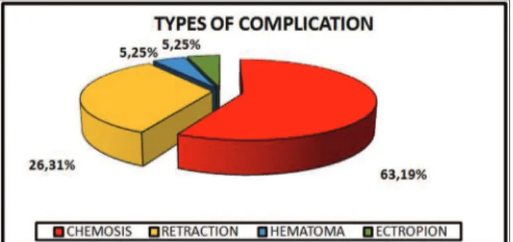
The third step consisted of a critical evaluation by three persons with varying experience on this topic: (1) healthcare professionals with experience in esthetic surgery

The incidence of cholesteatoma varies worldwide, depending on each population. Bezold 2 has suggested that auditory canal dysfunction causes retraction of the tympanic

the 5-HT2A receptor is mostly related with serotonin (5- HT) excitation within the representative motor nucleus of upper airways, lack of common polymorphisms or

To describe the impact the tongue electrotactile sti- mulation has on the balance control of patients who have CNS dysfunctions and did not obtain satisfactory results with

The left cerebral hemisphere was the most affected (6 cases), followed by the right cerebral hemisphere (4 cases) and the left cerebellar hemisphere (3 cases).. cases) and