INTERRELATIONSHIP BETWEEN RADIOLOGIC
FINDINGS AND PROGNOSIS OF EPILEPSY IN
CHILDREN WITH NEUROCYSTICERCOSIS
Lisiane Seguti Ferreira
1, Verônica A. Zanardi
2, Li Min Li
1, Marilisa M. Guerreiro
1ABSTRACT - Introduction: Epileptic manifestations of Neurocysticercosis (NC) appear to depend on number and localization of the cysts. The objective of this study was to investigate the relationship between CT findings, number of parasites and the evolutive stage of the cysts, and the prognosis of epilepsy in children with NC. Method: We studied 28 patients with the parenchymal form of NC, considering: epilepsy duration; seizure frequency before and after AED treatment; seizure control; number of AED and recurrence after AED withdrawal. Clinical information was crossed with the number of lesions and disease activity in univariate comparison. Results: The analysis of the clinical data in relation to the number of lesions and disease activity showed no statistical difference among the variables (p>0.05). Conclusion: We conclude that the course of epilepsy due to NC in childhood cannot be based exclusively on the number or stage of the parasites.
KEY WORDS: neurocysticercosis, childhood epilepsy, radiology, prognosis.
Interrelação entre achados radiológicos e o prognóstico da epilepsia em crianças com neurocisticercose Interrelação entre achados radiológicos e o prognóstico da epilepsia em crianças com neurocisticercoseInterrelação entre achados radiológicos e o prognóstico da epilepsia em crianças com neurocisticercose Interrelação entre achados radiológicos e o prognóstico da epilepsia em crianças com neurocisticercoseInterrelação entre achados radiológicos e o prognóstico da epilepsia em crianças com neurocisticercose
RESUMO - Introdução: As manifestações epilépticas da neurocisticercose (NC) parecem depender do número e localização das lesões. O objetivo desse estudo foi investigar a relação entre os achados de CT, número de parasitas e estágio evolutivo dos cistos, com o prognóstico da epilepsia em crianças. Método: Nós estudamos 28 pacientes com a forma parenquimatosa da NC, considerando: duração da epilepsia; freqüência de crises antes e após o tratamento com DAE; aquisição de controle; número de drogas e recorrência. Esses aspectos foram correlacionados com o número de lesões e atividade da doença em análise estatística univariada. Resultados: A análise dos dados clínicos em relação ao número de lesões e atividade da doença não revelou diferenças estatisticamente significativas (p>0,05). Conclusão: Concluímos que o curso da epilepsia por NC na infância não pode se basear exclusivamente no número ou estágio dos parasitas.
PALAVRAS-CHAVE: neurocisticercose, prognóstico, epilepsia na infância, radiologia.
Departamentos de Neurologia1 e Radiologia2, Universidade Estadual de Campinas (UNICAMP), Campinas SP, Brasil. Dr. Seguti received a
scholarship from FAPESP.
Received 7 June 2001, received in final form 17 September 2001. Accepted 2 October 2001.
Dra. Marilisa M. Guerreiro - Departamento de Neurologia FCM / UNICAMP - Caixa Postal 6111 - 13083-970 Campinas SP – Brasil. FAX: 19 3871 6715. E-mail: [email protected]
Neurocysticercosis (NC) is a common health prob-lem in developing countries. It affects patients of all ages and it is endemic in adults as well as among children in Latin America1-3. Epilepsy is the most
im-portant clinical manifestation. It occurs in 70-90% of all children with the parenchymal form of the dis-ease, usually being its primary presentation4,5 the
pa-thophysiology of the seizures due to NC is not com-pletely understood yet. In active and transitional forms, seizures may be the consequence of compres-sion or inflammatory reaction. In inactive form, pe-rilesional gliosis is probably the cause of the seizures. Chronic inflammatory reaction sometimes takes sev-eral years to disappear and it may have an impor-tant role in the pathophysiology of focal epilepsy in
NC6,7. Epileptic manifestations appear to depend on
number and localization of the cysts8. Nevertheless,
some studies have not shown any difference both in seizure frequency and in clinical or electroencepha-lographic characteristics in patients with a single le-sion compared to those with multiple lele-sions 9,10.
The objective of this study was to investigate the relationship between CT findings, number of para-sites and the evolutive stage of the cysts, and the prognosis of epilepsy in children with NC.
METHOD
pa-tients with NC were followed at the Pediatric Epilepsy Out-patient Clinic at the University of Campinas from January 1983 to January 1999. Five patients had a single seizure with follow-up of at least 12 months. Eight patients had the encephalitic form of NC. These 13 patients were exclu-ded because single seizure is not epilepsy, and the ence-phalitic form is a more severe presentation of NC with a tendency for permanent neurological sequelae and severe epileptic condition than the one habitually found in pa-tients with epilepsy resulting from NC.
We obtained all information from the remaining 28 patients on revision of their medical records, complemen-ted with direct interview with patients and guardians whe-never possible. A semi-structured protocol was filled in for every patient, considering: epilepsy duration (this was defined as the period between the first seizure up to the moment that antiepileptic drugs (AED) were withdrawn or until the last appointment for patients with persistent seizures, regardless of any remission period during the follow-up); seizure frequency before AED treatment was classified according to the total number of seizures quan-tified as: A<10, B = 10 to 50, C>50; seizure frequency after AED introduction (same classification as above) ; zure control was defined as one year without having sei-zures; number of AED to obtain seizure control; recurrence after AED withdrawal (the policy of AED withdrawal was carried out after two years of seizure-freedom).
All 28 patients had computerized tomography scan (CT) at the time of the diagnosis in our center. All exams were revised by one of us (VAZ), a neuroradiologist with experience with NC. Lesions were counted and classified into three groups: five or less, between six and 10, and more than 10. Patients with a single calcification were excluded from this research. The disease activity was clas-sified as active (appears on CT as hipodense cyst without enhancement), transitional (there is a ring or nodular con-trast enhancement), and inactive (calcified lesions on CT) based on the viability of the parasite as proposed by Carpio et al.12.When lesions in different stages were found in the
same patient, they were classified according to the most active lesion detected.
The clinical information listed above was crossed with the number of lesions and disease activity in univariate comparison using Kruskal-Wallis with post hoc pairwise comparison or Fisher exact test, and significance was as-sumed when p<0.05.
RESULTS
Twenty-eight patients (16 girls, mean age = 7.2 years, mean follow-up of 64.5 months) had paren-chymal form of NC and normal neurological exami-nation and were the subjects of this study.
Concerning the evolutive stage of the parasite, 17 patients were at inactive phase, six at transitional phase and five children had active lesions on CT. Regarding number of lesions, 18 patients had five or less than five lesions, four had between six and 10 lesions, and six had more than 10 cysts.
The statistical analysis of clinical data in relation to the number of lesions and disease activity showed the following.
Epilepsy duration: mean duration of epilepsy was 7.2 years (range from 2.3 to 14.1 years). We did not find any statistical difference when we compared mean scores of epilepsy duration with stage Wallis test p=0.20) and number of lesions (Kruskal-Wallis test p=0.18). Multiple cysts, regardless of stage, did not influence epilepsy duration and had the same behavior as a few lesions.
Seizure frequency: 12 patients had less than 10 seizures, 10 children had seizures between 10 and 50, and six had more than 50 seizures. We did not find statistical difference when we compared in univaried analysis seizure frequency with stage and number of lesions (Table 1).
Seizure frequency after AED treatment: 15 pa-tients had less than 10 seizures, nine had seizures between 10 and 50, and four had more than 50 sei-zures. There was no statistical difference comparing
Table 1. Seizure frequency.
Number of lesions Disease activity
Seizure frequency ≤ 5 5-10 > 10 Active Inactive Transitional
A (< 10) 10 1 1 1 7 4
35.7% 3.5% 3.5% 3.5% 25% 14.3%
B (10-50) 5 2 3 3 5 2
17.9% 7.1% 10.7% 10.7% 18% 7.1%
C (> 50) 3 1 2 1 5 0
10.7% 3.5% 7.1% 3.5% 18%
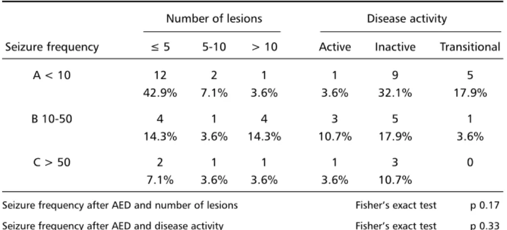
Table 2. Seizure frequency after AED introduction.
Number of lesions Disease activity
Seizure frequency ≤ 5 5-10 > 10 Active Inactive Transitional
A < 10 12 2 1 1 9 5
42.9% 7.1% 3.6% 3.6% 32.1% 17.9%
B 10-50 4 1 4 3 5 1
14.3% 3.6% 14.3% 10.7% 17.9% 3.6%
C > 50 2 1 1 1 3 0
7.1% 3.6% 3.6% 3.6% 10.7%
Seizure frequency after AED and number of lesions Fisher’s exact test p 0.17 Seizure frequency after AED and disease activity Fisher’s exact test p 0.33
Table 3. Number of AEDs.
Number of lesions Disease activity
Number of drugs ≤ 5 5-10 > 10 Active Inactive Transitional
1 8 1 0 1 7 1
28.6% 3.6% 3.6% 25% 3.6%
2 6 0 4 2 4 4
21.4% 14.3% 7.1% 14.3% 14.3%
≥3 4 3 2 2 6 1
14.3% 10.7% 7.1% 7.1% 21.4% 3.6%
Number of AED and number of lesions Fisher’s exact test p 0.06 Number of AED and disease activity Fisher’s Exact Test p 0.52
Table 4. Seizure control.
Number of lesions Disease activity
Seizure control ≤ 5 5-10 > 10 Active Inactive Transitional
Yes 17 3 4 3 15 6
n = 24 60.7% 10.7% 14.3% 10.7% 53.6% 21.4%
No 1 1 2 2 2 0
n = 4 3.6% 3.6% 7.1% 7.1% 7.1% 0
Control seizure and number of lesions Fisher’s exact test p 0.16 Control seizure and disease activity Fisher’s exact test p 0.40
in univaried analysis seizure frequency after AED with stage and number of lesions (Table 2).
Number of AED: nine patients received only one AED during their treatment, 10 received two AED, and nine received three or more AED, either isolated or in association. There was no statistical difference comparing number of AED with stage and number of lesions (Table 3).
Seizure control: 24 patients obtained control of
their epilepsy. The mean time to obtain control was 16.1 months. We did not find any difference when we compared seizure control with the stage and number of lesions (Table 4).
DISCUSSION
Neuroimaging has attained enormous progress during last decade. CT is very helpful in NC because it is a safe, precise and noninvasive method with more than 95% accuracy to define number, localiza-tion and evolutive stages of the parasites, especially in the parenchymal form of the disease. In develop-ing countries where MRI machines are not always available, and considering the fact that calcifications are the main radiologic finding in NC, CT is still the most performed and useful examination13-16.
In this study, we tried to correlate CT findings (num-ber and evolutive stages of lesions) with prognostic factors such as epilepsy duration, seizure frequency before and after AED introduction, number of AED, seizure control and seizure recurrence. In univaried analysis, we did not find any correlation among them. When isolated, neither number of parasites nor their stages are predictive factors of outcome. In this se-ries, we observed seizure-free patients with multiple lesions in contrast to patients with refractory epi-lepsy with few lesions.
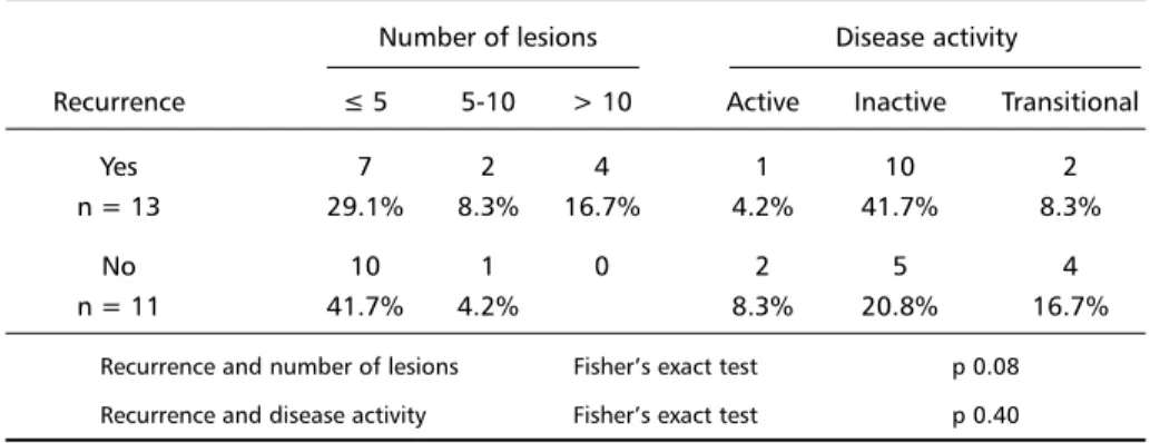
One limitation of our research is the low number of patients, which is due to the rigid inclusion crite-ria. However, out of 12 analyzed factors, only two showed borderline findings: number of lesions with recurrence (p=0.08) and number of lesions with number of AED (p=0.06). If number of patients were higher, there would be a chance that statistical analy-sis could show different data of the two variables described above. Some authors found that patients with calcified lesions in large number have a worse prognosis, while other authors have different point of view9,17,18.
The poor understanding of the pathophysiology of the seizures due to NC parallels difficulties in ex-plaining the variability of clinical manifestations7,19.
Particularly in childhood, presentation may vary from
“benign” to severe epileptic syndromes, such as Len-nox-Gastaut syndrome10,20,21. Some factors that may
contribute to explain this variability are: spontane-ous resolution of the lesions; persistence of perilesio-nal edema around calcified lesions; unpredictable evolution of the parasite that allows the coexistence of different forms in the same subject; the immune response of the host; and, the environment in which the child lives that may be responsible for new in-festations15,22-25.
The severity of clinical manifestation has also been correlated to HLA antigens in the surface of the para-sites, which suggests a genetic influence in the pre-sentation of NC. Del Bruto25 found higher rates of
HLA 28 in patients with NC when compared to con-trols. Another antigen, DQW2, may be related to the resistance of the disease. Therefore, an individual predisposition to develop parenchymal NC is likely to occur and could in part explain the variability of the clinical expression.
We conclude that the course of epilepsy due to NC in childhood cannot be based exclusively on the number or stage of the parasites. Patients with mul-tiple lesions will not necessarily present more sei-zures or need more drugs than the others. Intrinsic characteristics of the parasites as well as immunege-netic aspects may play important role in the expla-nation of the pleomorphic and unpredictable course of the clinical picture.
Acknowledgements - Acknowledgements - Acknowledgements - Acknowledgements -
Acknowledgements - The authors thank Mrs. Cleide Moreira Silva for statistical support.
REFERENCES
1. Román GI. Neuroepidemiologia de la cisticercosis. In Guzmán TAA (ed). Cisticercosis del sistema nervoso. Guayaquil: Offset Abad, 1999:21-29. 2. Agapejev S. Epidemiology of neurocysticercosis in Brasil. Rev Inst Med
Trop São Paulo 1996;38:207-216.
3. White AC Jr. Neurocysticercosis: a common cause of neurological dis-ease worldwide. Clin Infect Dis 1997;24:101-115.
Table 5. Recurrence.
Number of lesions Disease activity
Recurrence ≤ 5 5-10 > 10 Active Inactive Transitional
Yes 7 2 4 1 10 2
n = 13 29.1% 8.3% 16.7% 4.2% 41.7% 8.3%
No 10 1 0 2 5 4
n = 11 41.7% 4.2% 8.3% 20.8% 16.7%
4. Manreza MLG, Diament A. Cisticercosis pediátrica en Sudamérica. In San Esteban JE, Flisser A, Astiazarán, AG (eds). Neurocisticercosis en la infancia. Mexico: Grupo Editorial Miguel Ángel Porrúa, 1997:39-52. 5. Kalra V, Sethi A . Childhood neurocysticercosis: epidemiology,
diag-nosis and course. Acta Paediatr Jpn 1992;34:365-370.
6. Carpio A, Escobar A, Hauser WA. Cysticercosis and epilepsy: a critical review. Epilepsia 1998;39:1025-1040.
7. Bittencourt PRM, Adamolekum B, Bharucha, et al. Epilepsy in tropics: II. Clinical presentations, pathophisiology, imunologic diagnosis, eco-nomics, and therapy. Epilepsia 1996;37:1128-1137.
8. Sotelo J, Del Bruto OH. Brain cysticercosis: review article. Arch Med Res 2000;31:3-14.
9. Monteiro L, Nunes B, Mendonça D. Spectrum of epilepsy in neurocys-ticercosis: a long term follow up of 143 patients. Acta Neurol Scand 1995;92:33-40.
10. Cukiert A, Puglia P, Scapolan HB, Vilela MM, Marino RJ. Congruence of the topography of intracranial calcifications and epileptic foci. Arq Neuropsiquiatr 1994; 52:289-293.
11. Del Bruto OH, Wadia NH, Dumas M, Cruz M, Tsang VCW, Schantz PM. Proposal of diagnostic criteria for human cysticecosis and neurocysticercosis. J Neurol Sci 1996;142:1-6.
12. Carpio A, Marcelo P, Santillan F, Alfonso E. A proposal for classifica-tion of neurocysticercosis. Can J Neurol Sci 1994;21:43-47.
13. Daras M, Tuckman AJ, Strobos RJ. Computed tomography in adult-onset epileptic seizures in a city hospital population. Epilepsia 1987; 28:519-522.
14. Minguetti G, Ferreira MVC. Computed tomography in neurocysticer-cosis. J Neurol Neurosurg Psychiatry 1983;46:936-942.
15. Garcia MR, Astiazarán AG, Franco FR. Neurocysticercosis in children: clinical experience in 122 patients. Chid’s Nerv Syst 1997;13:608-612. 16. Guerreiro MM, Facure NO, Guerreiro CAM. Aspectos da tomografia
computadorizada craniana na neurocisticercose na infância. Arq Neuropsiquiatr 1989;47: 153-158.
17. Del Bruto OH. Prognostic factors for seizure recurrence after with-drawal of antiepileptic drugs in patients with neurocysticercosis. Neu-rology 1994;44:1706-1709.
18. Del Bruto OH. Neurocisticercosis en niños: análisis clínico, radiológico y factores prognósticos en 54 pacientes. Rev Neurol 1997;25:1681-1684. 19. Manreza MLG. Epilepsia e neurocisticercose. In Guerreiro CAM (ed).
Epilepsia. São Paulo: Lemos Editorial, 2000:255-264.
20. Sakamoto AC, Bustamante VCT, Garzon E, et al. Cysticercosis and epilepsy. In Kotagal P, Luders HO (eds). The epilepsies: etiologies and prevention. New York: Academic Press, 1999:275-282.
21. Agapejev S, Padula NAMR, Morales NMO, Lima MMF. Neurocisticerco-se e síndrome de Lennox-Gastaut. Arq Neuropsiquiatr 2000;58: 538-547. 22. Nash TE, Patronas NJ. Edema associated with lesions in
neurocysticer-cosis. Neurology 1999;53:777-781.
23. Miller B, Grinnell V, Goldberg MA, Heiner D. Spontaneous radiographic disappearance of cerebral cysticercosis: three cases. Neurology 1983;33: 1377-1379.
24. White AC Jr. Neurocysticercosis: updates on epidemiology, pathogen-esis, diagnosis and management. Annu Rev Med 2000;5:187-206. 25. Del Brutto OH, Granados G, Talamas O, Sotelo J, Gorodesky C.