ORIGINAL ARTICLE
Pleurodesis induced by intrapleural injection of silver nitrate
or talc in rabbits: can it be used in humans?*
FRANCISCO S. VARGAS1, LEILA ANTONANGELO2, MARCELO A.C. VAZ3, EVALDO MARCHI3, VERA LUIZA CAPELOZZI4, EDUARDO H. GENOFRE5(TE SBPT), LISETE R. TEIXEIRA6(TE SBPT)
Objective: Objective:Objective:
Objective: To evaluate the pleuropulmonary alterations caused by intrapleural injection of silver nitrate or talc in an experimental model, in order to consider its use in human beings. Method:Method:Method:Method: 112 rabbits were randomly selected to receive intrapleural 0.5% silver nitrate or 400 mg/kg talc slurry in 2 ml saline. Eight rabbits of each group were sacrificed after 1, 2, 4, 6, 8, 10, or 12 months. Regarding the pleural cavity, the
degree of macroscopic pleurodesis (adherences) and microscopic alterations, represented by inflammation and pleural fibrosis, were analyzed. The parenchyma was evaluated regarding the degree of alveolar collapse, intra-alveolar septum edema, and cellularity, on a 0 to 4 scale. Results: IResults: IResults: Intrapleural injection of silver nitrate Results: I produced earlier and more intense pleurodesis than talc slurry injection. The parenchymal damage was more
evident with silver nitrate, considered as moderate, and limited to the first evaluation (after one month). From the second month on and throughout the entire one-year follow-up, the parenchymal damage was similar with both substances, only the pleural adherences were more intense with silver nitrate. Conclusions:Conclusions:Conclusions: Conclusions: Intrapleural silver nitrate produces better and longer-lasting than intrapleural talc injection. The parenchymal
alterations, although discreet, are more pronounced when silver nitrate is used, but minimal after two months, and similar to those produced by talc injection during the entire one-year observation period. These effects on the pulmonary parenchyma do not contraindicate the use in humans. Thus, the use of intrapleural silver nitrate to produce fast and effective pleurodesis can be considered in patients in which pleural cavity
symphysis is desired. (J PneumolJ PneumolJ PneumolJ Pneumol 2003;29(2):57-63)
Key words – Pleurodesis. Talc. Silver nitrate. Pleural
effusion.
Acronym used in this work
TNF – Tumor necrosis factor
* Work performed at the Pleura Laboratory (Laboratório de Pleura) – Division of Respiratory Diseases – Instituto do Coração (InCor), Faculdade de Medicina da Universidade de São Paulo (São Paulo University School of Medicine).
1. Full Professor of Pneumology. 2. Assistant Professor.
3. PhD, Assistant Professor of Pneumology. 4. Associate Professor.
5. Graduate Student of the Discipline of Pneumology. SBPT Certified Specialist. 6. Assistant Professor of Pneumology. SBPT Certified Specialist.
Mailing Address – Francisco S. Vargas, Rua Itapeva, 500, cj. 4C – 01332-000 – São Paulo, SP, Brazil. Tel./fax
(55) (11) 3082-7040; e-mail: [email protected]
Support from Fundação de Amparo à Pesquisa do Estado de São Paulo (Fapesp) and Conselho Nacional de Desenvolvimento Científico e Tecnológico (CNPq).
Received for publication on 08/18/02. Received for publication on 08/18/02. Received for publication on 08/18/02.
I
NTRODUCTIONThe ideal agent to produce pleurodesis has not been identified so far. Now, at the beginning of the 21st
century, the most frequently used agent is talc. Although it is considered the agent of choice to produce pleurodesis in patients with pneumothorax or pleural effusion due to cancer, it certainly cannot be considered ideal. Talc has achieved such a degree of popularity as a result of its effectiveness in experimental studies(1) and its large distribution throughout the world (2). Moreover, talc seems to be more effective(3) and less expensive (2) than tetracycline and its derived products and than chemotherapic drugs. It should be mentioned that its administration does not interfere with posterior chemotherapy(4). However, there is serious concern regarding its safety. There is evidence that the intrapleural administration of talc induces respiratory distress syndrome in up to 8% of patients, with a mortality rate around 1%(5-7).
Recent experimental studies have confirmed that silver nitrate is an effective agent to produce pleurodesis, and can even replace talc(8). It was successfully used in the past in patients with pneumothorax(9), being gradually abandoned in the 1980s, without any clear justification. Apparently, it was due to aspects regarding its morbidity, believed to be a consequence of the high concentrations used (1-10%). However, in the animal model used by us, low concentrations (0.5%) produced effects which were comparable to those obtained with tetracycline(9) and superior to those observed with talc(10); therefore, we have proposed its use in humans. On the other hand, since silver nitrate is a substance with irritative and caustic properties (pH @ 5.5), a potential aggression with consequent damage to the pulmonary parenchyma should be considered before using it in humans.
Thus, the objective of this work was to evaluate the alterations of the pleura (representing the effectiveness in producing pleurodesis) and of the parenchyma (reflecting the aggression and the undesired pulmonary damage) which occurred during the 12 months of follow-up after intrapleural talc (400mg/kg) or silver nitrate (0.5%) injection. The rate between effectiveness (pleural) and morbidity (pulmonary) will allow or not to indicate silver nitrate as a pleural sclerosing agent, either as an alternative substance or as a substitute for talc. The use of silver nitrate would allow to prevent the undesired effects caused by talc, among which the respiratory distress syndrome, that stands out for its importance.
M
ATERIAL AND METHODSWhite rabbits (from New Zealand) weighing between 2.0 and 2.5 kg were superficially anesthetized by intramuscular administration of ketamine hydrochloride (35mg/kg) and xylazine hydrochloride (5mg/kg). The right hemithorax was prepared for operation, after washing and iodine asepsis. The procedure began with a 2cm skin incision, above the seventh or eighth intercostal space, at half distance between the spine and the sternum, allowing to dissect the musculature, exposing the parietal pleura. Then, under direct vision, a 25 gauge needle was inserted in the pleural space, allowing to inject the sclerosing agent; finally, the muscles and the skin were sutured. After the operation, the rabbits were monitored, to observe the presence of any possible clinical manifestations such as pain (moaning), tachypnea, and agitation. The rabbits showing distress received intramuscular dipyrone. The study was approved by the Ethics Committee of “Instituto do Coração” (Heart Institute) (InCor) of the University of São Paulo School of Medicine.
The animals were divided into two groups of 56 rabbits. One group received 0.5% silver nitrate, and the other one 400mg/kg talc, both in a total volume of 2ml saline. The talc used had no trace of asbestos, and its particles had a mean diameter of 25.4@m. Eight rabbits of each group were sacrificed, respectively, one, two, four, six, eight, 10, and 12 months after intrapleural administration, by a lethal pentobarbital injection, given in the marginal vein of the ear. The thorax was removed as a block, and the lungs expanded by intratracheal injection of 10% formalin. After tracheal ligation, the block containing the lungs was immersed in 10% formalin for more than 48 hours.
The necropsy of the animals was performed by an investigator who was unaware of the group each rabbit belonged to (in some animals the talc was visible in the pleural cavity). The pleural spaces were carefully exposed by making bilateral incisions in the diaphragm and in the ribs, approximately along the medium clavicular line. Thus, the sternum and part of the costal framework were removed, allowing to evaluate the lungs and the pleural cavities. The left hemithorax was used as control, and the existence of systemic effects on the liver and spleen was investigated.
The macroscopic pleurodesis evaluation was semiquantified on a 0 to 4 scale, where 0 is normal (without adherences), 1 represents less than three adherences, 2 means more than three localized adherences, 3 stands for the presence of disseminated adherences, and 4 characterizes the complete obliteration of the pleural space as a result of adherences.
fibrosis were microscopically evaluated, without knowing to which group the animal belonged. Inflammation and fibrosis were evaluated as: absent (0), minimal (1), discreet (2), moderate (3), and pronounced (4).
Parenchymal alterations were microscopically characterized by the degree of alveolar collapse, of edema (proteinaceous and amorphous material inside the air spaces), and of cell infiltrate (cellular density in the alveoli). This analysis was made by two observers who were unaware of the treatment undergone by the animals. These parameters were subjectively semiquantified using a score (0: absent; 1: minimal; 2: discreet; 3: moderate; and 4: severe), according to the extent and intensity of the lesions found in the pulmonary tissue.
Statistical Analysis Statistical Analysis Statistical Analysis Statistical Analysis
All data are expressed as mean ± standard deviation (SD). The means of the different macroscopic and microscopic scores in the two groups were compared using the unpaired t test. When the data did not show a normal distribution, Mann-Whitney’s non-parametric test was used to
compare the values in the two groups. Differences were considered significant whenever p < 0.05.
R
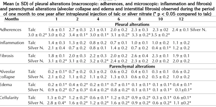
ESULTSThe intrapleural injection of silver nitrate determined a significantly more intense pleurodesis than the one produced by the administration of talc (Table 1). The mean score obtained after 0.5% silver nitrate (3.19 ± 0.21) was significantly higher (p < 0.001) than the one obtained after 400mg/kg talc (2.20 ± 0.35). The mean scores observed in all evaluations (except the one made at two months of the study) were significantly higher (p < 0.05) in the group that had received silver nitrate (Figure 1). Similarly, in this group, the mean scores observed in all evaluations were always equal to or higher than 3, reflecting an effective pleurodesis, whereas in the group that had received talc this value was always lower than 3, characterizing non-effective pleurodesis (Figure 1). Differences regarding pleurodesis were also observed qualitatively: the group that had received silver nitrate presented a homogeneous distribution of adherences all over the pleural cavity, whereas the group to which talc was administered showed a predominance of adherences in the ventral (anterior) portion, bringing evidence of a gravity-dependent distribution.
TABLE 1 TABLE 1TABLE 1 TABLE 1
Mean (± SD) of pleural alterations (macroscopic: adherences, and microscopic: inflammation and fibrosis) Mean (± SD) of pleural alterations (macroscopic: adherences, and microscopic: inflammation and fibrosis) Mean (± SD) of pleural alterations (macroscopic: adherences, and microscopic: inflammation and fibrosis) Mean (± SD) of pleural alterations (macroscopic: adherences, and microscopic: inflammation and fibrosis) and parenchymal alterations (alveolar collapse and edema and inter
and parenchymal alterations (alveolar collapse and edema and interand parenchymal alterations (alveolar collapse and edema and inter
and parenchymal alterations (alveolar collapse and edema and interstitial fibrosis) observed during the period stitial fibrosis) observed during the period stitial fibrosis) observed during the period stitial fibrosis) observed during the period of one month to one year after intrapleural injection of talc or silver nitrate (* p < 0.05 compared to talc) of one month to one year after intrapleural injection of talc or silver nitrate (* p < 0.05 compared to talc)of one month to one year after intrapleural injection of talc or silver nitrate (* p < 0.05 compared to talc) of one month to one year after intrapleural injection of talc or silver nitrate (* p < 0.05 compared to talc)
Months 1 2 4 6 8 10 12
Pleural alterations
Adherences Talc 1.6 ± 0.1 2.7 ± 0.3 2.1 ± 0.1 2.0 ± 0.2 2.3 ± 0.3 2.3 ± 02 2.4 ± 0.1 Silver N.
3.0 ± 0.2* 3.0 ± 0.2 *3.4 ± 0.1**3.0 ± 0.1**3.1 ± 0.2**3.3 ± 0.2* 3.5 ± 0.2*
Inflammation Talc 1.4 ± 0.3 1.3 ± 0.2 0.8 ± 0.2 0.7 ± 0.1 1.0 ± 0.1 1.0 ± 0.2 1.1 ± 0.2
Silver N. 2.1 ± 0.4 0.7 ± 0.2 0.8 ± 0.1 1.4 ± 0.2 0.7 ± 0.2 *0.4 ± 0.1* 1.2 ± 0.2
Fibrosis Talc 1.8 ± 0.1 2.0 ± 0.3 2.2 ± 0.3 2.0 ± 0.2 2.6 ± 0.4 2.3 ± 0.1 1.9 ± 0.1
Silver N. *3.1 ± 0.2* 3.1 ± 0.2 *3.2 ± 0.2* 2.4 ± 0.2 2.3 ± 0.2 2.0 ± 0.2 2.0 ± 0.2 Parenchymal alterations
Alveolar Talc *0.2 ± 0.1* 0.7 ± 0.2 0.3 ± 0.2 0.6 ± 0.2 0.4 ± 0.1 0.3 ± 0.1 0.6 ± 0.2
collapse Silver N. 2.1 ± 0.2 1.1 ± 0.2 1.1 ± 0.2 1.3 ± 0.3 0.6 ± 0.2 0.5 ± 0.2 1.0 ± 0.2
Edema Talc *0.2 ± 0.1**0.4 ± 0.2**0.2 ± 0.1**0.7 ± 0.1**0.1 ± 0.1**0.1 ± 0.1* 0.4 ± 0.1*
Silver N. *0.9 ± 0.2**0.7 ± 0.3**0.4 ± 0.2**0.8 ± 0.2**0.1 ± 0.1**0.1 ± 0.1* 0.1+0.1*
Cellularity Talc 1.3 ± 0.2* *1.2 ± 0.2**0.6 ± 0.1**1.2 ± 0.2**0.9 ± 0.2**0.3 ± 0.1**0.6 ±0.1*
Figura 1. Alterações pleuro--pulmonares observadas no hemitórax injetado com talco ou nitrato de prata.
(* p < 0,05
(* p < 0,05 ––Nitrato de Prata x Talco)Nitrato de Prata x Talco)
Alterações pleurais Pleural alterations
0 1 2 3 4
Months s
c o r e
1 2 4 6 8 10 12
Adherences
* * * *
* *
1 2 4 6 8 10 12
Fibrosis * *
1 2 4 6 8 10 12
Inflammation
*
12 0
1 2 3
4 Alveolar Collapse
1 2 4 6 8 10
*
Intersticial edema
1 2 4 6 8 10 12
Cellularity
12
1 2 4 6 8 10
* s
c o r e
Months Alterações
Parenchymal alterationsparenquimatosas
Figure 1 – Pleuropulmonary alterations observed in the hemithorax injected with talc or silver nitrate (* p < 0.05 – silver nitrate x talc)
Regarding the pleural surface, inflammation and fibrosis were studied microscopically. The inflammatory alterations were discreet in both groups; the mean score obtained was lower than 2 (Figure 1). However, the intrapleural injection of silver nitrate produced a more intense degree of fibrosis than the talc injection (Figure 1). The degree of pleural fibrosis observed during the first four months of the study was significantly higher (p < 0.05) after administration of 0.5% silver nitrate (score > 3) than after talc (score < 3). As from the sixth month and up to one year of study, the intensity of fibrosis was similar (p > 0.05) in both groups (Table 1, Figure 1).
All lungs examined presented alterations in the configuration of the air spaces (alveolar collapse), in alveolus-capillary permeability (edema), and in cellular afflux (inflammatory reaction). The mean of the scores which evaluated alveolar collapse was higher after silver nitrate, even though statistical differences were observed only in the analysis performed one month after the procedure (p < 0.05). Edema was detected in minimal amounts, similar in both groups, during the entire study period.
The cellular infiltrate present in the pulmonary parenchyma one month after the approach was significantly larger (p < 0.05) in the group that had received silver nitrate. After this evaluation, cellularity decreased progressively, and remained low throughout the entire study period. As from the second month, the inflammatory reaction present in the pulmonary parenchyma was minimal and similar with both sclerosing agents (Table 1, Figure 1).
The pleural and parenchymal alterations present in the left hemithorax (pleural cavity that was not
approached, considered as control) were minimal, negligible, showing that there was no pulmonary effect caused by the agents injected into the contralateral pleural space.
Intrapleural injection of either silver nitrate and talc did not cause distress to any one of the animals. The rabbits rapidly recovered and started to take in adequate amounts of food and water, returning to their normal activities. None of the animals needed additional medication.
D
ISCUSSIONThe mechanisms responsible for producing pleurodesis are not entirely known yet. It is believed that the first events involved, from the injection of the sclerosing agent to the symphysis of the pleural membranes, are represented by the lesion, the decortication of the mesothelial cells (11), and by the formation of an exsudative pleural effusion (12). The repair process is complex and involves several stages, which include inflammatory response to aggression, regeneration of damaged cells, migration of connective tissue cells, extracellular matrix synthesis and formation of collagen, and, consequently, of scars, represented by the adherences(13).
Several factors should be considered from instillation of the sclerosing agent to pleural symphysis. The degree of the lesion is important, because it is probably proportional to the effectiveness of pleurodesis and to the capacity of the mesothelial cells to secrete or break down collagen(14). It has been suggested that the duration, extension, and intensity of the inflammation may influence the final result, on the grounds that, in rabbits, pleurodesis produced by talc is inhibited by the use of corticoids(15) and by intrapleural administration of tumor necrosis factor (TNF)-blocking antibodies (16). Actually, no ideal sclerosing agent is known so far; all substances currently in use present disadvantages. Talc is effective; however, there are serious restrictions regarding its safety (5-7). Antineoplastic drugs, including bleomycine, are expensive and less effective than the other substances used (3). Tetracycline is no longer for sale and therefore unavailable, and its derived drugs are expensive and not very effective.
Silver nitrate is a rather promising agent, it is inexpensive and easy to find all over the world. In our animal model, it produced a more effective and longer-lasting pleurodesis than talc, that persisted during the entire year of follow-up.
Studies with a temporal approach regarding the effectiveness of pleurodesis are scarse, in both humans and experimental animals. Sassoon et al.(17)
followed-up rabbits which had received 10mg/kg of intrapleural minocycline during six months, and found no decrease in the intensity of pleurodesis during this period of time. It should be pointed out that, in that study, the score found after six months from the procedure was discreet (score 2), whereas the score found in the present study with silver nitrate, after the same period of time and up to one year of evolution, was higher (score 3). This means that pleurodesis after minocycline was ineffective after six months from the intrapleural injection and strongly effective after silver nitrate, during one-year follow-up.
Although it was pointed out that none of the studied rabbits died, consideration should be given to the fact that silver nitrate, being caustic, can damage the
pulmonary parenchyma, compromising its function. Thus, we should evaluate the intensity of the pulmonary lesion caused by intrapleural injection of silver nitrate.
We have characterized in this research the alterations which occurred in lung architecture one month after intrapleural injection of talc or silver nitrate, and made a temporal follow-up of their behavior during one year. The results obtained clearly show that both agents determined only discreet parenchymal alterations during the 12 months of follow-up.
Pulmonary structure alterations following intrapleural injection of a sclerosing agent are predominantly characterized by alveolar collapse. This behavior was more evident after silver nitrate than after talc, and was observed only one month after the intrapleural injection. As from the second month, the alveolar collapse decreased and became minimal and similar to the one observed with talc. Our hypothesis is that an association occurs between this collapse and the change in viscoelastic and mechanical properties of the chest wall, especially of the pleural surface. The earlier and more intense fibrosis produced by silver nitrate apparently reduces pulmonary compliance, diminishing pulmonary expansion and, consequently, the air spaces.
The second mechanism to be considered concerns the alteration in capillary permeability, which is probably caused by inflammation and favors the diffusion of fluid into the alveolar spaces. The resulting pulmonary edema is, however, minimal and can be histologically visualized during the first six months of follow-up. From the viewpoint of its evolution, this edema is similar with both agents, being slightly more evident after silver nitrate (Figure 1).
A similar behavior was observed regarding the inflammatory picture. The evaluation made one month after the intrapleural injection showed a moderate cellular afflux after silver nitrate and a minimal one after talc. As from the second month, both agents determined inflammatory reactions of similar intensity. Possibly the aggression to the visceral pleural membrane can explain the more intense reaction after silver nitrate.
11 months of follow-up. Our hypothesis is that the early-developing alveolar collapse could be caused by the alteration of the mechanical properties of the lung, mainly by the reduction of pulmonary compliance resulting from the formation of strong adherences. Finally, it should be stressed that the alterations of the pulmonary architecture are reversible and not intense, with a clear tendency to normalize over time.
These results enable us to consider the use of silver nitrate as a sclerosing agent to produce collapse of the pleural membranes, in order to control pneumothorax or recurrent pleural effusion. Presently, a proposition regarding a new therapeutic option or the resuming of a previously used method that was abandoned for undefined reasons should be based on experimental studies. Particularly in this work, the model used mimics partially the usual methodology applied in humans.
The most important difference concerns pleural cavity drainage, which is routine and necessary in humans. Drainage was not performed in this model, which might allow to question its results. However, the presumptive effect of the missing chest drain would be on the reduction in pleurodesis intensity, which would obviously be expressed in both groups. So, even considering that the methodology applied was not the same as performed on humans, we can accept the obtained effectiveness rates, mainly when we compare different agents.
Since silver nitrate does not determine either systemic side effects or repercussions on pleural or contralateral pulmonary architecture, the effects to be considered concern the affected hemithorax which is
the target of pleurodesis. Thus, some particular characteristics of silver nitrate as pleural symphysis inductor can be considered:
a) Silver nitrate is inexpensive, easy to manipulate, and can be found everywhere in the world, even in less developed areas;
b) The use of silver nitrate prevents the most undesired side effect of talc: the respiratory distress syndrome;
c) Pleurodesis obtained with silver nitrate is more intense than the one obtained with talc, which allows to assume that it is more effective and, consequently, that it controls the underlying pleural picture better;
d) Pleurodesis resulting from intrapleural administration of silver nitrate occurs earlier and lasts longer than the one caused by talc;
e) The damage to the pulmonary parenchyma is discreet with both agents, although more evident with silver nitrate, and limited to the first month.
Thus, we suggest silver nitrate as pleurodesis-inducing agent when faster and longer-lasting control of pleural disease is desired. Until there is more experience available, we propose that its use should be limited to the control of malignant pleural effusion, avoiding its indication for the control of recurrent pneumothorax. In humans(18,19), its effectiveness has been confirmed by previous works carried out between the 1960s and the 1980s and by a current prospective study, in which control of pleural malignant effusion was found in more than 90% of the evaluated patients (20).
R
EFERENCES1. Xie C, Teixeira LR, Wang N, McGovern JP, Light RW. Serial observations after high dose talc slurry in the rabbit model for pleurodesis. Lung 1998;176:299-307.
2. Kennedy L, Rush VW, Strange C, Ginsberg RJ, Sahn AS. Pleurodesis using talc slurry. Chest 1994;106:342-6.
3. Walker-Renard PB, Vaughan LM, Sahn AS. Chemical pleurodesis for malignant pleural effusions. Ann Intern Med 1994;120:56-64.
4. Adler RH, Sayer I. Treatment of malignant pleural effusion: a method using tube thoracostomy and talc. Ann Thorac Surg 1976;22:8-15.
5. Milanez Campos JR, Werebe EC, Vargas FS, Jatene FB, Light RW. Respiratory failure due to insufflated talc. Lancet 1997;349:251-2.
6. Campos JRM, Vargas FS, Teixeira LR, Werebe EC, Cardoso P, Jatene FB, et al. Thoracoscopy talc poudrage: a 15 year experience. Chest 2001;119:801-6.
7. Rehse DH, Aye RW, Florence MG. Respiratory failure following talc pleurodesis. Am J Surg 1999;177:437-40.
8. Vargas FS, Teixeira LR, Silva LMMF, Carmo AO, Light RW. Comparison of silver nitrate and
tetracycline as pleural sclerosing agents in rabbits. Chest 1995;108:1080-3.
9. Wied U, Andersen K, Schultz A, Rasmussen E, Watt-Boolsen S. Silver nitrate pleurodesis in spontaneous pneumothorax. Scand J Thorac Cardiovasc Surg 1981;15:305-7.
10. Vargas FS, Teixeira LR, Vaz MAC, Carmo AO, Marchi E, Cury PM, et al. Silver nitrate is superior to talc slurry in producing pleurodesis in rabbits. Chest 2000;118:808-13.
11. Kennedy L, Harley LA, Shan AS, Strange C. Talc slurry pleurodesis. Pleural fluid and histologic analysis. Chest 1995;107:1707-12.
12. Sahn SA, Good JT. The effect of common sclerosing agents on the rabbit pleura space. Am Rev Respir Dis 1981;124:65-7.
13. Cotran R, Kumar V, Collins T. Robbins pathologic basis of disease. 6th ed. Pennsylvania: WB Saunders 1999;89-134.
15. Xie C, Teixeira LR, McGovern JP, Light RW. Systemic corticosteroids decrease the effectiveness of talc pleurodesis. Am J Respir Crit Care Med 1998;157:1441-4.
16. Cheng DS, Rogers J, Wheeler A, Parker R, Teixeira L, Light RW. The effects of intrapleural polyclonal anti-tumor necrosis factor alpha (TNF) Fab fragments on pleurodesis in rabbits. Lung 2000;178:19-30. 17. Sassoon CSH, Light RW, Vargas FS, Gruer SE, Wang
NS. Temporal evolution of pleural fibrosis induced by intrapleural minocycline injection. Am J Respir Crit Care Med 1995;151:791-4.
18. Wied U, Andersen K, Schultz A, Ramussen E, Watt-Boolsen S. Silver nitrate pleurodesis in spontaneous pneumothorax. Scand J Thorac Cardiovasc Surg 1981;15:305-7.
19. Hopkirk JAC, Pullen MJ, Fraser JR. Pleurodesis: the results of treatment for spontaneous pneumothorax in the Royal Air Force. Aviat Space Environ Med 1983;54:158-60.