Ar
ti
cle
0103 - 5053 $6.00+0.00
*e-mail: [email protected]
Conformational Analysis of Phloroglucinols from
Hypericum Brasiliense
by using
X-ray Diffraction and Molecular Modeling
Kátia Z. Leal,*,a,b Julliane D. Yoneda,c Eric B. Lindgren,a Carlos B. Pinheiro,d Arthur L. Corrêaa and Hildegardo S. Françae
aDepartamento de Físico-Química, Instituto de Química, Universidade Federal Fluminense,
24020-150 Niterói-RJ, Brazil
bPrograma de Pós-Graduação em Química, Instituto de Química, Universidade Federal Fluminense,
24020-150 Niterói-RJ, Brazil
cPólo Universitário de Volta Redonda, Universidade Federal Fluminense,
27255-125 Volta Redonda-RJ, Brazil
dDepartamento de Física, Universidade Federal de Minas Gerais,
31270-901 Belo Horizonte-MG, Brazil.
eDepartamento de Tecnologia Farmacêutica, Faculdade de Farmácia, Universidade Federal
Fluminense, 24241-000 Niterói-RJ, Brazil
Neste trabalho, veriicou-se a aplicabilidade de uma metodologia computacional para se predizer a estrutura de compostos orgânicos com atividade biológica. Para isso, selecionaram-se três loroglucinóis, e compararam-selecionaram-se suas conformações obtidas por modelagem molecular e por difração de raios X. Os resultados mostraram que as conformações obtidas por análise conformacional com o método AM1 seguidas de otimização de geometria utilizando o método DFT (B3LYP/6-31G(d,p)) estão em boa concordância com os dados obtidos experimentalmente por difração de raios X, indicando que a metodologia empregada parece ser uma ótima ferramenta para predizer preferências conformacionais desta classe de compostos.
In this work we intend to verify the applicability of a computational methodology to predict structural features of organic compounds with biological activity. We selected three phloroglucinols and compared their calculated conformational data with their X-ray crystallographic structure. The results showed that conformations obtained by conformational analysis with the AM1 method followed by geometry optimization by using the DFT B3LYP/6-31 G(d,p) basis set are in very good agreement with X-ray data, indicating that the methodology employed here seems to be a very useful tool in order to predict the conformational preference for this class of compounds.
Keywords: conformational analysis, X-ray, molecular modeling, phloroglucinols
Introduction
Despite the development of organic synthesis, biotechnology and combinatorial chemistry, natural plants are still a great source of bioactive compounds. However, just 8% of the Brazilian lora has been already studied in the search for new bioactive substances.1,2
In the last decades several antibacterial compounds are being less effective in the treatment of infectious diseases due to multi-resistant bacteria.3 In this context
medicinal plants are extremely important in the search of new molecules as therapeutic alternatives. The genus Hypericum seems to be a good choice for this
problem. It is constituted by lavonoids, xantones and phloroglucinols with considerable pharmacological and biological effects.4,5 Phloroglucinol derivatives have been
high antibacterial activity.6-9 The Hypericum brasiliense
presents three phloroglucinols in its constitution and they are active against bacteria.10,11 These phloroglucinols are
the japonicin A, the uliginosin B and the isouliginosin B (Figure 1).
The study of conformations of bioactive compounds is very important in the design of new drugs, since that conformational proile may have direct implications in their activity. When the conformational properties of a drug are known to play an important role in establishing its therapeutic value, any newly designed analogue should have similar conformational properties to enhance the probability that it will bind to the receptor target.
Nowadays computational chemistry is a very important tool in drug design. It can lead for example, to the determination of physical chemical properties and to conformational proiles of biomolecules. However, success in using conformational proiles in the study of biochemical interactions depends on how well computer simulation is able to reproduce the structural features of the species involved.12-17
In this work we intend to verify the applicability of a computational methodology to predict structural features of organic compounds with biological activity. We selected the phloroglucinols 1, 2 and 3 and compared their calculated
conformational data with X-ray experimental results.
Experimental
The phloroglucinols used in this work were isolated from the Hypericum brasiliense.4,5 The structure of the
compounds was established by comparing its NMR data (¹H and ¹³C spectra) with that from literature.4
Samples of japonicin A (1) and isouliginosin B (3)
suitable for X-ray diffraction experiments were obtained by slow solvent evaporation at room temperature of solutions
containing hexane. All attempts to obtain single crystals of uliginosin B (2) by slow solvent evaporation failed.
Uliginosin B (2) data collection and data reduction was
performed in a two fold-twinned sample crystallized after evaporation of acetonitrile solution.
X-ray diffraction data collections were performed on a Oxford-Diffraction GEMINI diffractometer (LabCri) using
graphite-Enhance Source MoKα radiation (λ = 0.71069 Å)
at 150(2) K. Data Integration and scaling of the relections were performed with the Crysalis suite.18 Final unit cell
parameters were based on the itting of all relections positions. Empirical multiscan absorption corrections using equivalent relections were performed with the program
SCALE3 ABSPACK.19
The structures of all compounds were solved by direct methods using the SHELXS program.20 For each compound,
the positions of all atoms could be unambiguously assigned on consecutive difference Fourier maps. Refinements were performed using SHELXL20 based on F2 through
full-matrix least square routine. For uliginosin B (2) the
Crysalis suite help in inding the twin law to generate a relection ile for reinement with the HKLF5 option of the SHELXL program.20 All hydrogen atoms were reined
with anisotropic atomic displacement parameters. During the reinements neither solvent nor disordered groups were identiied in the crystals.
Except for those in the hydroxyl groups, the hydrogen atoms in the compounds were added in the structure in idealized positions and further reined according to the riding model.21 Owing the high X-ray diffraction data
quality the coordinates of the H atoms of the hydroxyl groups in the japonicin A (1) and in isouliginosin B (3) were
freely reined. The hydrogen atom from hydroxyl group bounded to C3 in the japonicin A (1) molecule as well as
those in the hydroxyl groups in the uliginosin B (3) were
identiied trough the electron density peaks observed in the Fourier difference maps nearby the oxygen atoms. These peaks were associated to H atoms and their coordinates were reined according to the riding model.21
The conformational search was done by the AM1 method using the Hyperchem 7.0 program.22 The geometry
of the conformer of lowest energy was fully optimized using the B3LYP/6-31G(d,p) basis set on the Gaussian 98
package of molecular orbital programs.23
Results and Discussion
X-ray results show that japonicin A (1) unit cell is
composed by eight molecules, uliginosin B (2) by four
molecules and isouliginosin B (3) by two molecules. In
all compounds the molecules are bonded by a series of Figure 1. Phloroglucinols from Hypericum brasiliense: japonicin A (1),
uliginosin B (2) and isouliginosin B (3) respectively.
O HO O OH O O OH HO 1 2
3 4 5
6 7 8 9 OH OH O O OH HO 1 2
3 4 5
6 7 8 9 O O 2' 3' 4' 5' 6' 7' 8' 9' 10' 11' 12' OH O O OH HO 1 2
3 4 5
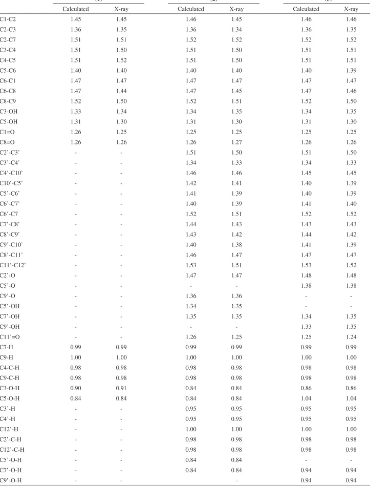
Table 1. Bond lengths (Å) calculated by the Gaussian 98 program and obtained by X-ray diffraction for the phloroglucinols studied
(1) (2) (3)
Calculated X-ray Calculated X-ray Calculated X-ray
C1-C2 1.45 1.45 1.46 1.45 1.46 1.46
C2-C3 1.36 1.35 1.36 1.34 1.36 1.35
C2-C7 1.51 1.51 1.52 1.52 1.52 1.52
C3-C4 1.51 1.50 1.51 1.50 1.51 1.51
C4-C5 1.51 1.52 1.51 1.50 1.51 1.51
C5-C6 1.40 1.40 1.40 1.40 1.40 1.39
C6-C1 1.47 1.47 1.47 1.47 1.47 1.47
C6-C8 1.47 1.44 1.47 1.45 1.47 1.46
C8-C9 1.52 1.50 1.52 1.51 1.52 1.50
C3-OH 1.33 1.34 1.34 1.35 1.34 1.35
C5-OH 1.31 1.30 1.31 1.30 1.31 1.30
C1=O 1.26 1.25 1.25 1.25 1.25 1.25
C8=O 1.26 1.26 1.26 1.27 1.26 1.26
C2’-C3’ - - 1.51 1.50 1.51 1.50
C3’-C4’ - - 1.34 1.33 1.34 1.33
C4’-C10’ - - 1.46 1.46 1.45 1.45
C10’-C5’ - - 1.42 1.41 1.40 1.39
C5’-C6’ - - 1.41 1.39 1.40 1.39
C6’-C7’ - - 1.40 1.39 1.41 1.40
C6’-C7 - - 1.52 1.51 1.52 1.52
C7’-C8’ - - 1.44 1.43 1.43 1.43
C8’-C9’ - - 1.43 1.42 1.44 1.42
C9’-C10’ - - 1.40 1.38 1.41 1.39
C8’-C11’ - - 1.46 1.47 1.47 1.47
C11’-C12’ - - 1.53 1.51 1.53 1.52
C2’-O - - 1.47 1.47 1.48 1.48
C5’-O - - - - 1.38 1.38
C9’-O - - 1.36 1.36 -
-C5’-OH - - 1.34 1.35 -
-C7’-OH - - 1.35 1.35 1.34 1.35
C9’-OH - - - - 1.33 1.35
C11’=O - - 1.26 1.25 1.25 1.24
C7-H 0.99 0.99 0.99 0.99 0.99 0.99
C9-H 1.00 1.00 1.00 1.00 1.00 1.00
C4-C-H 0.98 0.98 0.98 0.98 0.98 0.98
C9-C-H 0.98 0.98 0.98 0.98 0.98 0.98
C3-O-H 0.90 0.91 0.84 0.84 0.86 0.86
C5-O-H 0.84 0.84 0.84 0.84 1.04 1.04
C3’-H - - 0.95 0.95 0.95 0.95
C4’-H - - 0.95 0.95 0.95 0.95
C12’-H - - 1.00 1.00 1.00 1.00
C2’-C-H - - 0.98 0.98 0.98 0.98
C12’-C-H - - 0.98 0.98 0.98 0.98
C5’-O-H - - 0.84 0.84 -
-C7’-O-H - - 0.84 0.84 0.94 0.94
weak hydrogen bonds and van deer Waals contacts. Crystal structure and reinement data for compounds (1), (2) and
(3) are given as Supplementary Information. Calculated and
experimental data are shown in Tables 1 and 2.
Structural parameters calculated by the DFT method on Tables 1 and 2 are in very good agreement with X-ray experimental data. Calculated bond lengths (Table 1) are within 0.03 Å from the experimental ones. Calculations also relect very well the hydrogen bond interactions (Table 2) which seem to be essential to keep the conformations of these phloroglucinols (Figure 2), since that these interactions correspond to hydrogen bonds of strong to moderate intensity according to Jeffrey,24 who deines that
distances between H…O in the range of about 1.2-1.5 Å are related to strong interaction, distances of about 1.5-2.2 Å correspond to a moderate interaction and distances of about 2.2-3.2 Å mean a weak one.
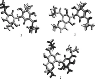
The conformations on Figure 2 reveals that this type of calculation should be extremely useful in predicting conformational preferences of similar structures since the geometries obtained experimentally by X-ray diffraction and theoretically by molecular modeling are very close (RMS = 0.016 Å for compound (1) and
0.014 Å for compounds (2) and (3)). It is important
to notice that although the structure in solution is the most important one when we are studying biological activity, in the case focused here, changes in conformation will probably be minor due to the several hydrogen bonds.
Conclusions
Calculations using the DFT B3LYP/6-31 G(d,p) basis set showed very good agreement between structural parameters calculated at this level and X-ray data, indicating that the methodology employed here seems to be a very useful tool in order to predict the conformational preference for this class of compounds.
Supplementary Information
CCDC 737455-737457 contains the supplementary crystallographic data for the structures in this paper. These data can be obtained free of charge from the Director, CCDC, 12 Union Road, Cambridge, CB2 1EZ, UK (fax: +44-1223-336033; e-mail: [email protected] or http://www.ccdc.cam.ac.uk).
Acknowledgements
EBL held a graduate fellowship from FAPERJ and ALC from CNPq.
Figure 2. Conformations (obtained by molecular modeling - in gray - and
by X-ray - in black) adopted by the phloroglucinols studied: japonicin A (1), uliginosin B (2) and isouliginosin B (3) respectively.
Table 2. Interatomic distances (Å) for hydrogen bonds calculated by the Gaussian 98 program and obtained by X-ray diffraction for the phloroglucinols studied
(1) (2) (3)
Calculated X-ray Calculated X-ray Calculated X-ray
C1-O...HO-C3 1.62 1.73 - - -
-C1-O...HO-C5 1.61 1.72 - - -
-C8=O...HO-C3 1.42 1.62 - - -
-C8=O...HO-C5 1.42 1.65 - - -
-C1-O...HO-C5’ 1.64 1.80
C3-OH...OH-C7’ 1.71 1.88
C8=O...HO-C5 1.42 1.65
C11’=O...HO-C7’ 1.44 1.68
C1-O...HO-C7’ - - - - 1.63 1.69
C2’-O...HO-C3 - - - - 1.75 1.90
C8=O...HO-C5 - - - - 1.41 1.41
References
1. Simões, C. M.; Farmacognosia da Planta ao Medicamento. 6th ed., Editora da UFSC: Santa Catarina, 2007.
2. Morais, S. K. R.; Teixeira, A. F.; Torres, Z. E. D. S.; Nunomura, S. M.; Yamashiro-Kanashiro, E. H.; Lindoso, J. A. L.; Yoshida, M.; J. Braz. Chem. Soc. 2009, 20, 1110.
3. Von Eiff, C.; Peters, G.; Heilmann, C.; Lancet Infect. Dis.2002,
2, 677.
4. França, H. S.; Kuster, R. M.; Riyo, P. N.; Oliveira, A. P.; Teixeira, L. A.; Rocha, L.; Quim. Nova2009, 35,1103.
5. França, H. S.; MSc Dissertation,Universidade Federal do Rio
de Janeiro, Brazil, 2005.
6. Jayasuriya, H.; Clark, A. M.; Mcchfsney, J. D.; J. Nat. Prod.
1991, 54, 1314.
7. Li-Hong, H.; Ching-Wan, K.; Vittal, J. J.; Keng-Yeow, S.;
Phytochemistry 2000, 53, 705.
8. Matsuhisa, M.; Shikishima, Y.; Takaishi, Y.; Honda, G.; Ito, M.; Takeda, Y.; Shibata, H.; Higuti, T.; Kodzhimatov, O. K.; Ashurmetov, O.; J. Nat. Prod. 2002, 65, 290.
9. Winkelmann, K.; Heilmann, J.; Zerbe, O.; Rali, T.; Sticher, O.;
J. Nat. Prod. 2001, 64, 701.
10. Rocha, L.; Marston, A.; Potterat, O.; Kaplan, A. C.; Stoeckli-Evans, H.; Hostettmann, K.; Phytochemistry 1995, 40, 1447.
11. Rocha, L.; Marston, A.; Potterat, O.; Kaplan, A. C.; Hostettmann, K.; Phytochemistry1996, 42, 185.
12. Leal, K. Z.; Seidl, P. R.; Yoneda, J. D.; dos Santos, C. V. B.; De Souza, M. C. B. V.; Ferreira, V. F.; J. Mol. Struct. 2005, 748,
137.
13. Braga, C. F.; Longo, R. L.; J. Braz. Chem. Soc. 2008, 19, 321. 14. Salum, L. B.; Dias, L. C.; Andricopulo, A. D.; J. Braz. Chem.
Soc. 2009, 20, 693.
15. Zanatta, N.; Borchhardt, D. M.; Carpes, A. D.; Marchi, T. M. ; Andricopulo, A. D. ; Salum, L. B.; Schetinger, M. R. C.; Bonacorso, H. G.; Martins, M. A. P.; Flores, A. F. C.; J. Braz. Chem. Soc. 2008, 19, 1118.
16. França, T. C. C.; Rocha, M. R. M.; Reboredo, B. M.; Rennó, M. N.; Tinoco, L. W.; Villar, J. D. F.; J. Braz. Chem. Soc. 2008,
19, 64.
17. Da Silva, J. B. P.; Ramos, M. N.; de Barros, B.; De Melo, S. J.; Falcão, E. P. S.; Catanho, M. T. J. A.; J. Braz. Chem. Soc.
2008, 19, 337.
18. Program CrysAlis-CCD and -RED, Oxford Diffraction Ltd., version 1.171.32.38.
19. SCALE3 ABSPACK scaling algorithm. CrysAlis RED, Oxford Diffraction Ltd., Version 1.171.32.38.
20. Sheldrick, G. M.; Schneider, T. R.; Methods Enzymol. 1997,
277, 319.
21. Johnson, C. K.; Crystallographic Computing, Ahmed, F.R., ed.; Copenhagen, Munksgaard, 1970.
22. HyperChem(TM), Hypercube, Inc., 1115 NW 4th Street, Gainesville, Florida 32601, USA.
23. Frisch, M. J.; Trucks, G. W.; Schlegel, H. B.; Gill, P. M. W.; Johnson, B. G.; Robb, M. A.; Cheeseman, J. R.; Keith, T. G.; Peterson, G. A.; Montgomery, J. A.; Raghavachari, K.; Al-Laham, M. A.; Zakrzewski, V. G.; Ortiz, J. V.; Foresman, J. B.; Cioslowski, J.; Stefanov, B. B.; Nanayakkara, A.; Challacombe, M.; Peng, C. Y.; Ayala, P. Y.; Chen, W.; Wong, N. W.; Andress, J. L.; Replogle, E. S.; Gomperts, R.; Martin, R. L.; Fox, D. L.; Binkley, J. S.; Defrees, D. J.; Baker, J.; Stewart, J. P.; Head-Gordon, M.; Gonzalez, C.; Pople, J. A.; Gaussian 98. Gaussian Inc.: Pittsburg, PA, 1998.
24. Jeffrey, G. A.; An Introduction to Hydrogen Bonding, Oxford University Press: New York, 1997, ch. 2.
Received: November 27, 2009
Su
pp
le
m
enta
ry
Inf
or
m
ati
on
0103 - 5053 $6.00+0.00
*e-mail: [email protected]
Conformational Analysis of Phloroglucinols from
Hypericum Brasiliense
by using
X-ray Diffraction and Molecular Modeling
Kátia Z. Leal,*,a,b Julliane D. Yoneda,c Eric B. Lindgren,a Carlos B. Pinheiro,d Arthur L. Corrêaa and Hildegardo S. Françae
aDepartamento de Físico-Química, Instituto de Química, Universidade Federal Fluminense,
00000-000 Niterói-RJ, Brazil
bPrograma de Pós-Graduação em Química, Instituto de Química, Universidade Federal Fluminense,
00000-000 Niterói-RJ, Brazil
cPólo Universitário de Volta Redonda, Universidade Federal Fluminense,
00000-000 Volta Redonda-RJ, Brazil
dDepartamento de Física, Universidade Federal de Minas Gerais,
00000-000 Belo Horizonte-MG, Brazil.
eDepartamento de Tecnologia Farmacêutica, Faculdade de Farmácia, Universidade Federal
Fluminense, 00000-000 Niterói-RJ, Brazil
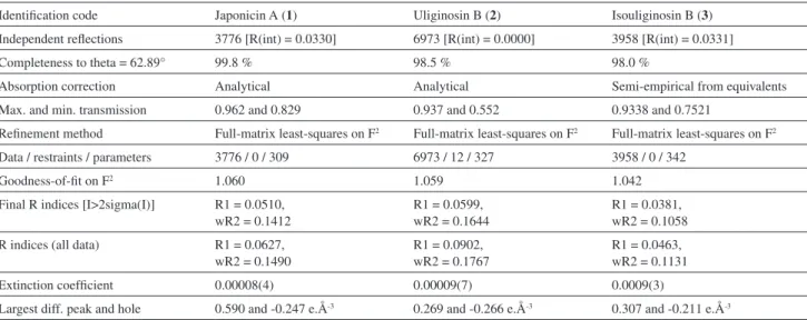
Table S1. Summary of the crystal structure data collection and reinement for japonicin A (1), uliginosin B (2) and isouliginosin B (3)
Identiication code Japonicin A (1) Uliginosin B (2) Isouliginosin B (3)
Empirical formula C25H32O8 C28H34O8 C28H34O8
Formula weight 460.51 498.55 498.55
Temperature 150(2) K 150(2) K 150(2) K
Wavelength 1.5418 Å 1.5418 Å 1.5418 Å
Crystal system Monoclinic Monoclinic Triclinic
Space group P 21/n P 21/n P -1
Unit cell dimensions a = 9.2461(4) Å
b = 17.8198(6) Å c = 28.8240(10) Å
α = 90°.
β = 90°.
γ = 90°
a = 10.4085(4) Å b = 9.1035(3) Å c = 27.5920(10) Å
α = 90.000(3)°.
β = 99.070(3)°.
γ = 90.000(3)°.
a = 10.4944(4) Å b = 10.7537(3) Å c = 12.3616(4) Å
α = 103.610(3)°.
β = 110.336(3)°.
γ = 93.969(3)°
Volume 4749.1(3) Å3 2581.76(16) Å3 1253.80(8) Å3
Z 8 4 2
Density (calculated) 1.288 Mg/m3 1.283 Mg/m3 1.321 Mg/m3
Absorption coeficient 0.792 mm-1 0.77 mm-1 0.793 mm-1
F(000) 1968 1064 532
Crystal size 0.39 x 0.11 x 0.06 mm3 0.49 x 0.14 x 0.03 mm3 0.38 x 0.22 x 0.09 mm3
Theta range for data collection 3.07 to 62.48°. 3.24 to 62.68° 3.97 to 62.89°
Index ranges -10<=h<=10,
-20<=k<=20, -33<=l<=33
-11<=h<=11, -10<=k<=10, -31<=l<=31
-12<=h<=11, -12<=k<=12, -14<=l<=14
Identiication code Japonicin A (1) Uliginosin B (2) Isouliginosin B (3)
Independent relections 3776 [R(int) = 0.0330] 6973 [R(int) = 0.0000] 3958 [R(int) = 0.0331]
Completeness to theta = 62.89° 99.8 % 98.5 % 98.0 %
Absorption correction Analytical Analytical Semi-empirical from equivalents
Max. and min. transmission 0.962 and 0.829 0.937 and 0.552 0.9338 and 0.7521
Reinement method Full-matrix least-squares on F2 Full-matrix least-squares on F2 Full-matrix least-squares on F2
Data / restraints / parameters 3776 / 0 / 309 6973 / 12 / 327 3958 / 0 / 342
Goodness-of-it on F2 1.060 1.059 1.042
Final R indices [I>2sigma(I)] R1 = 0.0510,
wR2 = 0.1412
R1 = 0.0599, wR2 = 0.1644
R1 = 0.0381, wR2 = 0.1058
R indices (all data) R1 = 0.0627,
wR2 = 0.1490
R1 = 0.0902, wR2 = 0.1767
R1 = 0.0463, wR2 = 0.1131
Extinction coeficient 0.00008(4) 0.00009(7) 0.0009(3)
Largest diff. peak and hole 0.590 and -0.247 e.Å-3 0.269 and -0.266 e.Å-3 0.307 and -0.211 e.Å-3
Uliginosin reinement performed on data obtained from a twinned sample. For japonicin A w=1/[\s2 (F
o2)+(0.0755P)2+3.4186P] where P=(Fo2+2Fc2)/3,
for uliginosin w=1/[\s2(F
o2)+(0.0574P)2+0.2333P] where P=(Fo2+2Fc2)/3 and for isouliginosin w=1/[\s2(Fo2)+(0.0642P)2^+0.4656P] where P=(Fo2+2Fc2)/3.