Article
Printed in Brazil - ©2016 Sociedade Brasileira de Química0103 - 5053 $6.00+0.00*e-mail: [email protected], [email protected]
Genome Mining of Endophytic Streptomyces wadayamensis Reveals High Antibiotic
Production Capability
Célio F. F. Angolini,*,a Ana B. Gonçalves,a Renata Sigrist,a Bruno S. Paulo,a Markiyan
Samborskyy,b Pedro L. R. Cruz,a Adriana F. Vivian,c Eduardo M. Schmidt,a Marcos N.
Eberlin,a Welington L. Araújod and Luciana G. de Oliveira*,a
aDepartamento de Química Orgânica, Instituto de Química, Universidade Estadual de Campinas,
13083-970 Campinas-SP, Brazil
bDepartment of Biochemistry, University of Cambridge, Tennis Court Road, Cambridge,
Cambridgeshire, CB2 1GA, England
cFaculdade de Ciências Farmacêuticas, Universidade Estadual de Campinas,
13083-970 Campinas-SP, Brazil
dDepartamento de Microbiologia, Instituto de Ciências Biomédicas, Universidade de São Paulo,
05508-900 São Paulo-SP, Brazil
The actinobacteria Streptomyces wadayamensis A23, an endophitic strain, was recently
sequenced and previous work showed qualitatively that the strain inhibits the growth of
some pathogens. Herein we report the genome analysis of S. wadayamensis which reveals
several antibiotic biosynthetic pathways. Using mass spectrometry, we were able to identify desferoxamines, several antimycins and candicidin, as predicted. Additionally, it was possible to confirm that the biosynthetic machinery of the strain when compared to identified known metabolites is far underestimated. As suggested by biochemical qualitative tests, genome encoded information reveals that the strain A23 has high capability to produce antibiotics.
Keywords: genome sequencing, genome mining, molecular networking, Streptomyces, antibiotic activity
Introduction
Streptomyces genera carry an extremely versatile group of biosynthetic machineries capable to produce complex molecules of medical, agricultural and economical significance. They are recognized as the most resourceful producers of molecules that present antibiotic activities.1
The mutualism existing among several actinobacteria and plants, ants and other organisms also seemed to be a key element to the capability to produce such small molecules.2,3
Since the pioneers work in partial genome sequencing to a more recent and wide investment on whole genome sequencing, it became clear that the ability of Streptomyces
and other actinobacteria to biosynthesize promising metabolites is underestimated.1,4 Recent developments on
next generation sequencing revealed a diverse plethora of
gene clusters encoding for cryptic targets. It became clear that by accessing sequences genome encoded it is possible to predict enzymatic functions and the hole of an entire gene cluster involved in secondary metabolites biosynthesis.
Genome-mining tools have therefore opened a new and innovative avenue for natural products research.5
When combined with bioinformatic tools, it contributes to envisage a global map of biosynthetic gene clusters (BGC). The possibility to connect genes to natural products enables to direct the discovery of novel molecules, for instance, metabolites as coelichelin,6 orfamide A7 and salinilactam8
were found by searching programs guided by the prediction of physical-chemical properties, anticipation of precursors incorporation, manipulation of higher levels and pathway specific regulators, inactivation of biosynthetic genes and analytical evaluation, and by in vitro reconstitution.9-18
Shotgun project of Streptomyces wadayamensis A23, an endophytic strain isolated from Citrus reticulata
(tangerine).19 Biochemical qualitative tests showed that
strain A23 inhibits the growth of Candida albicans
pathogen, Bacillus megaterium, Neisseria meningitides
strains, and multiresistant Staphylococcus aureus.20 Herein
we report our preliminary results generated after genome annotation and mining of the WGS project. Annotation was critical to predict that S. wadayamensis has high capability to biosynthesize antibiotics. Experimental evaluation combined to powerful analytical tools revealed the production of several bioactive metabolites as anticipated by mining.
Experimental
Chemical and samples
Water was purified using a Milli-Q water purification system (Millipore). Beef extract, yeast extract, peptone and agar were purchased from Oxoid (Hampshire, UK). HPLC grade methanol was purchased from Tedia Brazil (Rio de Janeiro, Brazil). FeCl3.6H2O, HCl, chromeazurol S,
hexadecyltrimethylammonium bromide (HTMS), PIPES, KH2PO4, NaCl, NH4Cl, KOH, glucose, D-mannitol, casein
hydrolisate, MgSO4.7H2O, CaCl2, MnSO4.H2O, H3BO3,
CuSO4.5H2O, ZnSO4.7H2O and Na2MoO4.2H2O were
purchased from Sigma-Aldrich. The actinobacterium strain A23, identified as Streptomyces wadayamensis, was isolated from a population existing in the plant tissue of
Citrus reticulata (tangerine).
Culture media
The media used in this work were vegetative N (peptone 5 g L-1, soluble starch 20 g L-1, meat extract 2 g L-1, yeast
extract 3 g L-1, soy bean meal 2 g L-1, CaCO
3 1 g L-1,
pH 7.0); A medium (peptone 4 g L-1, meat extract 4 g L-1,
yeast extract 2 g L-1, soybean meal 2 g L-1, maltose 20 g L-1,
dextrine 10 g L-1, pH 7.0); INA (soy bean meal 15 g L-1,
CaCO3 5 g L-1, glycerol 30 mL L-1, NaCl 2 g L-1, pH 7.0);
RA3 (peptone 2 g L-1, yeast extract 4 g L-1, malt extract
10 g L-1, glucose 10 g L-1, glycerol 5 mL L-1, MgCl 2.6H2O
2 g L-1, pH 7); GPMY (potato starch 20 g L-1, yeast extract
5 g L-1, malt extract 5 g L-1, glycerol 20 mL L-1, pH 7.0)
AFMS (dextrose 20 g L-1, yeast extract 2 g L-1, soybean
meal 8 g L-1, CaCO
3 4 g L-1, NaCl 1 g L-1, pH 7.3); GYM
(yeast extract 4 g L-1, malt extract 10 g L-1, glucose 4 g L-1,
pH 7.0) and M8 (soluble starch 20 g L-1, meat extract
2 g L-1, yeast extract 2 g L-1, glucose 10 g L-1, CaCO
3 3 g L-1,
pH 7.0).
Fourier transfor m ion cyclotron resonance mass spectrometry (FT-ICR-MS) sample preparation
A single colony of Streptomyces wadayamensis was selected and added to 1 mL of vegetative N medium (seed). The cell was grown for 24 h at 30 oC and 250 rpm. After this
time, the seed medium was added in 100 mL of the culture medium described above. Streptomyces wadayamensis
was cultivated for 7 days in 500 mL Erlenmeyer flasks on reciprocal shaker at the same conditions. The medium was centrifuged and the mycelium was separated. The broth was extracted three times with ethyl acetate. The organic layer was evaporated. 1 mg of the culture extract was dissolved in 1 mL of methanol and centrifuged for 10 min at 13,000 rpm to remove any precipitate. This solution was used as stock solution from which aliquots of 100 µL were removed and diluted in 1 mL of methanol.
FT-ICR-MS analysis
T h e e q u i p m e n t w a s a n LT Q F T U l t r a 7 T (ThermoScientific) equipped with a nano-electrospray ionization (ESI) source (TriVersa NanoMate 100 system). Ionization was carried out both on the negative and positive ion mode on an m/z between 100 and 2000. The spectra generated were analyzed with the Xcalibur software.
Desor ption electrospray ionization imaging mass spectrometry (DESI-IMS) sample preparation
For microbial IMS sample preparation, a 0.25 µL sample from a Streptomyces spore solution was inoculated on thin-layered agar. The antibiotic assay medium 2 (1.5 g L-1 beef extract, 3.0 g L-1 yeast extract, 6 g L-1 peptone
and 1.5% agar) was prepared by first placing a sterile microscope slides in a Petri dish, followed by the pouring of agar medium. The inoculated medium was incubated at 30 °C, and the Petri dish was sealed with parafilm to minimized premature dehydration. After the incubation period (48 h), the microscope slides were removed from the Petri dish, photographed and put in a vacuum desiccator for complete agar dehydration at room temperature.
DESI-IMS analysis
temperature of 320 °C, 100 V S-lens, 160 psi ultra-pure nitrogen nebulizing gas pressure and a sprayed solvent of methanol in a 3.0 µL min-1 flow rate. Images were collected
scaning from m/z 133-1500 with a step size of 200 mm, a scan rate of 741 µm sec-1 and a pixel size of 200 × 200 µm.
MS and tandem mass spectrometry (MS/MS) for network analyses
For analysis of Streptomyces wadayamensis A23 metabolite profiles, cells cultures and medium were removed from microscope slides and extract with 0.5 mL methanol after DESI-IMS analysis. The extract were centrifuged for solid parts removal, and the remaining solution was analyzed by direct infusion ESI in a Thermo Scientific Q Exactive Hybrid Quadrupole-Orbitrap Mass Spectrometer. The MS/MS experiments were all conducted with 3.5 kV spray voltage, inlet capillary temperature of 320 °C, 100 V S-lens in a 3.0 µL min-1 syringe flow rate.
Data analysis
The DESI-IMS data was converted into imaged files using the Firefly data conversion software (version 2.1.05)21
and viewed using the BioMAP software (version 3.8.04).22
For molecular network creation, the tandem mass data (.raw) were converted in mzXML using msconvert from ProteoWizard and the GNPS automated platform23 in
default settings.24 In addition, Cytoscape 2.825 was used
for molecular network visualization.
Results and Discussion
As previously described, the genome draft of
S. wadayamensis A23 has near to 7 million base pairs, encoding 6006 protein-coding sequences with 73.5% GC content.19 The Illumina shotgun library produced
7.8 million reads in a total of 2.4 GB data assembled to generate 180 contigs. Genome annotation was carried out using a customized pipeline and revealed several putative secondary metabolites encoded by S. wadayamensis.
The input of the genome data into the antibiotics & Secondary Metabolite Analysis SHell (antiSMASH) server (version 3.0.5)26 resulted in an output predicting
the presence of 32 gene clusters codifying for secondary metabolites biosynthesis. Biosynthetic systems as terpene, nrps, bacteriocin-terpene, t1-pks, t3-pks, bacteriocin, nrps-t1pks, tiopeptide-lantipeptide, lantipeptide-nrps-t1-pks, siderophore, ectoine, lassopeptide, lantipeptide and other clusters are apparently encoded in S. wadayamensis
genome (Table S1). Crossing the output results from antiSMASH27 within the MIBiG (Minimum Information
about a Biosynthetic Gene cluster) pipeline,28 it was
possible to predict more accurately certain metabolites and/or the most likely class that the gene cluster fits as presented in Table 1.
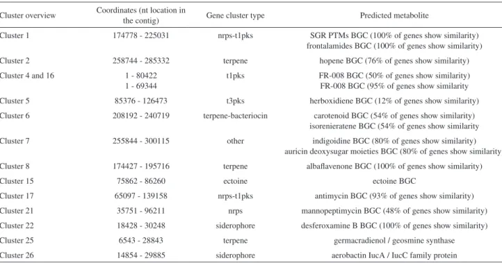
Table 1. Most significant gene clusters encoded in Streptomyces wadayamensis genome (antiSMASH-MIBiG output)a
Cluster overview Coordinates (nt location in
the contig) Gene cluster type Predicted metabolite
Cluster 1 174778 - 225031 nrps-t1pks SGR PTMs BGC (100% of genes show similarity)
frontalamides BGC (100% of genes show similarity)
Cluster 2 258744 - 285332 terpene hopene BGC (76% of genes show similarity)
Cluster 4 and 16 1 - 80422
1 - 69344
t1pks FR-008 BGC (50% of genes show similarity) FR-008 BGC (95% of genes show similarity
Cluster 5 85376 - 126473 t3pks herboxidiene BGC (12% of genes show similarity)
Cluster 6 208192 - 240719 terpene-bacteriocin carotenoid BGC (54% of genes show similarity) isorenieratene BGC (54% of genes show similarity
Cluster 7 255844 - 300115 other indigoidine BGC (80% of genes show similarity)
auricin deoxysugar moieties BGC (80% of genes show similarity)
Cluster 8 174427 - 195716 terpene albaflavenone BGC (100% of genes show similarity)
Cluster 15 75862 - 86260 ectoine ectoine BGC
Cluster 17 65097 - 139158 nrps-t1pks antimycin BGC (93% of genes show similarity)
Cluster 21 35751 - 96211 nrps mannopeptimycin BGC (48% of genes show similarity)
Cluster 22 18428 - 30248 siderophore desferoxamine B BGC (100% of genes show similarity)
Cluster 25 6543 - 28843 terpene germacradienol / geosmine synthase
Cluster 26 14854 - 29885 siderophore aerobactin IucA / IucC family protein
However, it is important to emphasize that, although the antiSMASH platform predicts 32 possible BGC, this actual number could be underestimated as the genome is organized in 180 contigs and some clusters are truncated (for example, both clusters 4 and 16 are complementary and correspond to the BGC of FR-008).
After in silico analysis, the metabolic profile of the ethyl acetate extracts from cultivation in several mediums was evaluated by FT-ICR-MS and Orbitrap in both the positive and negative ion modes using a m/z range between 100 and 2000. To identify the most common ions present in the extracts, we performed a manual search in the dictionary of natural products (DNP),29 with near to 226000 structures
from wide-ranging sources.
Although there is a difference in intensity for the signals in the chromatographic profile (Figure S1), almost the same set of ions were found in the conditions tested for fermentation (data not shown).
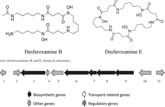
A careful evaluation of A medium pointed to an ion of m/z 601.3552 with attributed formula C27H48N6O9.
Among the structures generated in the DNP database, we identified nocardamine, also known as desferoxamine E (Figure 1), a siderophore with high affinity for iron, which forms a complex that is reabsorbed by the cell via the ATP-dependent transport system.30 We had already
observed the production of siderophores by qualitative tests on agar plates containing chrome azurol sulfonate.20
Additionally, we performed direct DESI-IMS on an agar culture and observed another siderophore of same family, that is, the desferoxamine B of m/z 561.3640 (Figure 1).31
Both siderophores predicted by the gene cluster 22 were therefore characterized by chemical/biological tests (chrome azurol) and MS/MS fragmentation patterns. The fragmentation of protonated desferoxamine E was mainly characterized by the neutral loss of a molecule of succinylcadaverine (200 Da, Figure S2).32
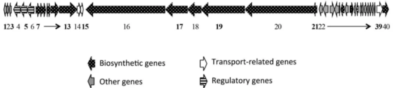
Desferoxamine (MIBiG BGC 0000941) is a highly conserved gene cluster in Streptomyces genera (Figure 2). The antiSMASH output for Streptomyces wadayamensis A23 resulted in 10 different sequences deposited in databases that possesses high similarity within our query sequence (Streptomyces albus J107,
Streptomyces sp. SM8, Streptomyces sp. PVA 94-07,
Streptomyces sp. GBA 94-10, Streptomyces mutabilis
TRM4554, Streptomyceswadayamensis A23 LGO A23 AS7,
Streptomyces sp. JS01, Streptomycesgriseus subsp. griseus NBRC 1335, Streptomyces griseus XylebKG-1). The gene cluster comprises a set of 5 main enzymes (5 to 9) with essential functions for the siderophore biosynthesis: siderophore biosynthetic enzyme (5), acetyltransferase (6), monooxygenase (7), pyridoxal-dependent decarboxylase (8) and siderophore-interacting protein (9).
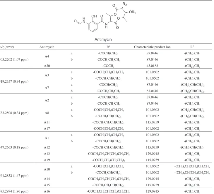
A total of 6 product ions related to antimycins were also identified, which are part of a family of polyketides-nonribosomal peptides already isolated from Streptomyces sp. exhibiting confirmed antifungal activity and predicted on gene cluster 17.33,34 The antimycins are biosynthesized by
a mixed cluster nrps-t1pks and the main differences among them are located in the chains R1 and R2 positions, which
corresponds to an aliphatic and fat acid chain, respectively,
Figure 1. Siderophore desferoxamines B and E chemical structures.
and characterize the main losses observed by MS/MS (Table 2).33,34 Most of the product ions from antimycins
fragmentation are common among all isoforms, e.g., m/z
263, 245, 219, 191 and 161, however and fortunately, there is often one fragment (R1, Figure 3A) which is characteristic for each antimycins variant. Therefore, based on these product ions, the production of antimycin variants such as A11, A13, A14, A16 and A20 could be excluded from our samples. As some variants have the same precursor ions and the same characteristic product ions, the fragmentation pattern may correspond, for instance, to A1a or A1b, A12 or A19, A8 or A17 (where R1 corresponds to distinct acyl
chains in the structural isomers), therefore it is not possible to distinguish between then by MS spectrometry and additional
analysis, such as NMR, would be necessary to discriminate the structural isomers.
The production of antimycins A1, A12 and/or A19 (C28H40N2O9) identified by the [M – H]– anion of m/z 547.2663; antimycin A4 (C25H34N2O9) of m/z 505.2202;
antimycin A3 and/or A7 (C26H36N2O9) of m/z 519.2357;
antimycin A2, A8 and/or A17 (C27H38N2O9) of m/z 533.2508;
antimycin A10 and/or A15 (C29H42N2O9) of m/z 561.2832
and antimycin A16 (C30H44N2O9) of m/z 575.2994 were
accordingly confirmed by A23 strain in the cultivation conditions (Figure 3 and Figure S3).
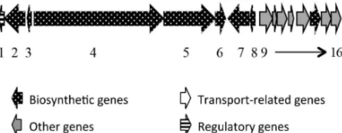
The gene cluster codifying for antimycins in
S. wadayamensis is 100% identical to the one described by Seipke et al.35 and fed in MIBiG platform (BGC0000958),
Table 2. The antimycins PKS-NRPS hybrids variants and their ESI(−)-MS/MS characteristics
m/z (error) Antimycin R1 Characteristic product ion R2
505.2202 (1.07 ppm) A4
a -COCH(CH3)2 87.0446 -(CH2)3CH3
b -COCH2CH2CH3 87.0446 -(CH2)3CH3
A20 -COCH3 43.0183 -(CH2)5CH3
519.2357 (0.94 ppm)
A3 a -COCH(CH3)CH2CH3 101.0602 -(CH2)3CH3
b -COCH2CH(CH3)2 101.0602 -(CH2)3CH3
A7 a -COCH(CH3)2 87.0446 -(CH2)2CH(CH3)2
b -COCH2CH2CH3 87.0446 -(CH2)2CH(CH3)2
533.2508 (0.34 ppm)
A2 a -COCH(CH3)2 87.0446 -(CH2)5CH3
b -COCH2CH2CH3 87.0446 -(CH2)3CH3
A8 a -COCH(CH3)CH2CH3 101.0602 -(CH2)2CH(CH3)2
b -COCH2CH(CH3)2 101.0602 -(CH2)2CH(CH3)2
A11 -COCH2CH2CH(CH3)2 115.0759 -(CH2)3CH3
A17 -COCH(CH3)CH2CH3 101.0602 -(CH2)4CH3
547.2663 (0.18 ppm)
A1 a -COCH(CH3)CH2CH3 101.0602 -(CH2)5CH3
b -COCH2CH(CH3)2 101.0602 -(CH2)5CH3
A12 -COCH2CH2CH(CH3)2 115.0759 -(CH2)2CH(CH3)2
A13 -COCH2CH2CH(CH3)CH2CH3 129.0915 -(CH2)3CH3
A19 -COCH(CH3)CH(CH3)2 115.0759 -(CH2)4CH3
561.2832 (1.47 ppm)
A10 a -COCH(CH3)CH2CH3 101.0602 -(CH2)3CH(CH3)CH2CH3
b -COCH2CH(CH3)2 101.0602 -(CH2)3CH(CH3)CH2CH3
A14 -COCH2CH2CH(CH3)CH2CH3 129.0915 -(CH2)3CH3
A15 -COCH2CH2CH(CH3)2 115.0759 -(CH2)5CH3
comprising the genes 1 to 16 in Figure 4. The modular polyketide synthase gene antD (gene number 5, module 3, Figure 4) carries a KS, AT, ACP and TE domain. According
to the differences observed in R1 chains, it is possible to infer that the AT specificity is highly atypical with the incorporation of several acyl-CoAs subunits.
The mining also indicated a possible metabolite described by the clusters 4 and 16 belonging to the FR-008 BGC, a complex polyene with high similarity to the candicidin gene cluster found in Streptomyces griseus
IMRU3570 (Figure 5).36 Chen et al.36 originally described
this metabolite and its gene cluster in 2003 and throughout this study it was possible to conclude that candicidin and FR-008 are identical compounds.
A meticulous search led us to find candicidin (m/z 1109.5784, C59H84N2O18, Figure 6) in the crude extract,
Figure 3. (A) ESI(−) FT-ICR-MS of Streptomyces A23 crude extract (AFMS medium) showing deprotonated antimycin analogues; (B) MS/MS fragmentation pattern of the deprotonated antimycin A1a (note that this fragmentation pattern could be expected for A1b, A12 and A19 as well).
which could be most likely, the compound responsible for
C. albicans growth inhibition.36
An indication for the production of candicidin in
S. wadayamensis was already anticipated in previous studies20 by the use of metabolic probes to promote
dereplication. The KS-AT domains from FscB and FscD (gene numbers 17 and 20, respectively, Figure 7) were identified using this methodology. The candicidin gene
cluster organization in the endophytic strain is 100% identical to the gene cluster description for FR-008 (BGC0000061 in MIBiG repository).
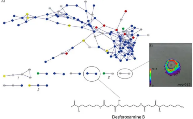
To enhance the A23 dereplication, we performed the molecular networking analysis of A23 cultivation extracts, but no further known molecules were found. Interestingly, we were able to cross several information found by FT-ICR-MS and DESI-IMS. As molecular networking also
Figure 5. Chemical structure of the complex polyene candicidin.
Figure 6. ESI(+) Orbitrap MS of Streptomyces A23 extract (A medium) showing the observed ions in the range m/z 1000-1200.
enables clustering families of structurally related natural products by MS/MS fragmentation pattern analysis, and DESI-IMS is able to correlate molecules of same spatial distribution during the microorganism growth, it is possible therefore to establish a correlation within a related set of compounds that could share chemical similarities.24,37 Using
molecular networking dereplication, we were able to find desferoxamine B (described in cluster 22) and their iron complex form (Figure 8),31 and we also tagged three other
clusters of potentially unknown natural products (cluster 1:
m/z 700.645; cluster 2: m/z 1193.38; cluster 3: m/z 912.627). IMS experiments revealed that metabolites possibly described by the gene clusters from Table 1 and Table S1 could be visualized over the agar media around the colony. A preliminary MS/MS analysis showed that the ion m/z 912 seems to be a nonribosomal linear peptide (NRP) not yet described (Figure S4). We failed to find a similar pattern in networking or in dictionary of natural products. In total, we found three different compounds (m/z 884, 898, and 912) differing in 14 u (CH2) between each other from which is
possible to observe a similar arrangement distribution over the agar media in IMS data.
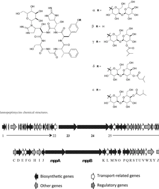
Finally, mining the genome revealed gene cluster 21, related to a NRPS responsible to mannopeptimycin biosynthesis (48% of the genes show similarity) produced by several strains of Streptomyces hygroscopicus. Mannopeptimycins are glycosylated cyclic hexapeptides
(glycopeptides) first described as a complex mixture of compounds named AC98 and showed effective activity against Gram-positive bacteria (Figure 9).38 Its activity was
also confirmed against methicillin resistant staphylococci and vancomycin resistance enterococci.39
The biosynthetic pathway for mannopeptimycins was described by Margavey et al.40 in 2006. Looking closely
and performing blast and protein function comparison, it is possible to verify that the main genes mppA and mppB
responsible for the hexapeptide core are 100% identical in S. wadayamensis A23 (genes 23 and 24 in Figure 10A) and S. hygroscopicus NRRL 30439, revealing similar operon type organization (Figure 10B). The most part of the right hand outside genes (except for mppM, mppN,
mppX, mppY, mppZ, and mppZ1) are also identical in both strains. mppM and mppN correspond to isovaleryl transferases, responsible for the isovaleryl incorporation in the terminal O-linked mannose in mannopeptimycins γ, δ, and ε. The absence of these genes in the BGC codified by
S. wadayamensis suggests that this strain could produce α
or β-mannopeptimycins. The genes mppX, mppY and mppZ
have unknown function and mppZ1 is a transcriptional regulator. The left hand side genes in Streptomyces wadayamensis codifies for a single major facilitator transporter (gene 22), while in the mannopeptimycin gene cluster used for comparison there are two genes (E and F) codifying for this function. In the MIBiG entry mppI and
mppH codifies for polyprenyl fosfo-mannosyl transferases,
mppJ for a methyltransferase, mppG for a polyprenyl mannose synthase, mppD for a sugar binding protein and
mppC for an acetyltransferase. For mannopeptimycin BGC from S. wadayamensis it is possible to describe several unknown function genes (labelled in grey) and two glucose specific phosphotransferase (genes 12 and 21) among others. Under the fermentation conditions we couldn’t identify the production of mannopeptimycin, but still the information is genome encoded and is under investigation.
All the molecules identified so far shows that Streptomyces
wadayamensis A23 has a high capability to produce
antibiotics. Still, we need to correlate the chemical entities with the biochemical activity prompted to inhibit the growth of pathogens as observed. Studies related to the unknowns metabolites produced by A23 strain are in progress.
Conclusions
The genome sequencing of Streptomyces wadayamensis
A23 combined with gene annotation and mass spectrometry pattern evaluation revealed the strain is a promising producer
Figure 9. Mannopeptimycins chemical structures.
of several secondary metabolites. As such tool improves the dereplication approach it was possible to observe the production of the siderophores deferoxamines B and E, several antimycins antibiotics and the polyene candicidin. These results open a wide field of research encompassing “from genome to natural products” as a promising strategy to find new biological and pharmacological targets.
Supplementary Information
Supplementary Information (LC, LC-MS and MS/MS data) is available free of charge at http://jbcs.sbq.org.br.
Acknowledgments
We gratefully acknowledge FAPESP (project grant 2014/12727-5 to L. G. O. and 2010/51677-2 to M. N. E.), PETROBRAS (grant 4712-0), and the University of Campinas. C. F. F. A. and B. S. P. acknowledges CNPq (studentships 162191/2015-4 and 130933/2015-5). A. B. G. is grateful for the PIBIC-SAE studentship. R. S. acknowledges FAPESP (studentships 2013/12598-8 and 2015/01013-4) and L. G. O. is also grateful for the grant support provided by IFS (Grant Support No. F/4735-1) and by the program For Women in Science (2008, Brazilian Edition).
References
1. Bentley, S. D.; Chater, K. F.; Cerdeño-Tárraga, A.-M.; Challis, G. L.; Thomson, N. R.; James, K. D.; Harris, D. E.; Quail, M. A.; Kieser, H.; Harper, D.; Bateman, A.; Brown, S.; Chandra, G.; Chen, C. W.; Collins, M.; Cronin, A.; Fraser, A.; Goble, A.; Hidalgo, J.; Hornsby, T.; Howarth, S.; Huang, C.-H.; Kieser, T.; Larke, L.; Murphy, L.; Oliver, K.; O’Neil, S.; Rabbinowitsch, E.; Rajandream, M.-A.; Rutherford, K.; Rutter, S.; Seeger, K.; Saunders, D.; Sharp, S.; Squares, R.; Squares, S.; Taylor, K.; Warren, T.; Wietzorrek, A.; Woodward, J.; Barrell, B. G.; Parkhill, J.; Hopwood, D. A.; Nature 2002, 417, 141. 2. Crawford, J. M.; Clardy, J.; Chem. Commun. 2011, 47, 7559. 3. Schmidt, E. W.; Nat. Chem. Biol. 2008, 4, 466.
4. Ikeda, H.; Ishikawa, J.; Hanamoto, A.; Shinose, M.; Kikuchi, H.; Shiba, T.; Sakaki, Y.; Hattori, M.; Ōmura, S.; Nat. Biotechnol.
2003, 21, 526.
5. Corre, C.; Challis, G. L.; Chem. Biol. 2007, 14, 7.
6. Challis, G. L.; Ravel, J.; FEMS Microbiol. Lett. 2000, 187, 111.
7. Gross, H.; Stockwell, V. O.; Henkels, M. D.; Nowak-Thompson, B.; Loper, J. E.; Gerwick, W. H.; Chem. Biol. 2007, 14, 53. 8. Udwary, D. W.; Zeigler, L.; Asolkar, R. N.; Singan, V.; Lapidus,
A.; Fenical, W.; Jensen, P. R.; Moore, B. S.; Proc. Natl. Acad. Sci. USA 2007, 104, 10376.
9. Tohyama, S.; Eguchi, T.; Dhakal, R. P.; Akashi, T.; Otsuka, M.; Kakinuma, K.; Tetrahedron 2004, 60, 3999.
10. McAlpine, J. B.; Bachmann, B. O.; Piraee, M.; Tremblay, S.; Alarco, A.-M.; Zazopoulos, E.; Farnet, C. M.; J. Nat. Prod.
2005, 68, 493.
11. Fischbach, M. A; Walsh, C. T.; Chem. Rev. 2006, 106, 3468. 12. Banskota, A. H.; Mcalpine, J. B.; Sørensen, D.; Aouidate, M.;
Piraee, M.; Alarco, A.-M.; Omura, S.; Shiomi, K.; Farnet, C. M.; Zazopoulos, E.; J. Antibiot. 2006, 59, 168.
13. Banskota, A. H.; McAlpine, J. B.; Sørensen, D.; Ibrahim, A.; Aouidate, M.; Piraee, M.; Alarco, A.-M.; Farnet, C. M.; Zazopoulos, E.; J. Antibiot. 2006, 59, 533.
14. Lin, X.; Hopson, R.; Cane, D. E.; J. Am. Chem. Soc. 2006, 128, 6022.
15. Scherlach, K.; Hertweck, C.; Org. Biomol. Chem. 2006, 4, 3517. 16. Wenzel, S. C.; Meiser, P.; Binz, T. M.; Mahmud, T.; Müller, R.;
Angew. Chemie Int. Ed. 2006, 45, 2296.
17. Song, L.; Barona-Gomez, F.; Corre, C.; Xiang, L.; Udwary, D. W.; Austin, M. B.; Noel, J. P.; Moore, B. S.; Challis, G. L.; J. Am. Chem. Soc. 2006, 128, 14754.
18. Liu, C.; Zhu, J.; Li, Y.; Zhang, J.; Lu, C.; Wang, H.; Shen, Y.;
ChemBioChem 2015, 16, 998.
19. de Oliveira, L. G.; Tormet Gonzalez, G. D.; Samborsky, M.; Marcon, J.; Araujo, W. L.; de Azevedo, J. L.; Genome Announc.
2014, 2, 1.
20. da Cruz, P. L. R.; Giarola, L. R.; Moraes, S. S.; da Silva, D. E. S. G.; Marcon, J.; Azevedo, J. L.; Araújo, W. L.; de Oliveira, L. G.; Quim. Nova 2015, 38, 333.
21. http://www.prosolia.com/products/firefly, accessed in June 2016.
22. http://www.imzml.org/, accessed in June 2016.
23. http://gnps.ucsd.edu/ProteoSAFe/static/gnps-splash.jsp, accessed in June 2016.
24. Watrous, J.; Roach, P.; Alexandrov, T.; Heath, B. S.; Yang, J. Y.; Kersten, R. D.; van der Voort, M.; Pogliano, K.; Gross, H.; Raaijmakers, J. M.; Moore, B. S.; Laskin, J.; Bandeira, N.; Dorrestein, P. C.; Proc. Natl. Acad. Sci. USA 2012, 109, E1743. 25. Shannon, P.; Markiel, A.; Ozier, O.; Baliga, N. S.; Wang, J. T.; Ramage, D.; Amin, N.; Schwikowski, B.; Ideker, T.; Genome Res. 2003, 13, 2498.
26. Weber, T.; Blin, K.; Duddela, S.; Krug, D.; Kim, H. U.; Bruccoleri, R.; Lee, S. Y.; Fischbach, M. a.; Müller, R.; Wohlleben, W.; Breitling, R.; Takano, E.; Medema, M. H.;
Nucleic Acids Res. 2015, 43, W237.
27. Medema, M. H.; Blin, K.; Cimermancic, P.; de Jager, V.; Zakrzewski, P.; Fischbach, M. a; Weber, T.; Takano, E.; Breitling, R.; Nucleic Acids Res. 2011, 39, W339.
28. http://mibig.secondarymetabolites.org/index.html, accessed in June 2016.
31. Angolini, C. F. F.; Vendramini, P. H.; Araújo, F. D. S.; Araújo, W. L.; Augusti, R.; Eberlin, M. N.; de Oliveira, L. G.; Anal. Chem. 2015, 87, 6925.
32. Ueki, M.; Suzuki, R.; Takamatsu, S.; Takagi, H.; Uramoto, M.; Ikeda, H.; Osada, H.; Actinomycetologica 2009, 23, 34. 33. Kim, H.; Esser, L.; Hossain, M. B.; Xia, D.; Yu, C.-A.; Rizo,
J.; van der Helm, D.; Deisenhofer, J.; J. Am. Chem. Soc. 1999,
121, 4902.
34. Atta, H. M.; Ahmad, M. S.; Aust. J. Basic Appl. Sci. 2009, 3, 126.
35. Seipke, R. F.; Barke, J.; Brearley, C.; Hill, L.; Yu, D. W.; Goss, R. J. M.; Hutchings, M. I.; PLoS One 2011, 6, e22028. 36. Chen, S.; Huang, X.; Zhou, X.; Bai, L.; He, J.; Jeong, K. J.;
Lee, S. Y.; Deng, Z.; Chem. Biol. 2003, 10, 1065.
37. Yang, J. Y.; Sanchez, L. M.; Rath, C. M.; Liu, X.; Boudreau, P. D.; Bruns, N.; Glukhov, E.; Wodtke, A.; de Felicio, R.; Fenner, A.; Wong, W. R.; Linington, R. G.; Zhang, L.; Debonsi, H. M.; Gerwick, W. H.; Dorrestein, P. C.; J. Nat. Prod. 2013, 76, 1686. 38. De Voe, S. E.; Kunstmann, M. P.; US pat. US3495004DA1970. 39. He, H.; Williamson, R. T.; Shen, B.; Graziani, E. I.; Yang, H.
Y.; Sakya, S. M.; Petersen, P. J.; Carter, G. T.; J. Am. Chem. Soc. 2002, 124, 9729.
40. Magarvey, N. A.; Haltli, B.; He, M.; Greenstein, M.; Hucul, J. A.; Antimicrob. Agents Chemother. 2006, 50, 2167.
Submitted: March 22, 2016
Published online: June 10, 2016