O
RIGINALA
RTICLE Revista Brasileira de FisioterapiaHematological changes produced by 1MHz
continuous ultrasound, applied during the acute
phase of iatrogenic muscle injury in rats
Alterações hematológicas provocadas pelo ultra-som de 1MHz na forma contínua
aplicadas no tratamento da fase aguda de lesão muscular iatrogênica em ratos
Plentz RDM1,2,3, Stoffel PB1, Kolling GJ4, Costa ST4,5, Beck C4, Signori LU1,2
Abstract
Background: The literature shows the beneficial effects of low-intensity ultrasound therapy on the healing process of several biological tissues. Objective: To evaluate the effects of continuous ultrasound (CUS) on the hematological dynamics of an acute inflammatory process in iatrogenic muscle injuries. Methods: Sixteen Wistar rats (350 to 400g) were divided into a control group (CG=8) and an experimental group (G1=8). The rats were submitted to a surgical incision on the lateral aspect of the right hind limb, in which the biceps femoris muscle was transversally injured. CUS (1MHz) was applied to the injury site at an intensity of 0.4W/cm2, for three minutes, one
hour after injury and also eight and 24hours after injury. At these times, blood was drawn by venipuncture of the retroorbital plexus,for analysis of red and white blood cells. Results: CUS reduced erythrocytes by 8% in the first blood collection (9.9±0.1 versus 7.8±0.1;
x105/mm3; p<0.001); it doubled the number of segmented neutrophils in the second collection (3,166.8±161.4 versus 6,426.2±306.0;
x103/mm3;p=0.008) and eosinophils in the third collection (2,883.6±99.0 versus 4,714.4±275.2; x103/mm3; p=0.011), when compared
to the CG. No differences between the groups were seen with regard to hematocrit, total leukocytes, rod neutrophils, monocytes or lymphocytes at the three times studied. Conclusions: Application of CUS for acute treatment of muscle injuries is contraindicated under this condition, because it promotes reductions in erythrocytes and increases in segmented neutrophils and eosinophils, thus favoring hemorrhage and increasing inflammatory process.
Key words: ultrasound therapy; rehabilitation; musculoskeletal system; wounds and injuries; inflammation; hematology.
Resumo
Contextualização: A literatura demonstra o efeito benéfico da terapia ultra-sônica de baixa intensidade sobre o processo de cicatrização de vários tecidos. Objetivo: Avaliar o efeito do ultra-som contínuo (USC) sobre a dinâmica hematológica do processo inflamatório agudo de lesão muscular iatrogênica. Materiais e métodos: Foram utilizados 16 ratos da raça Wistar (350 a 400g), divididos em grupo controle (GC=8) e grupo experimental (G1=8), submetidos à incisão cirúrgica na face lateral do membro posterior direito, onde o músculo
bíceps femoral foi lesionado transversalmente. O USC (1MHz) foi aplicado sobre o local da lesão a uma intensidade de 0,4W/cm2,
durante três minutos, na 1ª, 8ª e 24ª hora após a lesão. Nestes períodos, foram realizadas as coletas de sangue por punção venosa do plexo retroorbital para as análises sangüíneas das séries brancas e vermelhas. Resultados: O USC diminui 8% dos eritrócitos na primeira coleta (9,9±0,1 versus 7,8±0,1; x105/mm3, p<0,001); dobrou os neutrófilos segmentados na segunda coleta (3.166,8±161,4
versus 6.426,2±306,0; x103/mm3 p=0,008) e os eosinófilos na terceira coleta (2.883,6±99,0 versus 4.714,4±275,2; x103/mm3 p=0,011)
em relação ao GC. Não se observaram diferenças entre os grupos no hematócrito, leucócitos totais, neutrófilos bastonetes, monócitos e linfócitos, nos três momentos estudados. Conclusões: A aplicação do USC no tratamento agudo de lesão muscular é contra-indicada nesta condição, pois promove a redução dos eritrócitos, aumento dos neutrófilos segmentados e dos eosinófilos, favorecendo a hemorragia e o aumento do processo inflamatório.
Palavras-chave: terapia por ultra-som; reabilitação; sistema musculosquelético; ferimentos e lesões; inflamação; hematologia.
Received: 21/04/2008 – Revised: 22/07/2008 – Accepted: 29/09/2008
1 Department of Physical Therapy, Universidade de Cruz Alta (Unicruz) – Cruz Alta (RS), Brazil 2 Cardiology Institute, Fundação Universitária de Cardiologia – Porto Alegre (RS), Brazil
3 Department of Physical Therapy, Universidade Federal de Ciências da Saúde de Porto Alegre (UFCSPA) – Porto Alegre (RS), Brazil 4 Department of Veterinary Medicine, Unicruz – Cruz Alta (RS), Brazil
5 Department of Animal Sciences, Centro de Educação Superior do Norte (CESNORT) – Palmeira das Missões (RS), Brazil
Correspondence to: Luis Ulisses Signori, Curso de Fisioterapia, Unicruz, Avenida General Osório, 1.323, Centro, CEP 98005-150, Cruz Alta (RS), Brasil, e-mail: [email protected]
Introduction
Muscle injuries are common during physical and sports activities1; these injuries occur due to several mechanisms,
including direct trauma (lacerations, contusions and tensions) and indirect causes (ischemia and neurological impairments)2,3.
herapeutic ultrasound is commonly recommended for treat-ment of musculoskeletal injuries, however scientiic evidence of its efectiveness is still controversial4-7.
Low-intensity ultrasound (US) is used during Physical herapy practice. Traditionally, US varies in frequency (1 to 3MHz), intensity and dosage (0.1 to 3W/cm2), application time
and type of wave (continuous and pulsed)8,9. he biophysical
efects of ultrasound are traditionally divided into thermal and mechanical (non-thermal). Baker, Robertson and Duck8 claim
it is inadequate to assume that the thermal efects correspond to exposure to the continuous wave and the mechanical efects to the pulsed wave because these efects occur simultaneously. However, the thermal and/or mechanical therapeutic efects are optimized according to the type of wave8. hey also depend
on the other parameters used and on the interaction of these parameters with the diferent biological tissues5,9,10.
US has the peculiarity of interacting with the circulatory system, causing coagulation changes due to ibrinolysis11,
thrombolysis12, change in vasomotor activity due to the
re-lease of nitric oxide13 and angiogenic stimulus14. However,
these answers have been described in speciic and controlled situations.
Muscle repair and remodeling occur in four interrelated and time-dependent stages: degeneration, inlammation, re-generation and ibrosis1. Immediately after the musculoskeletal
injury, exudates are formed in the space between the muscle ibers, where ibroblasts and macrophages are activated to produce additional chemotactic signals (growth factor, cytok-ines, and chemokines) for inlammatory cell circulation1 and
satellite cell activation15. he injured myoibrils sufer necrosis
and self-digestion16. he fast degeneration of these myoibrils
activates the inlammation phase and contributes to tissue remodeling15. Inlammation is the most important phase in the
muscle remodeling process, when therapeutic interventions should limit the area afected by the hematoma and excessive inlammation17. he functional damage is associated with the
spatial and temporal distribution of the inlammatory cells, as well as to the type and magnitude of the response18.
Low-intensity US (<1W/cm2) is commonly used to
accel-erate the tissue remodeling process after muscle injury19. his
therapy is indicated because it decreases the size of the dam-aged area, and increases collagen deposition and elastic resis-tance20. However, the biological mechanisms of its efects have
yet to be fully understood19. It is known that the damaged area
becomes a source of physical and chemical signals which mod-ify the hematological concentrations of the white blood cells (leukocytes) and red blood cells (erythrocytes)1,15. Recent data
of this present research group suggest that pulsed US within 24 hours of the muscle injury promotes a reduction in total leukocytes as well as in segmented neutrophils and monocyte cells. his data suggests that pulsed US promotes inhibition of white blood cell proliferation21 therefore more speciic studies
are required to better understand this interaction.
To date, there have been no experimental studies that evaluate the efect of continuous ultrasound (CUS) on hemato-logical dynamics of iatrogenic acute muscle injuries. he aim of this study was to evaluate the efect of CUS on the hematologi-cal dynamics of diferent types of leukocytes and erythrocytes in this condition.
Methods
Animals
Animal manipulation was according to the animal testing guide, and this study was approved by the Research Ethics Committee of Universidade de Cruz Alta (Unicruz), protocol number 002/2008. All of the animals were maintained on a 12-hour dark/light cycle at 20 to 24ºC and relative humidity of approximately 50%. Food and water were ad libitum dur-ing the entire experimental protocol. he animals’ maturation time was 29 weeks. Sixteen mature Wistar rats (weighing 350 to 400g) were used in this study. he rats were randomized to the control group (submitted to the injury protocol and the therapeutic procedure with the ultrasound equipment turned of; CG=8) and the experimental group (submitted to the CUS therapeutic protocol; G1=8). he groups were submitted to a surgical incision on the lateral aspect of the right hind limb ac-cording to the injury protocol.
Injury protocol
he animals were anesthetized with a combination of xylazine (7mg/kg) and ketamine (70mg/kg) administered in-traperitoneally. A longitudinal surgical incision was made on the skin of the right hind limb to facilitate the subcutaneous tissue rupture and to provide easy access to the medium por-tion of the biceps femoris muscle. Its ibers were transversally incised in approximately 50% of the volume. Later, the skin lesion was closed by surgical suture. his muscle was chosen due to its easy access in rats and adequate distance from bone structures which could indirectly interfere in the therapeutic US stimulus.
Ultrasound treatment
After the surgery, the rats were treated with CUS which was applied directly to the injured area. The ultrasound equipment was the AVATAR V (model 9075 Biosistemas Equipamentos Eletrônicos Ltda, Amparo, São Paulo, Brazil), calibrated by the manufacturer before the study, through the radiant force method. In this method the ultrasound energy emanated from the transducer is applied to a sub-merged cone in water (target) and the mechanical energy (ultrasound) is ‘weighted’, and then converted into its ther-mal equivalent (Watts). The US was applied continuously for three minutes at a frequency of 1MHz and intensity of 0.4W/cm², using a 3cm-diameter head (number TR3CCE02) with an effective radiating area (ERA) of 5cm2. The
treat-ment head was moved over the injured area in a circular motion corresponding to 1/3 of its radius22. The procedure
took place immediately after the surgery, on the 8th and on
the 24th hour after the injury protocol. The animals of the
CG were manipulated in the same way, but with the equip-ment switched off.
Hematological preparation and measures
Blood samples were collected through venipuncture of the right retroorbital plexus, with the aid of a micro-hematocrit capillary tube, previously heparinized, and conditioned in ependorff tubes with anticoagulants23. The
samples were collected one hour, eight hours and 24 hours after the injury.
A Neubauer chamber and a macrodilution technique were used to determine the number of leukocytes per millili-ter (mL) of blood. To achieve that, 20 μL of blood were diluted in 4mL of Türk liquid and the number of leukocytes in the four wide angle squares was counted. his number was then multiplied by 50, and the results were given in μL. Before the cell count, the Neubauer chamber was placed for ive minutes inside an inverted Petri dish containing a moist cotton ball to allow cell sedimentation. To observe the morphology and do the diferential count of the leukocytes, for which the exam-iners were blind, a smear of blood was made on a slide and received a Romanowsky stain. After being washed and dried at room temperature, the slide was examined under an opti-cal microscope. One hundred cells were counted according to the Shilling zigzag technique, and the values were expressed in x10³/mm³.
A Neubauer chamber and a macrodilution technique were also used to determine the number of erythrocytes per millili-ter (mL) of blood. Marcano liquid was used as diluent for the erythrocyte counting. Four mL of the diluent to 20 μL of blood
were used, and the erythrocytes in the ive middle squares of the central square were counted. hen, this number was mul-tiplied by 10,000 and the values were expressed in x105/mm³.
In the hematocrit determination, the microhematocrit tube was illed with blood up to approximately 3/4 of its capacity and one of its ends was sealed with the aid of a Bunsen burner. hen, the capillary tube was placed in a microcentrifuge for ive minutes at 3,000 rpm, and the reading was carried out on the appropriate card.
Statistical analyses
he data are shown as mean and standard deviation. Two-way repeated measures analyses of variance (ANOVA), fol-lowed by Bonferroni post hoc tests were used to compare the hematological changes between groups. he α level considered for analyses was set at 0.05.
Results
he hematocrit demonstrated a progressive reduction (p<0.001). For G1 this reduction was only observed in the last collection, and for CG this reduction happened on the eighth and on the 24th hour. here were no diferences between groups (p=0.076) or in the interaction between them (p=0.077), according to Table 1.
he erythrocytes showed a reduction of approximately 8% in the irst blood collection for G1 (CUS versus controls; p<0.001). CG showed reduction compared to the irst hour, on the second and third collections of approximately 27% (p=0.011), and, in G1, this variable did not change over time (Table 1). he modiication in the interaction between groups (p<0.001) represents the maintenance of erythrocyte concen-trations in G1 and the reduction in CG (Figure 1).
he total leukocytes did not change in both groups during the experimental protocol (Table 1). G1 increased the blood concentrations of segmented neutrophils in the eighth hour (CUS versus controls; p=0.008) and remained unaltered over time. Conversely, CG values changed over time (p=0.045), and in the last collection, these values represented approxi-mately half of the irst hour (Figure 2). he young neutro-phils (rods) and the monocytes (p=0.014, unconirmed by the Bonferroni test) did not change during the experimental protocol (Table 1).
The eosinophils in G1, in the last collection, increased three times when compared to CG (CUS versus controls; p=0.011). Regarding the first and the second collections (time p=0.015), this concentration increased 225 and 360% respectively (Table 1). The changes in the group interaction
(p=0.011) demonstrate that, while CG data remained con-stant in the last collection, the values increased in G1 (Figure 3).
The lymphocytes showed no difference between groups or over the course of the experimental protocol (time). How-ever, the interaction (p<0.001) changed, with a decrease in G1 values in the second collection compared to the other times (Table 1).
Discussion
The main result of this study was the demonstration, for the first time in the literature, that CUS treatment, ap-plied to an acute inflammatory process in iatrogenic muscle injuries, promotes: erythrocyte reduction in the first hour; increase in parts of the leukocytes represented by the seg-mented neutrophils in the eighth hour and increase in parts
Erythrocytes
1sth 8thh 24thh
6 7 8 9 10 11
Control
Continuous ultrasound
*
#
#
Collections
Values x
1
0
5/m
m
3
Figure 1. Behavior of erythrocytes during experimental protocol. Values
are presented as mean ± standard error (x105/mm3). For the comparisons
among the groups using the two-way ANOVA with repeated-measures (group p<0.001; time p=0.011; interaction p<0.001) followed by the Bonferroni post hoc test. *p<0.05 variation among the groups; #p<0.05 variation over time versus 1st hour; CUS: Continuous ultrasound; Control:
group submitted to the procedure with US equipment switched off.
Segmented Neutrophilis
1sth 8thh 24thh
0 1,000 2,000 3,000 4,000 5,000 6,000 7,000
8,000 Control
Continuous ultrasound
*
#
Collections
Values x
1
0
3/m
m
3
Figure 2. Behavior of segmented neutrophils during experimental
protocol. Values are presented as mean and standard error (x103/mm3).
For the comparisons among the groups using two-way ANOVA with repeated-measures (group p<0.001; time p=0.011; interaction p<0.001) followed by the Bonferroni post hoc test. *p<0.05 variation among the groups; #p<0.05 variation over time versus 1st hour; CUS=continuous
ultrasound; Control=group submitted to the procedure with US equipment switched off.
Hematological
variable Unit
Group Collections ANOVA p Value
n (9) 1st hour 8th hour 24th hour Group Time Interaction
Hematocrit % Control 48.8±0.4 43.9±0.4
# 37.5±0.3#†
0.076 <0.001 0.077 CUS 48.0±0.2 46.8±0.4 41.7±0.3#†
Erythrocytes x105/mm3 Control 9.9±0.1 7.3±0.04
# 7.0±0.06#
<0.001 0.011 <0.001 CUS 7.8±0.1* 7.4±0.07 7.2±0.05
Leukocytes x103/mm3 Control 9075.5±137.4 8642.1±161.4 8133.1±185.1 0.425 0.602 0.638
CUS 9840.0±487.9 8837.4±330.3 9690.9±337.0 Segmented
Neutrophils x10
3/mm3 Control 5204.5±105.3 3166.8±161.4 2883.6±99.0 #
0.008 0.045 0.157 CUS 4879.0±214.4 6426.2±306.0* 4714.4±275.2
Young
Neutrophils (Rods) x103/mm3
Control 37.6±6.6 32.2±4.8 68.6±8.5
0.093 0.328 0.042 CUS 81.8±5.9 160.2±19.2 45.5±4.9
Monocytes x103/mm3 Control 464.1±30.7 822.5±35.7 864.8±35.2 0.144 0.014 0.840
CUS 287.9±19.3 704.7±77.8 526.0±26.4
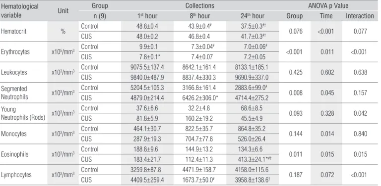
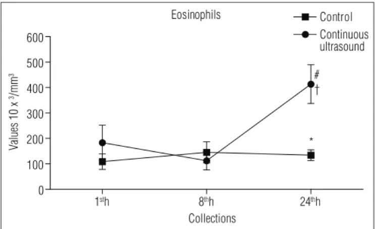
Eosinophils x103/mm3 Control 188.8±9.6 144.9±13.2 134.3±6.6 0.011 0.015 0.015
CUS 183.4±21.7 112.4±11.3 413.3±24.1*#†
Lymphocytes x103/mm3 Control 3259.8±87.8 4471.9±158.7 4158.0±115.6 0.187 0.072 <0.001
CUS 4409.5±259.4 1673.7±50.0# 3958.8±138.6†
Table 1. Hematological variables after muscle lesion and application of continuous ultrasound.
Values are presented as mean±standard error. CUS=continuous ultrasound; Control=group submitted to the procedure with the US equipment switched off. p=evaluation of the compari-sons among the groups using two-way ANOVA with repeated measures followed by the Bonferroni post hoc test. *p<0.05 variation among the groups; #p<0.05 variation over time versus 1st hour; †p<0.05 variation over time versus 8th hour.
of the leukocytes represented by the eosinophils in the 24th hour.
CUS produced greater erythrocyte reduction in the irst hour after muscle injury, possibly due to its thermal efect10,
which stimulates more extensive hemorrhage. Because eryth-rocytes are the most abundant cells in the blood, this hemor-rhage causes erythrocyte reduction. he endothelial injury sets of a sequence of events, starting with the platelet deposition which leads to white thrombus formation and temporarily obstructs the endothelial injury24. his thrombus is quickly
in-iltrated by ibrin, where the erythrocytes are captured, and the red thrombus is formed. he red thrombus is the main reason for the occlusion of the ruptured blood vessel25, a mechanism
that was probably present in this study.
Another aspect to be considered is that the erythrocytes decreased over time only in CG (in the eighth and in the 24th hour), which might have been induced by the successive blood collections. his reinforces the fact that the hemorrhage in-duced by CUS represented the diference in the irst hour. he hematocrit decreased over time in both groups which rein-forces the hypothesis that successive collections and hemor-rhage combine to reduce erythrocytes.
In addition to the thermal efects, other mechanisms by which CUS may have induced the hemorrhage are described in the literature, such as nitric oxide release which induces dependent endothelial vasodilation13, ibrinolysis11 and
thrombolysis12. hese phenomena may also be involved in
this answer.
Because neutrophils are the most abundant of the white blood cells, a signiicant number of them is passively collected by the temporary thrombus when a vessel is ruptured26,27. In the
early phase after the musculoskeletal tissue injury, polymor-phonuclear leukocytes (neutrophils) are the most abundant cells in the injured area1, and one day after the injury, those cells
will constitute 50% of the cells that migrated to that area27. his
phenomenon can explain the segmented neutrophil reduction in CG during the course of the experiment.
After this passive overflowing, neutrophils migrate to the surface of the injury to produce a barrier against the inva-sion of microorganisms and to promote active recruitment of more neutrophils from adjacent uninjured vessels26,27.
CUS changed this response, and in the eighth hour, the sys-temic concentration of segmented neutrophils was higher than in CG.
Later, the beginning of the inlammatory reaction is in-tensiied with the satellite cells and the necrotic tissues of the muscle ibers. hey are stimulated by the local release of cy-tokines (IL-6; IL-1β) and cellular growth factors (TNF-α; FGF; IGF) which act on the chemotaxis, increasing the response and overlow of inlammatory cells1. his mechanism is optimized
by the thermal efect of CUS. Another aspect to be considered is that there was no segmented neutrophil reduction in the irst hour in the experimental group. he increase in systemic con-centration of segmented neutrophils in the eighth hour and its slower reduction in the 24th hour suggest a pro-inlammatory systemic response of this therapy.
he polymorphonuclear leukocytes (or neutrophils) are progressively replaced by monocytes1, which are very
abun-dant in the injured area between the second and the ifth day28.
herefore, their blood concentrations must change before this period. he results observed in the present study suggest that there were no changes in the systemic concentrations of these cells during the experimental protocol, however the 24-hour time frame may not have been enough to alter this variable.
According to the basic inlammation principles, the mono-cytes are transformed into macrophagomono-cytes which, then, actively begin the proteolysis and phagocytosis of the necrotic material by releasing lysosomic enzymes29. he phagocytosis
of macrophagocytes is a process remarkably speciic to ne-crotic material, such as the preserved cylinders of the basal membrane which surrounds the necrosis area of the injured myoibrils that survived the macrophage attack. hey serve as scafolding in which viable satellite cells begin to form new myoibrils1.
he monocytes not only help the neutrophils eliminate microorganisms through phagocytosis but also present their peptides through the major histocompatibility complex to the auxiliary T cells. hus, the phagocytosis of these cells acts as a link between the innate and the adaptive immune systems28.
However, the lymphocytes, at the end of the repair phase,
Eosinophils
1sth 8thh 24thh
0 100 200 300 400 500 600
Control Continuous ultrasound
* # †
Collections
Values
10
x
3/m
m
3
Figure 3. Behavior of eosinophils during experimental protocol.
Values are presented as mean and standard error (x103/mm3). For the
comparisons among the groups using two-way ANOVA with repeated-measures (group p<0.001; time p=0.011; interaction p<0.001) followed by the Bonferronipost hoc test. *p<0.05 variation among the groups; #p<0.05 variation over time versus 1st hour; †p<0.05 variation in the
time versus 8th hour. CUS=continuous ultrasound; Control=group
submitted to the procedure with the equipment switched off.
1. Järvinen TA, Järvinen TL, Kääriäinen M, Kalimo H, Järvinen M. Muscle injuries: biology and treatment. Am J Sports Med. 2005;33(5):745-64.
2. Crisco JJ, Jokl P, Heinen GT, Connell MD, Panjabi MM. A muscle contusion injury model. Biomechanics, physiology, and histology. Am J Sports Med. 1994;22(5):702-10.
3. Huard J, Li Y, Fu FH. Muscle injuries and repair: current trends in research. J Bone Joint Surg Am. 2002;84-A(5):822-32.
4. Harris GR, Susman JL. Managing musculoskeletal complaints with rehabilitation therapy: summary of the Philadelphia Panel evidence-based clinical practice guidelines on musculoskeletal rehabilitation interventions. J Fam Pract. 2002;51(12):1042-6.
5. Rantanen J, Thorsson O, Wollmer P, Hurme T, Kalimo H. Effects of therapeutic ultrasound on the regeneration of skeletal myofibers after experimental muscle injury. Am J Sports Med. 1999;27(1): 54-9.
6. Warden SJ. A new direction for ultrasound therapy in sports medicine. Sports Med. 2003;33(2):95-107.
7. Wilkin LD, Merrick MA, Kirby TE, Devor ST. Influence of therapeutic ultrasound on skeletal muscle regeneration following blunt contusion. Int J Sports Med. 2004;25(1):73-7.
8. Baker KG, Robertson VJ, Duck FA. A review of therapeutic ultrasound: biophysical effects. Phys Ther. 2001;81(7):1351-8.
9. O’Brien WD Jr. Ultrasound-biophysics mechanisms. Prog Biophys Mol Biol. 2007;93(1-3):212-55.
10. Johns LD. Nonthermal Effects of Therapeutic Ultrasound: The Frequency Resonance Hypothesis. J Athl Train. 2002;37(3):293-9.
11. Kashyap A, Blinc A, Marder VJ, Penney DP, Francis CW. Acceleration of fibrinolysis by ultrasound in a rabbit ear model of small vessel injury. Thromb Res. 1994;76(5):475-85.
constitute the most abundant subsystem27; they are attracted
to the injured area in equal number to the monocytes and, from the 14th day, the leukocytes predominate the area30. he
changes in lymphocyte interaction for G1 represent natural os-cillations in the counting of these cells in repeated evaluations, because they are not accompanied by diferences between groups or diferences in the time.
Inlammatory mediators make the uninjured capillary vessels dilate which slows down blood circulation and allows leukocyte margination and connection to adhesion molecules expressed in the endothelial cells. he eosinophils appear in the inal phases of repair and may be related to the production of growth factors31. he results of the present study suggest that
after 24 hours of muscle injury treated with CUS, there was a nearly three-fold increase in the cellular concentration of eo-sinophils, however these values are still within physiological parameters.
he results of this research regarding CUS application dif-fer from the pulsed US study21 that suggested that this form of
application decreases white cells (total leukocytes, segmented neutrophils and monocytes). In this study there were an in-crease in white blood cells and a dein-crease in red blood cells. A combination of factors, including the type of examined tis-sue and injury, and the US application (continuous or pulsed), intensity, and frequency of treatment, can explain the diferent results from other studies21,32.
he limitations of this research reside in the absence of his-tological and histochemical tissue analyses, which would have
allowed the comparison between the systemic hematological data and the tissue data, as well as the quantiication of the injured area through ultrasound.
Conclusions
he present experimental study demonstrates that CUS use (on the 1st, 8th and 24th hour, with a three minutes ap-plications and 0,4W/cm2 intensity) in the acute phase of
iatrogenic muscle injury promotes changes in hematological dynamics. hese changes are characterized by erythrocyte reduction and an increase in segmented neutrophils and eo-sinophils. hese modiications suggest increased hemorrhage and an ampliied inlammatory response of the muscle and conirm the contraindication of CUS application on acute muscle injury.
Acknowledgments
To Adão Saurin, undergraduate Physical herapy student, and to Danielli Maria Donadel, undergraduate veterinary stu-dent, who collaborated in the data collections; to laboratory technician Jéssica Arsand of the Veterinary Medicine course, who helped to process the hematological analyses; and to the employees of the Unicruz vivarium, Roberto Machado Moraes and Giovane Lopes Seccon.
493
12. Riggs PN, Francis CW, Bartos SR, Penney DP. Ultrasound enhancement of rabbit femoral artery thrombolysis. Cardiovasc Surg. 1997;5(2):201-7.
13. Sugita Y, Mizuno S, Nakayama N, Iwaki T, Murakami E, Wang Z et al. Nitric oxide generation directly responds to ultrasound exposure. Ultrasound Med Biol. 2008;34(3):487-93.
14. Barzelai S, Sharabani-Yosef O, Holbova R, Castel D, Walden R, Engelberg S et al. Low-intensity ultrasound induces angiogenesis in rat hind-limb ischemia. Ultrasound Med Biol. 2006;32(1):139-45.
15. Merly F, Lescaudron L, Rouaud T, Crossin F, Gardahaut MF. Macrophages enhance muscle satellite cell proliferation and delay their differentiation. Muscle Nerve. 1999;22(6):724-32.
16. Lille ST, Lefler SR, Mowlavi A, Suchy H, Boyle EM Jr, Farr AL et al. Inhibition of the initial wave of NF-kappaB activity in rat muscle reduces ischemia/ reperfusion injury. Muscle Nerve. 2001;24(4):534-41.
17. Worrell TW. Factors associated with hamstring injuries. An approach to treatment and preventative measures. Sports Med. 1994;17(5):338-45.
18. Douglas MR, Morrison KE, Salmon M, Buckley CD. Why does inflammation persist: a dominant role for the stromal microenvironment? Expert Rev Mol Med. 2002;4(25):1-18.
19. Hill GE, Fenwick S, Matthews BJ, Chivers RA, Southgate J. The effect of low-intensity pulsed ultrasound on repair of epithelial cell monolayers in vitro. Ultrasound Med Biol. 2005;31(12):1701-6.
20. Byl NN, McKenzie A, Wong T, West J, Hunt TK. Incisional wound healing: a controlled study of low and high dose ultrasound. J Orthop Sports Phys Ther. 1993;18(5):619-28.
21. Costa ST, Lauxen J, Sturzenegger TM, Signori LU, Plentz RDM. Efeito anti-inflamatório do ultra som de 1 MHz na dinâmica Hematológica. Rev Bras Fisioter. 2006;10(Supl 1):60-1.
22. Chang CJ, Hsu SH, Lin FT, Chang H, Chang CS. Low-intensity-ultrasound-accelerated nerve regeneration using cell-seeded poly(D,L-lactic acid-co-glycolic acid) conduits: an in vivo and in vitro study. J Biomed Mater Res B Appl Biomater. 2005;75(1):99-107.
23. Chen J, Yang WL, Li G, Qian J, Xue JL, Fu SK et al. Transfection of mEpo gene to intestinal epithelium in vivo mediated by oral delivery of chitosan-DNA nanoparticles. World J Gastroenterol. 2004;10(1):112-6
24. Lefkovits J, Plow EF, Topol EJ. Platelet glycoprotein IIb/IIIa receptors in cardiovascular medicine. N Engl J Med. 1995;332(23):1553-9.
25. Davies MJ. A macro and micro view of coronary vascular insult in ischemic heart disease. Circulation. 1990;82(3 Suppl):II38-46.
26. Foxman EF, Campbell JJ, Butcher EC. Multistep navigation and the combinatorial control of leukocyte chemotaxis. J Cell Biol. 1997;139(5):1349-60.
27. Engelhardt E, Toksoy A, Goebeler M, Debus S, Bröcker EB, Gillitzer R. Chemokines IL-8, GROalpha, MCP-1, IP-10, and Mig are sequentially and differentially expressed during phase-specific infiltration of leukocyte subsets in human wound healing. Am J Pathol. 1998;153(6):1849-60.
28. DiPietro LA. Wound healing: the role of the macrophage and other immune cells. Shock. 1995;4(4):233-40.
29. Farges MC, Balcerzak D, Fisher BD, Attaix D, Béchet D, Ferrara M et al. Increased muscle proteolysis after local trauma mainly reflects macrophage-associated lysosomal proteolysis. Am J Physiol Endocrinol Metab. 2002;282(2):E326-35.
30. Blotnick S, Peoples GE, Freeman MR, Eberlein TJ, Klagsbrun M. T lymphocytes synthesize and export heparin-binding epidermal growth factor-like growth factor and basic fibroblast growth factor, mitogens for vascular cells and fibroblasts: differential production and release by CD4+ and CD8+ T cells. Proc Natl Acad Sci U S A. 1994;91(8):2890-4.
31. Wong DT, Donoff RB, Yang J, Song BZ, Matossian K, Nagura N et al. Sequential expression of transforming growth factors alpha and beta 1 by eosinophils during cutaneous wound healing in the hamster. Am J Pathol. 1993;143(1):130-42.
32. Karnes JL, Burton HW. Continuous therapeutic ultrasound accelerates repair of contraction-induced skeletal muscle damage in rats. Arch Phys Med Rehabil. 2002;83(1):1-4.