Retention of oral microorganisms on conventional and
resin-modified glass-ionomer cements
Retenção de microrganismos bucais em cimentos de ionômero de
vidro convencionais e modificados por resina
Denise PEDRINI*
Elerson GAETTI-JARDIM JÚNIOR* Andréia Coelho de VASCONCELOS**
PEDRINI, D.; GAETTI-JARDIM JÚNIOR, E.; VASCONCELOS, A. C. de. Retention of oral microorganisms on conventio-nal and resin-modified glass-ionomer cements.Pesqui Odontol Bras, v. 15, n. 3, p. 196-200, jul./set. 2001. Secondary caries are a worldwide public and socioeconomic problem. The placement of restorations can lead to the de-velopment of environmental conditions favorable to microbial colonization, especially on the tooth/restoration interfa-ce, which is a predisposing factor for secondary caries. The aim of this study was to evaluate microbial retention on conventional (Chelon-Fil and Vidrion R) and resin-modified (Vitremer and Fuji II LC) glass-ionomer cements,in situ, using a hybrid composite resin (Z100) as a control. Twelve volunteers wore Hawley appliances with specimens made of all tested filling materials for 7 days. The specimens were then removed from the appliances and transferred to tubes containing 2.0 ml of Ringer-PRAS. Microorganisms from the samples were inoculated onto blood agar and Mitis Saliva-rius Bacitracin agar and incubated under anaerobiosis (90% N2, 10% CO2), at 37°C, for 10 and 2 days, respectively.
The resin-modified glass-ionomer cements and the composite resin retained the same levels of microorganisms on the-ir surfaces. The resin-modified glass-ionomers retained lessmutansstreptococci than the composite resin and con-ventional glass-ionomer cements. The concon-ventional glass-ionomer cements retained lessmutansstreptococci than the composite resin, but that difference was not statistically significant.
UNITERMS: Dental caries; Dental plaque; Glass-ionomer cements.
INTRODUCTION
The colonization of surfaces of restorations by cariogenic bacteria can contribute to increase the incidence of secondary caries25, which is one of the
major reasons for replacement of restorations18.
This fact is even more complicated when the resto-ration is placed on cervical areas, where the reten-tion of bacterial biofilm can also compromise gingival health.
Due to their physical, chemical and biological properties, glass-ionomer cements have been used as lining, luting and filling materials, especially for restoring cervical areas6. Resin-modified
glass-ionomer cements have shown improved properties, including faster setting, as well as less sensitivity to hydration and dehydratation9.
The surfaces of conventional glass-ionomer ce-ments remain relatively rough, even with careful
polishing6,20. This might enhance the accumulation
of bacterial biofilm on glass-ionomer restorations, leading to gingival inflammation6.
The bacterial plaque located on the surface of glass-ionomer fillings shows a number of microor-ganisms similar to that of the plaque observed on composite resin restorations, however, the levels of
mutansstreptococci are lower24. This phenomenon
may be related to fluoride release and to the effect of this ion on many metabolic processes in bacteria26.
There appear to be few reports on the effects of resin-modified glass-ionomer cements – which permit an improvement of finishing and polishing16– on the surrounding microbiota,
espe-cially regarding the levels of mutans streptococci. Therefore, the aim of this study was to evaluate, in situ, microbial retention on conventional and resin-modified glass-ionomer cements.
MATERIALS AND METHODS
Dental materials
Two conventional glass-ionomer cements, Chelon-Fil (Espe GmbH, Germany) and Vidrion R (SS White, Brazil), and two resin-modified glass-ionomer cements, Vitremer (3M Dental Prod-ucts, Brazil) and Fuji II LC (GC, Japan), were stud-ied. The hybrid composite resin Z100 (3M Dental Products, Brazil) was used as a control.
Specimens were prepared with each material according to their manufacturers’ instructions. The materials were placed into cavities (diameter of 5 mm, depth of 1mm) on the outer surface of the palate of Hawley appliances. Twelve specimens
were prepared with each material. The
glass-ionomer specimens were covered with the light-curing bonding resin Probond (Dentsply, Brazil) and stored at 100% relative humidity after preparation, in order to avoid changes on their surfaces. After 24 hours, all specimens were polished11using sequential Sof-Lex discs (3M
Den-tal Products, Brazil)20. An acrylic net was used to
cover the specimens so as to avoid the autocleaning produced by the movements of the tongue, thus, simulating the conditions of interproximal and cervical areas of teeth.
Experimental design
Twelve 17-23-year-old volunteers participated in this study. All were students of the School of Dentistry of Araçatuba - UNESP. All students re-ceived oral hygiene instructions (flossing and tooth brushing) before the experimental essay.
The volunteers wore the Hawley appliances for 7 days. During the experimental period, the volun-teers were allowed to brush their teeth with the Hawley appliances removed from their mouth, but were not allowed to use a toothpaste or any chemi-cal compound with fluoride or other substance
with antimicrobial properties. During the meals, the Hawley appliances were stored at 100% hu-midity.
After this period, the specimens on the Hawley appliances were removed and transferred to tubes containing 2.0 ml of pre-reduced anaerobically sterilized Ringer solution21 under CO
2flux.
All samples were taken to the laboratory, sub-jected to serial ten-fold dilutions29in Ringer-PRAS,
and cultured onto brain heart infusion agar (Difco) supplemented with yeast extract (0.5%), hemin (0.5 mg/ml), menadione (5 mg/ml) and 5% defibrinated sheep blood (blood agar) – for the iso-lation of fastidious microorganisms and determi-nation of total bacterial count –, as well as on Mitis Salivarius Bacitracin agar10(MSB agar) – to recover
mutans streptococci, in duplicate. The blood agar
and MSB agar plates were incubated under anaerobiosis (90% N2, 10% CO2), at 37ºC, for 10
and 2 days, respectively.
Identification ofmutansstreptococci was car-ried out through the analysis of the morphology of cells (Gram) and colonies, as well through the analysis of the fermentation of manitol and sorbitol4. The differences in the levels of mutans
streptococci and in the total number of viable bac-terial cells on conventional glass ionomer cement, resin-modified glass-ionomer cement and compos-ite resin were determined through the Kruskal-Wallis test.
RESULTS
The results are presented in Tables 1 and 2. Ta-ble 1 presents the total number of microorganisms recovered from the samples, whereas Table 2 shows the levels of mutans streptococci isolated from the specimens.
The resin-modified glass-ionomer cements and the composite resin showed lower levels of
micro-TABLE 1 -Number of microorganisms recovered from the glass-ionomer cements and the composite resin.
Material Mean levels (103 CFU)
Z100 51.54 a
Fuji II LC 80.05 ab
Vitremer 69.83 ab
Vidrion R 271.15 b
Chelon-Fil 157.52 b
Different letters indicate statistical difference at the level of 1%.
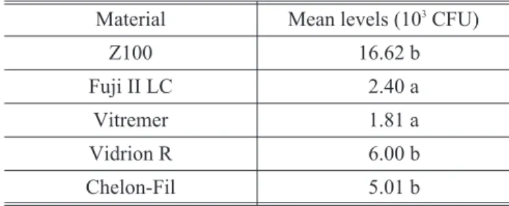
TABLE 2 -Levels of mutansstreptococci on the tested materials.
Material Mean levels (103 CFU)
Z100 16.62 b
Fuji II LC 2.40 a
Vitremer 1.81 a
Vidrion R 6.00 b
Chelon-Fil 5.01 b
organisms than the conventional glass-ionomer cements, even though statistical significance was observed only between the composite resin and the conventional glass-ionomer cements (p = 0.01). The resin-modified glass-ionomer cements pre-sented lower numbers ofmutansstreptococci than the other tested materials (p = 0.05). There were no significant differences between conventional glass-ionomer cements and the composite resin.
DISCUSSION
Although the clinical use of glass-ionomer ce-ments has increased due to their properties, such as fluoride release and adhesion to dental tissues6,7,19, these materials have deficiencies. For
instance, their surface is rough in comparison to that of composite resins, which can enhance mi-crobial retention and plaque accumulation6,22.
The resinous content of resin-modified glass-ionomer cements, while improving their physical properties15, can also reduce plaque
accu-mulation, preventing secondary caries. The num-ber of microorganisms recovered from resin-modi-fied ionomer specimens was similar to that recovered from composite resin samples, and it was lower than the microbial levels found on con-ventional glass-ionomer cements. However, the differences between conventional and resin-modi-fied glass-ionomer cements were not statistically significant (Table 1).
The glass-ionomer cements, particularly the resin-modified ionomers (Table 2), showed smaller numbers ofmutansstreptococci than the compos-ite resin, and these results are in agreement with those presented by SVANBERGet al.24 (1990) and
BERG et al.2 (1990). According to BERG et al.2
(1990), the number of mutansstreptococci on re-storative materials stabilized three months after the treatment, probably due to the decrease of flu-oride release from ionomer restorations. However, VAN DIJKENet al.28(1991) did not report these
dif-ferences.
The glass-ionomer cements can interfere with the growth of cariogenic bacteria, as shown by
BERGet al.2(1990) and GARIBet al.8(1993). The
presence of a less cariogenic microflora on these cements might be due to fluoride release3,
suggest-ing that the resinous content of resin-modified glass-ionomers fillings does not interfere substan-tially with fluoride release5,27.
Since the levels of fluoride release in glass-ionomer cements decreased over time1,7, the
effects of these materials on the microbiota might vary at the same extension. However, even in sublethal concentrations, fluoride is able to pro-duce remarkable effects on the acidogenicity and adhesion ability of mutans streptococci13,23. It can
reduce the production of glucan polymers30, inhibit
glycolysis14 and other metabolic pathways,
pre-venting the colonization of dental tissues17, and
af-fecting the activity of enzymes associated with the citoplasmatic membrane12. Therefore, it is possible
thatmutans streptococci isolated from specimens
made of conventional and resin-modified glass-ionomers present lower metabolic activity than those strains isolated from specimens made of hybrid resin (Z100).
The lack of significant differences between the conventional glass-ionomer cements and the com-posite resin (Z100) as to the levels ofmutans strep-tococci might reflect the greater roughness of con-ventional ionomers, although careful polishing was carried out6,20. That roughness might decrease
the advantageous effects produced by fluoride re-lease.
Interactions between restorative materials and oral microflora can produce different environ-ments, which could lead to conditions suitable to the development of dental caries, or maintenance of oral health. These factors must be considered in the choice of a dental material.
CONCLUSIONS
On the basis of this study, it can be concluded that:
1. Conventional glass-ionomer cements (Vidrion R and Chelon-Fil) retained a greater number of microorganisms than the hybrid resin (Z100). 2. The levels of microorganisms retained on the
surface of resin-modified glass-ionomers (Vitre-mer and Fuji II LC) were similar to those obser-ved on composite resin (Z100) samples.
3. Resin-modified glass-ionomers (Vitremer and Fuji II LC) retained less mutans streptococci than the resin (Z100) and the conventional glass-ionomer cements (Vidrion R and Che-lon-Fil).
ACKNOWLEDGMENTS
PEDRINI, D.; GAETTI-JARDIM JÚNIOR, E.; VASCONCELOS, A. C. de. Retenção de microrganismos bucais em cimentos de ionômero de vidro convencionais e modificados por resina. Pesqui Odontol Bras, v. 15, n. 3, p. 196-200, jul./set. 2001.
A cárie secundária representa problema de saúde pública e socioeconômico no mundo. A restauração de dentes aco-metidos por cárie pode criar condições favoráveis à proliferação microbiana na superfície do material restaurador ou na interface dente/restauração, criando ambiente propício para o estabelecimento de cárie secundária. O objetivo des-te estudo foi avaliar a capacidade de redes-tenção de placa bacdes-teriana em cimentos de ionômero de vidro convencionais (Chelon-Fil e Vidrion R) e modificados por resina (Vitremer e Fuji II LC) e de resina composta híbrida (Z100), utilizada como controle. Nos testes de retenção de microrganismos,in situ, 12 voluntários utilizaram, por 7 dias, placa de Haw-ley contendo corpos-de-prova de todos os materiais. A seguir, os corpos-de-prova foram transferidos para tubos contendo 2,0 ml de Ringer-PRAS e os microrganismos presentes em sua superfície foram cultivados em placa com ágar-sangue e ágar Mitis Salivarius Bacitracina, os quais foram incubados, a 37ºC, em anaerobiose (90% N2, 10%
CO2), por 10 e 2 dias, respectivamente. Os ionômeros modificados por resina retiveram quantidade de bactérias
simi-lar àquela mostrada pela resina testada. Os ionômeros modificados por resina também apresentaram menor número de estreptococos do grupomutansdo que a resina e os cimentos ionoméricos convencionais. Os ionômeros de vidro convencionais apresentaram menor número de estreptococos do grupomutansque a resina, sendo que essa diferença não foi estatisticamente significativa.
UNITERMOS: Cárie dentária; Placa dentária; Cimentos de ionômero de vidro.
BIBLIOGRAPHIC REFERENCES
1. ARAUJO, F. B.; GARCÍA-GODOY, F.; CURY, J. A.; CONCEIÇÃO, E. N. Fluoride release from fluori-de-containing materials. Oper Dent, v. 21, n. 5, p. 185-190, Sept./Oct. 1996.
2. BERG, J. H.; FARRELL, J. E.; BROWN, L. R. Class II glass ionomer/silver cermet restorations and their effect on interproximal growth of mutans streptococci. Pediatr Dent, v. 12, n. 1, p. 20-23, Feb. 1990.
3. CARDOSO, D. C.; MARTÍNEZ, B. G.; BAÑOS, I. A. et al.
Efecto del fluoruro liberado a partir de ionómero de vi-drio sobreStreptococcus mutans.Rev ADM, v. 51, n. 5, p. 285-287, Sept./Oct. 1994.
4. COLLINA, E.; MOREIRA, M.; BARBOSA, A. D. Comparação da ação do verniz fluoretado Duraphat e do cariostático Bioride (diamino fluoreto de prata 12%), sobre a conta-gem de Streptococcus do grupo mutans, em crianças com dentição decídua. Rev ABO Nac, v. 8, n. 1, p. 14-20, fev./mar. 2000.
5. DUNNE, S. M.; GOOLNIK, J. S.; MILLAR, B. J.; SEDDON, R. P. Caries inhibition by a resin-modified and a conven-tional glass ionomer cement, in vitro. J Dent, v. 24, n. 1-2, p. 91-94, Jan./Mar. 1996.
6. FORSS, H.; SEPPÄ, L.; ALAKUIJALA, P. Plaque accumula-tion on glass ionomer filling materials.Proc Finn Dent Soc, v. 87, n. 3, p. 343-350, 1991.
7. FORSTEN, L. Short- and long-term fluoride release from glass ionomers and other fluoride-containing filling ma-terials in vitro. Scand J Dent Res, v. 98, n. 2, p. 179-185, Apr. 1990.
8. GARIB, T. M.; ROSA, O. P. S.; ROCHA, R. S. S. Ação antimi-crobiana de cimentos de ionômero de vidro restaurado-res. Rev Fac Odontol Bauru, v. 1, n. 1-4, p. 1-5, jan./dez. 1993.
9. GLADYS, S.; van MEERBEEK, B.; BRAEM, M.et al. Com-parative physico-mechanical characterization of new hybrid restorative materials with conventional
glass-ionomer and resin composite restorative mate-rials.J Dent Res, v. 76, n. 4, p. 883- 894, Apr. 1997. 10. GOLD, O. G.; JORDAN, H. V.; van HOUTE, J. A selective
medium for Streptococcus mutans. Arch Oral Biol, v. 18, n. 11, p. 1357-1364, Nov. 1973.
11. GUIDE to the use of glass ionomer filling materials. Fédéra-tion Dentaire InternaFédéra-tionale. Technical Report nº 27.
Int Dent J, v. 37, n. 3, p. 183-184, Sept. 1987. 12. HAMILTON, I. R. Biochemical effects of fluoride on oral
bacteria.J Dent Res, v. 69 (special issue), p. 660-667, Feb. 1990.
13. IZAGUIRRE-FERNANDEZ, E. J.; EISENBERG, A. D.; CURZON, M. E. Interaction of zinc with fluoride on growth, glycolysis and survival ofStreptococcus mutans
GS-5.Caries Res, v. 23, n. 1, p. 18-25, 1989.
14. KAUFMANN, M.; BARTHOLMES, P. Purification, characte-rization and inhibition by fluoride of enolase from Strep-tococcus mutansDSM 320523.Caries Res, v. 26, n. 2, p. 110-116, 1992.
15. KIM, Y. G.; HIRANO, S.; HIRASAWA, T. Physical properties of resin-modified glass-ionomers.Dent Mater J, v. 17, n. 1, p. 68-76, Mar. 1998.
16. LEINFELDER, K. F. Glass ionomers are still necessary.
Esthet Dent Update, v. 5, n. 6, p. 151, 1994.
17. MEURMAN, J. H. Effect of sodium and amine fluoride tre-atment on adsorption and ultrastructure ofS. mutans
and S. sanguis. Scand J Dent Res, v. 95, n. 5, p. 389-396, Oct. 1987.
18. MJÖR, I. A. Frequency of secondary caries at various ana-tomical locations. Oper Dent, v. 10, n. 3, p. 88-92, Summer 1985.
19. MOUNT, G. J. Restoration with glass-ionomer cement: re-quirements for clinical success.Oper Dent, v. 6, n. 2, p.59-65, Spring 1981.
(Doutorado) - Faculdade de Odontologia de Araraquara, Universidade Estadual Paulista.
21. SLOTS, J. Selective medium forActinobacillus actinomyce-temcomitans.J Clin Microbiol, v. 15, n. 4, p. 606-609, Apr. 1982.
22. SMALES, R. J. Plaque growth on dental restorative materi-als.J Dent, v. 9, n. 2, p. 133-140, June 1981. 23. SVANBERG, M.; KRASSE, B.; ÖRNERFELDT, H. O.Mutans
streptococci in interproximal plaque from amalgam and glass ionomer restorations. Caries Res, v. 24, n. 2, p. 133-136, 1990.
24. SVANBERG, M.; MJÖR, I. A.; ORSTAVIK, D.Mutans strep-tococci in plaque from margins of amalgam, composite, and glass-ionomer restorations.J Dent Res, v. 69, n. 3, p. 861-864, Mar. 1990.
25. TAYLOR, M. J.; LYNCH, E. Microleakage. J Dent, v. 20, n. 1, p. 3-10, Feb. 1992.
26. TEN CATE, J. M. Current concepts on the theories of the
mechanism of action of fluoride.Acta Odontol Scand, v. 57, n. 6, p. 325-329, Dec. 1999.
27. TORABZADEH, H.; ABOUSH, Y. E. Y.; LEE, A. R. Compara-tive assessment of long-term fluoride release from light-curing glass-ionomer cements.J Dent Res, v. 73, n. 4, p. 853, Apr. 1994.
28. van DIJKEN, J.; PERSSON, S.; SJÖSTRÖM, S. Presence of
Streptococcus mutansand lactobacilli in saliva and on enamel, glass ionomer cement, and composite resin surfaces.Scand J Dent Res, v. 99, n. 1, p. 13-19, Feb. 1991.
29. YAMAMOTO, K.; NODA, H.; KIMURA, K. Adherence of oral streptococci to composite resin restorative materials.J Dent, v. 17, n. 5, p. 225-229, Oct. 1989.
30. ZAMECK, R. L.; TINANOFF, N. Effects of NaF and SnF2on
growth, acid and glucan production of several oral streptococci.Arch Oral Biol, v. 32, n. 11, p. 807-810, 1987.