• Nenhum resultado encontrado

Mat. Res. vol.6 número1

N/A
N/A
Protected

Academic year: 2018

Share "Mat. Res. vol.6 número1"

Copied!
6
0
0

Texto

(1)

*e-mail: rgpalma@forp.usp.br

Microhardness of Esthetic Restorative Materials at Different Depths

Regina Guenka Palma-Dibba*, Alessandra Elias Palmaa, Edmir Matsonb,

Michelle Alexandra Chinelattia, Renata Pereira Ramosa

aDepartment of Restorative Dentistry,

Ribeirão Preto School of Dentistry, University of São Paulo, Ribeirão Preto - SP, Brazil

bDepartment of Restorative Dentistry, Endodontics and Dental Materials,

School of Dentistry, University of São Paulo, São Paulo - SP, Brazil

Received: April 26, 2002; Revised: October 8, 2002

The aim of this study was to analyze the microhardness of two resin-modified glass ionomer cements (Vitremer; Fuji II LC); two polyacid-modified composite resins (Freedom; F2000) and a hybrid composite resin (Prodigy), at different depths from the upper surface. Six hemi-cylinders (3 mm height; 3 mm radius) per tested material were obtained. Vickers Hardness was determined using a micro-indentation tester. For each hemi-cylinder, three indentations were taken at one of the following depths: 0.4, 1.0, 2.0 and 2.6 mm. For each material, microhardness average was calculated. Statistical analysis was performed using two-way ANOVA and Tukey test Fuji II LC and Vitremer showed no statistically significant difference among hardness means recorded at the four analyzed depths. The hybrid and the polyacid-modified composite resins showed significant decrease microhardness with increasing depth. It may be concluded that for the RMGIC, microhardness was not affected at depths up to 2.6 mm. On the other hand, both hybrid and PMCRs should de better placed in increments not thicker than 2 mm to achieve optimal hardness through-out the restoration.

Keywords: composite resin, glass ionomer cement, compomer, microhardness

1. Introduction

The conventional glass-ionomer cements (GIC) were first described in the 1970’s as materials consisting of a base, usually an ion-leachable glass powder, which is combined with a polyacrylic acid or its copolymers. Several features contributed to the wide acceptance of GIC in dental prac-tice, including biocompatibility1,2, good adhesion to tooth

substrates2, ability to take up and release fluoride1-4,

mini-mal shrinkage on setting2 and a coefficient of thermal

ex-pansion similar to that of tooth structure5. However, the

prob-lems associated to conventional glass-ionomer cements in-clude early low mechanical strengths, limited aesthetics re-sulting from moisture sensitivity and susceptibility to frac-ture and dehydration, especially during the initial stages of setting reaction6-8. These characteristics may lead to

de-creased physical properties, thereby limiting the applica-bility9 of glass ionomer cements to certain clinical

situa-tions.

(2)

86 Palma-Dibb et al. Materials Research

are responsible for the post-hardening of these cements10-12.

Recently, another category of light-cured restorative materials, which contain GIC components and release fluo-ride, was introduced to market. These materials, termed “polyacid-modified composite resins” (PMCR) or “compomers” may not be classified as conventional glass-ionomer cements, as they are presented as one-component resin-based products and do not have an acid-base reaction which occurs without photoactivation13 and diffusion of

water, and, when set, do not exhibit the typical properties of true GIC.

Compared to conventional GIC,the hybridglass ionomer cements and compomers are claimed to possess improved physical properties4,7, decreased sensitivity to water loss and

absorption, enhanced esthetics and longer working-time11.

Nevertheless, it has also been reportedthat these materials may undergo alterations, depending on light-curing proc-ess, that could affect the extent of polymerization at differ-ent depths.

Since the introduction of light-cured restorative materi-als, there has been concern about the depth of appropriate cure throughout the restoration. Recent studies have indi-cated that the resin materials themselves or the light sources used to activate curing process may not provide optimal polymerization in the deepest areas of the restoration11. The

restoration may be mechanically compromised by the in-complete cure of the mass of the restorative material at the bottom surface9,11. Moreover, unreacted components may

leach from the restoration, increasing the potential for an adverse biocompatibility reaction11.

The polymerization depth of visible light-activated ma-terials is directly related to the thickness of the material and is also influenced by light intensity14,15. In addition, other

factors also affect this property such as the measurement method, matrix/mould composition and dimensions of light source and its distance to the tested surface10,16,17.

Moreo-ver, it has been widely reported that the top surface presents higher hardness than the bottom surface of restorations14,18.

However, the different types of restorative materials currently available may yield different performances with respect to the depth of cure. Therefore, the purpose of this investigation was to assess the microhardness of two poly-acid-modified composite resins, two resin-modified glass ionomer cements, and a hybrid composite resin, at different depths from the top surface.

2. Material and Methods

The tested materials with their compositions, specifica-tions and manufacturers are listed on Table 1.

Thirty specimens were prepared using a black poly-urethane cylindrical split mould with 3 mm in height and 6 mm in diameter (Fig. 1a). The cylindrical mould was placed in a metallic clamping apparatus (Fig. 1b and Fig. 1c) and bisected by a stainless steel sheet, thus forming a separated cavity (Fig. 1d). The goal of using the stainless steel sheet was to obtain a smooth, flattened surface on which Vickers hardness was assessed. The cavity formed was then slightly filled in a single incrementwith the following ma-terials: two resin-modified glass ionomer cements Vitremer (3M Dental Products, St Paul, MN 55144) and Fuji II LC (GC Corporation, Tokyo, Japan); two polyacid-modified composite resins Freedom (Southern Dental Industries, Bayswater, Victoria, Australia) and F2000 (3M Dental Prod-ucts, St Paul, MN 55144); and a hybrid composite resin Prodigy (Kerr Corporation, Orange, CA 92667). The RMGIC components (powder and liquid) were disposed, mixed according to the manufacturers’ instructions and in-jected into the matrix cavity with a Centrix injector (Cavifil Injector - Vivadent, Schaan, Liechtenstein) to prevent bub-bles, voids and air entrapment. After insertion, the incre-ment was covered and gently pressed by a clear polyester matrix strip and cured for 60 s using a visible light-curing unit with a 450 mW/cm2 output (XL 3000, 3M

Den-tal Products, St Paul, MN 55144).

The split mould was removed from the clamping device thereby providing a hemi-cylinder with the same

dimen-Table 1. Tested materials

Material (Shade) Manufacturer Batch number Type of Material Powder/Liquid Ratio (g) Vitremer (A3) 3 M Dental Products Powder-19970717 Resin-modified 2.5/1

Liquid-199770709 glass ionomer cement

Fuji II LC (A3) GC Corporation Powder-070181 Resin-modified 3.2/1 Liquid-260181 glass ionomer

F2000 (A3) 3M Dental Products 23038 Polyacid-modified -composite resin

Freedom (A3) Southern Dental Industries 2256 Polyacid-modified -composite resin

(3)
(4)

88 Palma-Dibb et al. Materials Research

sions of the bisected cavity (3 mm in height and 6 mm in diameter) (Fig. 1e). Six specimens per material were ob-tained. The hemi-cylinders were stored for 24 h inside a lightproof container with distilled water at 37 °C. After-wards, Vickers hardness was measured on the surface in contact with the stainless steel sheet using a micro-indenta-tion tester (Shimadzu Micro Hardness Testers HMV-2, Shimadzu Corporation, Kyoto, Japan) with a 100 g load applied for 45 s. The specimens were individually fixed in a clamping apparatus and positioned in such a way that the test surface was kept perpendicular to the indentator (Fig. 1f). Measurements were accomplished at depths of 0.4 mm, 1.0 mm, 2.0 mm and 2.6 mm from the upper sur-face of specimen. In each sample, three indentations equally spaced over a circle and not closer than 1 mm to the adja-cent indentations or the margin of the specimen were taken at the all predetermined depths (Fig. 1g) and the averaged was calculated. Then, for each tested material, a micro-hardness mean value was determined at the specified depths of cure.

Statistical analysis was performed using two-way ANOVA using a factorial design with material and depth as independent variables. Means were compared to Tukey in-terval calculated at the 0.05 significance level. A software* was used to tabulate and analyze data.

3. Results

Microhardness means recorded at the different depths of cure and standard deviation are shown in Table 2. Statis-tical analysis of the data revealed significant difference (p < 0.05) among the experimental groups. Comparing the materials, regardless of the depth of cure, Fuji II LC (50.37 Hv) and Prodigy (47.11 Hv) showed statistical simi-larity and yielded the highest means. On the other hand, Freedom (30,32 Hv) presented the lowest overall microhardness mean.

With respect to the depth of cure, regardless of the tested material, it was noticed that the 0.4 mm was statistically different (p < 0.05) to the other depths. At 2.6 mm, the low-est average was observed.

Analyzing the interaction of both variables, it was ob-served that for both resin-modified glass ionomer cements no statistically significant difference was found among microhardness means recorded at all the analyzed depths of cure (Table 2). The polyacid-modified and the hybrid composite resins showed a significant decrease in microhardness with the increase of depth of cure, mainly after 2.0 mm depth. Among these materials, the lowest mean was found for Freedom at 2.6 mm depth.

4. Discussion

The analysis of data demonstrated that for both RMGIC (Vitremer and Fuji II LC), statistically similar microhardness means were observed at all the analyzed depths, possibly due to the acid-base reaction and chemical-free radical po-lymerization that continue after the removal of the light-source and therefore ensure a complete hardening of the material. Nevertheless, statistically significant difference (p < 0.05) was found when the overall results of the RMGIC were compared one to another, regardless of the depth of cure: Vitremer (35.09 Hv) and Fuji II LC (50.37 Hv). A possible explanation for such performances would be as-cribed to differences in formulation, since these materials can be assigned to two different subgroups19: those, such as

Fuji II LC, in which polymerizable monomers like HEMA are merely blended with a polyalkenoic acid liquid, and those, such as Vitremer, in which in addition to the simple mixture of HEMA with a polyalkenoic acid, the latter itself

Figure 2. Distribution of Vivkers hardness (Hv) at the analyzed depths of cure.

*GMC Statistical Software- v.2002 – Prof. Geraldo Maia Campos “Estatística Prática para Docentes e Pós-Graduandos”

(Online version) - http://www.forp.usp.br/restauradora/gmc/gmc.html

Table 2. Means (Hv) and standard deviation of the experimental groups

Depth (mm) Restorative Materials

Vitremer Fuji II LC Freedom F2000 Prodigy

0.4 37.66 (4.14) 50.13 (8.10) 36.78 (3.07) 50.47 (3.68) 56.57 (7.09) 1.0 36.59 (3.17) 48.02 (6.36) 33.48 (4.09) 43.07 (8.17) 49.23 (6.56) 2.0 34.62 (2.56) 50.90 (4.92) 28.84 (6.52) 42.06 (4.41) 44.27 (7.10) 2.6 31.48 (3.63) 52.41 (8.29) 22.15 (4.52) 37.49 (6.70) 38.37 (2.41)

X 35,09 50,37 30,32 43,28 47,11

(5)

is also modified by the attachment of polymerizable meth-acrylate side groups19. Furthermore, the great diversity in

particle-size distribution among the hybrid materials indi-cates that the manufacturers may fill the matrix in different ways. Some of them add a great amount of small particles to large particles, while another manufacturers opt for many large particles added to a smaller amount of small particles, as in Fuji II LC. For Vitremer, the amount of small and large particles is more equally distributed. Although the optimal packing of filler particles in hybrid restorative materials does not yet appear to have been established, the differences be-tween the materials may have influenced the better perform-ance of Fuji II LC over Vitremer.

Studies9,20 have indicated that glass-ionomer cements

hardness usually increases in a humid environment and with time. When the storage media is either water or saliva, the surface hardness is not affected or is slightly decreased after initial setting.However, when the materials are contaminated with saliva before set, it may result in significant alteration of microhardness. In the conducted research, specimens were stored in distilled water for 24 h previously to microhardness indentation test and the analysis of the results disclosed that both RMGICs showed statistically similar hardness at al-most all tested depths. Conversely, it has been reported that the storage of polyacid-modified composite resins in either aqueous solution or saliva (58,3 Hv) led to significant de-crease in microhardness when the results were compared to those recorded after dry storage (95,1 Hv), thus indicating a significant surface degradation18,21.

The findings of the reported study disclosed that microhardness means recorded for the resin-modified glass ionomer cements were comparable to those registered for the hybrid composite resin and considerably higher than those recorded for the polyacid-modified composite resins. Likewise, an earlier investigation10 also compared different

esthetic restorative materials and found that the tested RMGICs yielded microhardness means equivalent to those of composite resins and higher than conventional glass ionomer cements.

In general, the polyacid modified and the hybrid com-posite resins showed significant decrease in microhardness with the increase of the depth of cure, and this drop was particularly evident at depths higher than 2 mm from the light source, what would compromise their clinical perform-ance. A possible explanation for such performance would be the fact that the polymerization of these materials rely exclusively upon light activation and thus require maximum proximity to the light source22. In addition to this,

light-cured composite resins require optimal light intensity, suf-ficient irradiation time and a maximal thickness to allow the appropriate penetration of light throughout the restora-tive material placed in cavity preparation23,24.

While the tested composite resins presented higher

microhardness at regions closer to the upper surface of the specimens, the resin-modified glass ionomer cements, es-pecially Fuji II LC, did not show a significant decrease in microhardness with the increase of depth of cure.

Further investigation is certainly required to establish the basis to yield an optimized and rational assessment of microhardness as well as to assess the influence of storage time on the properties of light-cured restorative materials, especially the resin-modified glass-ionomer cements.

4. Conclusions

Based on the findings of the reported research, and within the limitations of an in vitro study, it may be concluded that for the resin-modified glass ionomer cements, micro-hardness was not affected at depths up to 2.6 mm, what leads to the assumption that these materials may be suc-cessfully placed into cavity preparations and further light-cured in a single increment. On the other hand, hybrid and polyacid-modified composite resins should be better placed in increments not thicker than 2 mm to achieve optimized hardness throughout the restoration.

References

1. Wilson, A.D.; Kent, B.E. British Dental Journal, v. 132, p. 133, 1972.

2. Navarro, M.F. de L.; Pascotto, R. Glass ionomer cement. São Paulo, Artes Médicas, p. 179, 1997.

3. Barnes, D.M.; Blank, L.W. Journal American Dental Association, v. 126, p. 1245, 1995.

4. Sidhu, S.K.; Watson, T.F. American Journal of Dentistry, v. 8, p. 59, 1995.

5. Abdalla, A.I.; Alhadainy, H.A.; Garcia-Godoy, F. Ameri-can Journal of Dentistry, v. 10, p. 19, 1997.

6. Goldman, M. J. Biomedical Materials Science, v. 10, p. 771, 1985.

7. Kerby, R.E.; Knobloch, L.; Thakur, A. Operative Den-tistry, v. 22, p. 79, 1997.

8. Mount, G.J.; Makinson, O.F. Operative Dentistry, v. 7, p. 134, 1982.

9. Dupuis, V.; Moya, F.; Payan, J.; Bartala, M. Biomaterials, v. 17, p. 71, 1996.

10. Kanchanavasita, W.; Anstice, H.M.; Pearson, G.J. Jour-nal of Dentistry, v. 26, p. 707, 1998.

11. Atmadja, G.; Bryant, R.W. Australian Dental Journal, v. 35, n. 3, p. 213, 1990.

12. Tyas, M.J. Operative Dentistry, v. 23, p. 77, 1998. 13. McLean, J.W.; Nicholson, J.W.; Wilson, A.D.

Quintes-sence International, v. 25, p. 587, 1994.

14. Correr Sobrinho, L.; De Goes, M.F.; Consani, S.; Sinhoreti, M.A.C. Journal of Materials Science: Mate-rials in Medicine, v. 11, p. 361, 1994.

(6)

90 Palma-Dibb et al. Materials Research

v. 66, p. 727, 1987.

16. Yearn, J.A. International Dental Journal, v. 35, p. 218, 1985.

17. Mitra, S.B. Proceedings of the 2nd International Sympo-sium on Glass ionomers. Philadelphia, PA: International Symposia in Dentistry, 1992.

18. Geurtsen, W.; Leyhausen, G.; Garcia-Godoy, F. Dental Materials, v. 15, p. 196, 1999.

19. Gladys, S.; Van Meerbeek, B.; Braem, M.; Lambrechts, P.; Vanherle, G. Journal of Dental Research, v. 76, n. 4, p. 883, 1997.

20. De Moor, R.J.G.; Veerbeeck, R.M.H. Biomaterials, v. 19, p. 2269, 1998.

21. Milleding, P; Ahlgren, F.; Wennerberg, A.; Örtengren, U.; Karlsson, S. International Journal of Prosthodon-tics, v. 1, p. 21, 1998.

22. Abate, P.F.; Polack, M.A.; Macchi, R.L. Quintessence International, v. 28, n. 5, p. 345, 1997.

23. Brosh, T.; Baharav, H.; Gross, O.; Laufer, B.Z. Journal of Prosthetic Dentistry, v. 77, p. 573, 1997.

Imagem

Table 1. Tested materials
Figure 1. Schematic representation of specimen preparation and microhardness indentations
Table  2. Means (Hv) and standard deviation of the experimental groups

Referências

Documentos relacionados

Ao preço de NCz$ 0,18, que representava o valor de comercialização na época da pesquisa, o &#34;coeficiente de eficiência&#34; nos grupos analisados está de acordo com a Tabela 6.

Nos anos 2000 ocorreram mudanças na sociedade brasileira a partir da ação do sujeito político feminista no campo e sua interação com o Estado em transformação. O feminismo no

There was an interaction between sexes and sedentary behaviors (p &lt; 0.001); therefore, re- gression analyses were performed separately for sex (Table 5). The crude analysis

Abstract: The purpose of this study was to evaluate the compressive strength of two glass ionomer cements, a conventional one (Vitro Fil  - DFL) and a resin-modiied material (Vi- tro

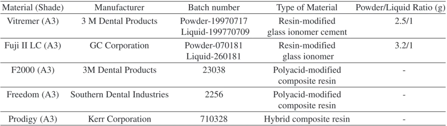
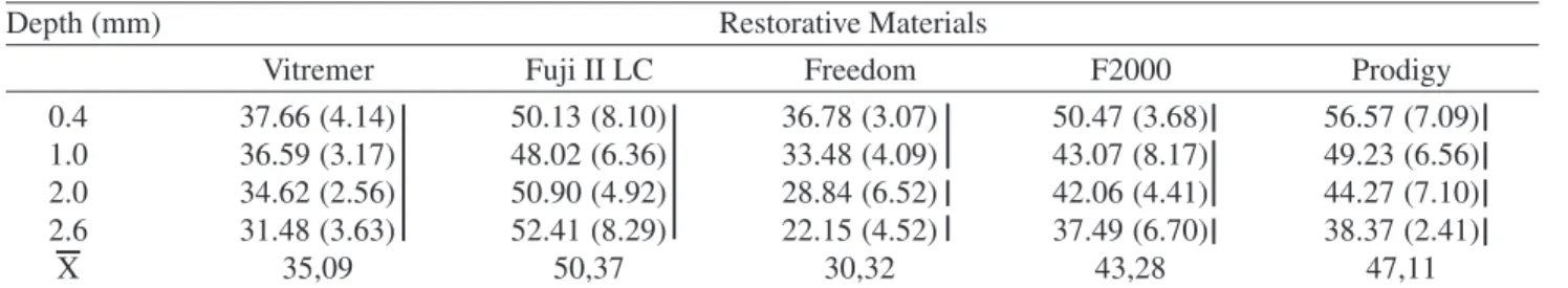
This study evaluated the surface microhardness and fluoride release of 5 restorative materials - Ketac-Fil Plus, Vitremer, Fuji II LC, Freedom and Fluorofil - in two storage

The aim of this study was to evaluate the microtensile bond strength (µTBS) of two substrates (enamel and dentin) considering two study factors: type of composite resin

The aim of this study was to evaluate the in vitro antibacterial and biofilm inhibition properties of glass ionomer restorative cements. Ketac Nano, Vitremer, Ketac Molar Easymix

This study has shown that the resin-based materials (polyacid modified and conventional composite resins) provided better marginal adaptation to both deciduous and immature