Myocardial Fibrosis in Patients with Hypertrophic Cardiomyopathy
and High Risk for Sudden Death
Afonso Akio Shiozaki*, Tiago Senra*, Edmundo Arteaga, Cristiane Guedes Pita, Martino Martinelli Filho, Luis
Francisco R Ávila, José Rodrigues Parga Filho, Charles Mady, Carlos Eduardo Rochitte
Instituto do Coração (InCor) do Hospital das Clínicas da Faculdade de Medicina da Universidade de São Paulo, São Paulo, SP - Brazil
* Both authors contributed equally as first authors to this work
Mailing address: Carlos E. Rochitte •
BInstituto do Coração - InCor - Setor de Ressonância Magnética e Tomografia Computadorizada Cardiovascular - Av. Dr. Enéas de Carvalho Aguiar, 44 - Cerqueira César - 05403-000 - São Paulo, SP - Brazil E-mail: [email protected]
Manuscript received November 17, 2008; revised manuscript received May 14, 2009; accepted July 1st, 2009.
Abstract
Background: The stratification of risk for sudden death in hypertrophic cardiomyopathy (HCM) continues to be a true challenge due to the great heterogeneity of this disease’s presentation, as most individuals remain asymptomatic during their entire lives and others present sudden death as first symptom. Recent studies have suggested that myocardial fibrosis may represent an important substrate for the malignant ventricular arrhythmias, that are responsible for the cases of sudden death related to this disease.
Objective: To assess the prevalence and quantification of myocardial fibrosis (MF) in hypertrophic cardiomyopathy (HCM) patients with implantablecardioverter - defibrillator (ICD) indicated due to their high risk or recovered from cardiac sudden death.
Methods: Twenty-eight HCM patients with ICD were submitted to multidetector computed tomography to assess myocardial fibrosis by delayed enhancement technique.
Results: Myocardial fibrosis was present in 96% of these HCM patients with (20.38 ± 15.55 g) comprising 15.96 ± 10.20% of the total myocardial mass. MF was observed in a significantly higher prevalence as compared to other classical risk factors for sudden death.
Conclusion: It is possible to conclude that there is a high prevalence of myocardial fibrosis in hypertrophic cardiomyopathy patients with high-risk or recovered from cardiac sudden death, like those with clinical indication to implantable cardioverter - defibrillator. The higher prevalence of myocardial fibrosis in comparison to classical risk factors of worse prognosis raise the hypothesis that the myocardial fibrosis may be an important substrate in the genesis of lifethreatening arrhythmias in these high risk HCM population. (Arq Bras Cardiol 2010; 94(4):502-506)
Key words: Hypertrophic cardiomyopathy; myocardial fibrosis; multidetector computed tomography.
The histopathological analysis of the hearts of HCM patients who died of sudden death demonstrated considerable presence of myocardial fibrosis19 (MF). This fibrosis may
be diagnosed by the non-invasive delayed enhancement technique by magnetic resonance20, which may constitute
an important substrate for malignant ventricular arrhythmias, as suggested by studies that correlated the presence of non-sustained ventricular tachycardia and the presence of MF diagnosed by magnetic resonance18,21-24.
In this manner, although the delayed enhancement magnetic resonance technique has showed to be an important tool for the assessment of MF in HCM20,25, it presents some
limitations, like in the assessment of patients who carry pacemakers and implantable cardioverter-defibrillator (ICD), because in such cases there are formal contraindications regarding the employment of magnetic resonance26.
Recently, the detection of MF in ischemic cardiomyopathy by multidetector computed tomography (MDCT) delayed enhancement technique was demonstrated27,28. Moreover, our
Introduction
Hypertrophic cardiomyopathy is considered to be the major cause of sudden cardiac death (SCD) among young adults1-4.
Despite the traditional clinical criteria of worse prognosis
5-12 and the genetic advances with regard to the discovery of
over 200 mutations that are responsible for the disease 13-18, the stratification of sudden death risk in hypertrophic
group was the first to demonstrate the MF can be assessment by MDCT delayed enhancement using a technique similar to that employed in ischemic patients, as the areas of MF evaluated by tomography presented an excellent correlation to the resonance29.
Thus, the objective of this paper was to investigate the prevalence of myocardial fibrosis in high risk HCM patients with ICD due to clinical indication by MDCT delayed enhancement technique.
Methods
Between October 2006 and December 2007, 30 hypertrophic patients with ICD by clinical indication, followed-up in the Unit of Cardiomyopathies and Artificial Cardiac Stimulation of Instituto do Coração (InCor-HC, FMUSP, São Paulo, Brazil), were consecutively referred to the assessment of myocardial fibrosis by MDCT. Of this sample, a patient with renal insufficiency and another one who refused to participate in the study were excluded. The 28 remaining patients who signed the informed consent were submitted to the analyses after approval by the institutional ethics committee.
Multidetector computed tomography
The assessment of MF by delayed enhancement MDCT was obtained using a tomograph with 64 columns of detector (Aquilion 64, Toshiba Medical Systems, Otawara, Japan). The images were acquired seven minutes after infusion of 150 ml of iodine contrast (Iopamiron 370, Shering AG, Germany), using the following protocol retrospectively synchronized with electrocardiogram: gantry rotation of 350 to 500 ms adjusted by cardiac frequency, as to allow a multisegmented reconstruction, collimation of 64 x 0.5 mm, tube voltage of 120kV, tube current of 500mA, helical-pitch 14.4 (or pitch factor of 0.225), and scanning field of view of 220 mm.
Clinical characteristics
The assessment of risk factors for sudden cardiac death was registered in compliance with a previously described guidance4, thus being considered: syncope, like episodes of
loss of consciousness of uncertain etiology occurred within a 12-month period before implantation of ICD30; recovery
from sudden death31; family history of sudden cardiac
death in first grade relatives younger than 40 years old32,33;
record of non-sustained ventricular tachycardia (NSVT) at Holter exam7,8,4,35, defined as three or more consecutive
ventricular extrasystoles with frequency equal or higher than 120 beats per minute during less than 30 seconds; maximal end diastolic wall thickness higher than 30 mm6,36 verified
at the echocardiogram. Moreover, other criteria were also analyzed: dilation of left ventricle was considered as end-diastolic diameter higher than 50 mm; left ventricle ejection fraction lower than 60%37; obstruction of left ventricle outflow
tract was considered as intraventricular gradient higher than 30 mmHg and, finally, atrial fibrillation was considered as clinical characteristics that could be associated with a worse
prognosis. Symptoms of cardiac heart failure were assorted in compliance with NYHA, and the use of antiarrhythmic drugs was also registered.
Images analyses
Data from images acquired by the tomography were reconstructed using a multisegmented image reconstruction algorithm, with a 1 mm thickness during diastolic phase (in the 75% phase of R-R interval). Axial images were processed using multiplanar reformation as to generate contiguous slices in short axis covering the whole extension of the left ventricle, from apex to basis, with a 10 mm thickness average reformation, a technique capable of reflecting the mean of all the pixels of the slice as to avoid the loss of any pixel that could represent myocardial fibrosis. Habitually, 12 slices in short axis were enough to cover the entire LV. All the segments involved by the artifacts caused by the ICD cables were excluded of the analysis, thus the inferior-septal wall was removed from all patients (Figure 1)38. The myocardial fibrosis areas were
defined aided by dedicated software (Image J, NIH, USA). Using the density of pixels in a histogram that comprised the graphical representation of the myocardial fibrosis areas as well as the myocardial without fibrosis areas, a semi-automatic threshold technique, that was able to distinguish the densities of the areas visually defined as presenting or not myocardial fibrosis, was applied.
Statistical analysis
Data were shown as mean ± standard deviation. The Shapiro-Wilk test was applied in order to confirm the normality distribution. The prevalence of traditionally accepted risk factors for sudden death and the presence of myocardial fibrosis was expressed in percentage and compared through chi-square or Fisher’s Exact test. Moreover, the prevalence of cardiac heart failure (CHF) functional class based on NYHA, intraventricular obstruction and atrial fibrillation of the sample was registered.
Results
Mean age was 38.5 ± 16.6 years old, and males constituted 46.4% of the studied population.
Among the criteria of classic sudden death risk, 18% of the patients recovered from sudden death, 68% presented history of syncope of uncertain etiology, 78% presented family history of sudden death, 32% presented NSVT, and 21% of the patients presented septal hypertrophy larger than 30 mm.
Besides, the presence of intraventricular obstruction was observed in 50% of the sample, atrial fibrillation in 32%, functional class II CHF in 39%, functional class III in only one patient, and functional class IV was not present in the studied population. The mean LV ejection fraction was 70.9 ± 12.4%, the diastolic diameter of the LV was 4.1 ± 0.5cm, and the mean size of the left atrium was 4.3 ± 0.6cm, as measured by the echocardiogram.
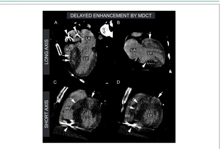
Figure 1 -Delayed enhancement (DE) by multidetector computed tomography (MDCT). Images of DE by MDCT of hypertrophic cardiomyopathy patients with ICD indicated by family sudden death (dad <40 years old), syncope, NSVT and septal hypertrophy higher than 30mm. Chart A and B - images of long axis, 4 chambers and 2
chambers, demonstrating an accentuated septal hypertrophy and myocardial ibrosis (white arrows). The metallic artifacts caused bay ICD cables are demonstrated by
the arrowheads. Chart C and D - images in short axis of the basal and medium portions of the myocardium, respectively, where it is possible to observe an accentuated
anterior and septal hypertrophy and myocardial ibrosis (white arrows). The metallic artifacts caused by ICD cables are predominantly present in the inferior-septal
segments, demonstrated by the arrowheads. All inferior-septal segments were removed and excluded of the myocardial delayed enhancement analysis to avoid their
presentation as confusing factors for myocardial ibrosis.
DELAYED ENHANCEMENT BY MDCT
L
O
N
G
AXI
S
SH
O
R
T
AXI
S
LA
LA RV
RV
RV LV
LV
LV
calcium channel antagonists, 39% of angiotensin-converting enzyme inhibitor or angiotensin II receptor antagonists, 14% were in use of spironolactone and 28% of acetylsalicylic acid or oral anticoagulation drugs.
Interestingly, 96.4% of individuals presented myocardial fibrosis by multidetector computed tomography delayed enhancement technique. There were no significantly static difference between the presence or absence of myocardial fibrosis analyses performed by two blind independent observers.
Myocardial fibrosis was present in all patients, except one, of the selected population, which presented high risk for sudden cardiac death, and appears to be more prevalent, even individually, than the other traditional risk factors of worse prognosis in this disease (Figure 2).
The model of delayed enhancement by MDCT was peculiar, characterized by its diffuse pattern in multiple focuses not respecting the coronary territory distribution, saving the subendocardium (Figure 1).
The mean mass of myocardial fibrosis was 20.38 ± 15.55 g, and the mean myocardial fibrosis percentage was 15.96 ± 10.20% of the left ventricle mass.
Figure 2 -Prevalence of myocardial ibrosis and other classic risk factors for
sudden death in hypertrophic cardiomyopathy patients with ICD who present high risk for sudden death.
Fibr osis
Fam ily h
isto ry
Sync ope
NSVT
Sudd en d
eath
Septu m h
yper trop
hy
Discussion
cardiomyopathy patients with ICD.
This kind of non-invasive analysis of myocardial fibrosis, performed exclusively by magnetic resonance up till now, could not be accomplished due to its formal contraindication to patients with ICD.
In this manner, tomography may be an important method for the diagnosis and assessment of myocardial fibrosis in patients to whom magnetic resonance is contraindicated.
Besides, the presence of myocardial fibrosis in all patients, except one, of this hypertrophic population of high risk for sudden death, or even recovered from sudden death, suggests that such fibrosis may be an important substrate for the genesis of complex ventricular arrhythmias which are responsible for sudden death, as also suggested by a study that demonstrated the presence of myocardial fibrosis in the hearts of hypertrophic cardiomyopathy patients who suddenly died19,39,40. Additionally, studies on magnetic resonance in
hypertrophic patients also demonstrated a higher prevalence of NSVT in patients with fibrosis as compared to those without myocardial fibrosis21,22.
The high prevalence of myocardial fibrosis in relation to all other classic criteria of worse prognosis observed in this paper may also suggest that fibrosis is a risk factor of higher sensitivity in comparison to the remaining criteria traditionally correlated to sudden death.
Apparently, evidence have been showing that myocardial fibrosis in HCM patients constitutes an important substrate for the arrhythmias responsible for sudden death in this disease21,22,25.
It is possible that, in the future, the non-invasive assessment of myocardial fibrosis - by resonance or tomography - be an important prognostic criteria in the complex determination of risk stratification in these patients, improving the indication of a more or less aggressive therapy, such as ICD implantation, and, at the same time, optimizing costs and avoiding the occurrence of obits secondary to this disease.
Limitations
The size of the studied sample is small. However, in this period, we recruited all HCM patients with ICD by clinical indication that were in follow-up in our ambulatory.
In this study, we did not use a control group due to HCM patients without ICD can be submitted to myocardial fibrosis investigation by magnetic resonance without ionizing radiation. Furthermore, the MDCT method for fibrosis was previously validated against magnetic resonance in ischemic diseases27,28
and in another published study carried out by our group29.
Thus, the assessed group was only submitted to MDCT with the purpose of assessing myocardial fibrosis because magnetic resonance is absolutely contraindicated when there is the presence of ICD.
The artifacts caused by ICD cables induced the exclusion of inferoseptal segments in majority of patients, and due to MF frequently occurs in these areas, probably we can underestimate the size of myocardial fibrosis.
Conclusion
This is a pioneer study with regard to the detection of myocardial fibrosis in hypertrophic cardiomyopathy patients with ICD.
We conclude that there is a high prevalence of myocardial fibrosis in high risk for cardiac sudden death hypertrophic cardiomyopathy patients with ICD. The higher prevalence of myocardial fibrosis in comparison to risk factors of worse prognosis, or even to recovery from sudden death, raise the hypothesis that myocardial fibrosis may be an important and potentially necessary substrate in the genesis of the arrhythmias that unleash sudden death.
Further studies with bigger samples and longer follow-up period could confirm if there is a direct association between malignant ventricular arrhythmias and myocardial fibrosis.
Acknowledgments
The authors thank the Fundação de Amparo à Pesquisa do Estado de São Paulo (FAPESP) number 07/58876-8, and the Zerbini Foundation. Doctor Afonso Akio Shiozaki was also supported by the Brazilian Society of Cardiology.
Potential Conflict of Interest
No potential conflict of interest relevant to this article was reported.
Sources of Funding
This study was funded by FAPESP.
Study Association
This article is part of the thesis of doctoral submitted by Afonso Akio Shiozaki, from Instituto do Coração - Hospital das Clínicas da Faculdade de Medicina da Universidade de São Paulo.
References
1. Elliott P, McKenna WJ. Hypertrophic cardiomyopathy. Lancet. 2004;363:1881-91.
2. Maron BJ, Shirani J, Poliac LC, Mathenge R, Roberts WC, Mueller FO. Sudden death in young competitive athletes. Clinical, demographic, and pathological profiles. JAMA. 1996;276(3):199-204.
3. M a r o n B J. S u d d e n d e a t h i n y o u n g a t h l e t e s . N E n g l J M e d . 2003;349(11):1064-75.
4. Maron BJ, McKenna WJ, Danielson GK, Kappenberger LJ, Kuhn HJ, Seidman CE, et al. American College of Cardiology/European Society of Cardiology clinical expert consensus document on hypertrophic cardiomyopathy. A report of the American College of Cardiology Foundation Task Force on Clinical Expert Consensus Documents and the European Society of Cardiology Committee for Practice Guidelines. J Am Coll Cardiol. 2003;42(9):1687-713.
patients. J Am Coll Cardiol. 2000;36(7):2212-8.
6. Spirito P, Bellone P, Harris KM, Bernabo P, Bruzzi P, Maron BJ. Magnitude of left ventricular hypertrophy and risk of sudden death in hypertrophic cardiomyopathy. N Engl J Med. 2000;342(24):1778-85.
7. Maron BJ, Savage DD, Wolfson JK, Epstein SE. Prognostic significance of 24 hour ambulatory electrocardiographic monitoring in patients with hypertrophic cardiomyopathy: a prospective study. Am J Cardiol. 1981;48(2):252-7.
8. McKenna WJ, England D, Doi YL, Deanfield JE, Oakley C, Goodwin JF. Arrhythmia in hypertrophic cardiomyopathy. I: Influence on prognosis. Br Heart J. 1981;46(2):168-72.
9. McKenna W, Deanfield J, Faruqui A, England D, Oakley C, Goodwin J. Prognosis in hypertrophic cardiomyopathy: role of age and clinical, electrocardiographic and hemodynamic features. Am J Cardiol. 981;47(3):532-8.
10. Sadoul N, Prasad K, Elliott PM, Bannerjee S, Frenneaux MP, McKenna WJ. Prospective prognostic assessment of blood pressure response during exercise in patients with hypertrophic cardiomyopathy. Circulation. 1997;96(9):2987-91.
11. Elliott PM, Gimeno B Jr, Mahon NG, Poloniecki JD, McKenna WJ. Relation between severity of left-ventricular hypertrophy and prognosis in patients with hypertrophic cardiomyopathy. Lancet. 2001;357(9254):420-4.
12. Cecchi F, Olivotto I, Montereggi A, Santoro G, Dolara A, Maron BJ. Hypertrophic cardiomyopathy in Tuscany: clinical course and outcome in an unselected regional population. J Am Coll Cardiol. 1995;26(6):1529-36.
13. Maron BJ, Niimura H, Casey AS, Soper MK, Wright GB, Seidman JG, et al. Development of left ventricular hypertrophy in adults in hypertrophic cardiomyopathy caused by cardiac myosin-binding protein C gene mutations. J Am Coll Cardiol. 2001;38(2):315-21.
14. Niimura H, Bachinski LL, Sangwatanaroj S, Watkins H, Chudlay AE, McKenna W, et al. Mutations in the gene for cardiac myosin-binding protein C and late-onset familial hypertrophic cardiomyopathy. N Engl J Med. 1998;338(18):1248-57.
15. Thierfelder L, Watkins H, MacRae C, Lamas R, McKenna W, Vosberg HP, et al. Alpha-tropomyosin and cardiac troponin T mutations cause familial hypertrophic cardiomyopathy: a disease of the sarcomere. Cell. 1994;77(5):701-12.
16. Watkins H, McKenna WJ, Thierfelder L, Suk HJ, Anan R, O’Donoqhue A, et al. Mutations in the genes for cardiac troponin T and alpha-tropomyosin in hypertrophic cardiomyopathy. N Engl J Med. 1995;332(16):1058-64.
17. Charron P, Dubourg O, Desnos M, Bennaceur M, Carrier L, Camfroux AC, et al. Clinical features and prognostic implications of familial hypertrophic cardiomyopathy related to the cardiac myosin-binding protein C gene. Circulation. 1998;97(22):2230-6.
18. Anan R, Greve G, Thierfelder L, Watkeins H, McKenna WJ, Solomon S, et al. Prognostic implications of novel beta cardiac myosin heavy chain gene mutations that cause familial hypertrophic cardiomyopathy. J Clin Invest. 1994;93(1):280-5.
19. Tanaka M, Fujiwara H, Onodera T, Wu DJ, Hamashima Y, Kawai C. Quantitative analysis of myocardial fibrosis in normals, hypertensive hearts, and hypertrophic cardiomyopathy. Br Heart J. 1986;55(6):575-81.
20. Choudhury L, Mahrholdt H, Wagner A, Choi KM, Elliott MD, Klocke FJ, et al. Myocardial scarring in asymptomatic or mildly symptomatic patients with hypertrophic cardiomyopathy. J Am Coll Cardiol. 2002;40(12):2156-64.
21. Adabag AS, Maron BJ, Appelbaum E, Harrigan CJ, Buros JL, Gibson CM, et al. Occurrence and frequency of arrhythmias in hypertrophic cardiomyopathy in relation to delayed enhancement on cardiovascular magnetic resonance. J Am Coll Cardiol. 2008;51(14):1369-74.
22. Dimitrow PP, Klimeczek P, Vliegenthart R, Pasowicz M, Oudkerk M, Podolec P, et al. Late hyperenhancement in gadolinium-enhanced magnetic resonance
imaging: comparison of hypertrophic cardiomyopathy patients with and without nonsustained ventricular tachycardia. Int J Cardiovasc Imaging. 2008;24(1):77-83.
23. Teraoka K, Hirano M, Ookubo H, Sasaki K, Katsuyama H, Amino M, et al. Delayed contrast enhancement of MRI in hypertrophic cardiomyopathy. Magn Reson Imaging. 2004;22(2):155-61.
24. Shirani J, Pick R, Roberts WC, Maron BJ. Morphology and significance of the left ventricular collagen network in young patients with hypertrophic cardiomyopathy and sudden cardiac death. J Am Coll Cardiol. 2000;35(1):36-44.
25. Moon JC, McKenna WJ, McCrohon JA, Elliott PM, Smith GC, Pennell DJ. Toward clinical risk assessment in hypertrophic cardiomyopathy with gadolinium cardiovascular magnetic resonance. J Am Coll Cardiol. 2003;41(9):1561-7.
26. Kanal E, Borgstede JP, Barkovich AJ, Bell C, Bradley WG, Etheridge S, et al. American College of Radiology White Paper on MR Safety: 2004 update and revisions. AJR Am J Roentgenol. 2004;182(5):1111-4.
27. Gerber BL, Belge B, Legros GJ, Lim P, Poncelet A, Pasquet A, et al. Characterization of acute and chronic myocardial infarcts by multidetector computed tomography: comparison with contrast-enhanced magnetic resonance. Circulation. 2006;113(6):823-33.
28. Lardo AC, Cordeiro MA, Silva C, Amado LC, George RT, Sallaris AP, et al. Contrast-enhanced multidetector computed tomography viability imaging after myocardial infarction: characterization of myocyte death, microvascular obstruction, and chronic scar. Circulation. 2006;113(3):394-404.
29. Shiozaki AA, Santos TS, Artega E, Rochitte CE. Images in cardiovascular medicine. Myocardial delayed enhancement by computed tomography in hypertrophic cardiomyopathy. Circulation. 2007;115(17):e430-e431.
30. Spirito P, Seidman CE, McKenna WJ, Maron BJ. The management of hypertrophic cardiomyopathy. N Engl J Med. 1997;336(11):775-85.
31. Cecchi F, Maron BJ, Epstein SE. Long-term outcome of patients with hypertrophic cardiomyopathy successfully resuscitated after cardiac arrest. J Am Coll Cardiol. 1989;13(6):1283-8.
32. Maron BJ. Hypertrophic cardiomyopathy. Lancet. 1997;350(9071):127-33.
33. Maron BJ. Hypertrophic cardiomyopathy: a systematic review. JAMA. 2002;287(10):1308-20.
34. Monserrat L, Elliott PM, Gimeno JR, Sharma S, Penas-Lado M, McKenna WJ. Non-sustained ventricular tachycardia in hypertrophic cardiomyopathy: an independent marker of sudden death risk in young patients. J Am Coll Cardiol. 2003;42(5):873-9.
35. Spirito P, Rapezzi C, Autore C, Bruzzi P, Bellone P, Ortolani P, et al. Prognosis of asymptomatic patients with hypertrophic cardiomyopathy and nonsustained ventricular tachycardia. Circulation. 1994;90(6):2743-7.
36. Spirito P, Maron BJ. Relation between extent of left ventricular hypertrophy and occurrence of sudden cardiac death in hypertrophic cardiomyopathy. J Am Coll Cardiol. 1990;15(7):1521-6.
37. Harris KM, Spirito P, Maron MS, Zenovich AG, Formisano F, Lesser JR, et al. Prevalence, clinical profile, and significance of left ventricular remodeling in the end-stage phase of hypertrophic cardiomyopathy. Circulation. 2006;114(3):216-25.
38. Ford WR, Menon V, Bhambhani A, Liyanage R, Khan MI, Jugdutt BI. Changes in myocardial density during postinfarction healing: effect on estimation of in vivo left ventricular mass by echocardiographic imaging. Can J Physiol Pharmacol. 1997;75(9):1075-82.
39. Basso C, Thiene G, Corrado D, Buja G, Melacini P, Nava A. Hypertrophic cardiomyopathy and sudden death in the young: pathologic evidence of myocardial ischemia. Hum Pathol. 2000;31(8):988-98.