Case 3 - Elderly Female Patient with Acute Myocardial Infarction
Presenting Sudden Shock during Thrombolytic Treatment
Márcio Sommer Bittencourt, Henrique Lane Staniak, Vera Demarchi Aiello
Instituto do Coração (InCor) HC-FMUSP, São Paulo, SP - BrazilMailing address: Vera D. Aiello •
InCor - Av. Dr. Enéas de Carvalho Aguiar, 44 - 05403-000 - São Paulo, SP E-mail: [email protected]
Key words
Myocardial infarction; shock/administration & dosage; myocardial ischemia.
Editor da Seção: Alfredo José Mansur ([email protected])
Editores Associados: Desidério Favarato ([email protected]) Vera Demarchi Aiello ([email protected])
The laboratory assessment disclosed levels of urea: e68 mg/dl, creatinine: 1.2 mg/dl, glycemia: 189 mg/dl and creatine kinase: 42 U/l. A diagnosis of ongoing myocardial infarction was achieved and TNK-tPA (Tecneplase - a genetically modified form of t-PA, with increased PAI-1 resistance, increased fibrin specificity and longer half-life) ficiwas administered as bolus of 40 mg with non-fractionated heparin as bolus of 6,060 UI/kg, followed by 1,000 UI/h IV. A residual retrosternal pain persisted.
One hour after the start of the treatment, the patient presented right hypochondrial pain, followed by cardiogenic shock. Volume replacement and dopamine were administered. The control electrocardiogram (April 7, 2001, 2: 17 PM) showed sinus tachycardia, HR of 110 bpm, QS complexes with ST-segment elevation in uI, III, aVF, and from V1 to V6 (Figure 3). A rescue angioplasty was indicated. However, the patient presented cardiac arrest with pulseless electrical activity, did not respond to the resuscitation maneuvers and died.
Clinical aspects
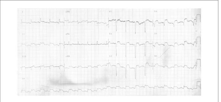
In a case such as the one described here, of a 72-year-old female patient, with history of coronary artery disease and myocardial revascularization carried out more than 10 years before, who sought emergency medical assistance due to typical precordial pain, the initial evaluation must always include an electrocardiogram (ECG). A normal ECG or an ECG with non-specific alterations or signs of ischemia would lead to an initial approach that would be different from the one described here. The admission ECG of this patient showed ST-segment elevation.
Considering that the patient had been submitted to a previous myocardial revascularization (MR), the correlation of the diagnostic findings of the ECG deserves special attention. First, the presence of chronic coronary artery disease, as well as the previous MR without the appropriate description of the grafts used in the surgery makes it difficult to establish the electrocardiographic correlation with the culprit artery, due both to the presence of bypasses and the possible existence of an extensive collateral network. It is also important to remember that, in spite of the native artery lesions, the probability of lesions in bypasses is of 38%1. Additionally,
the history of chronic coronary disease in elderly diabetic women and the QS in the anterior wall raises the possibility of previous myocardial infarction in the anterior wall, and, as a consequence, the possible correlation of the ST-segment elevation corresponding with the presence of aneurysm in the same wall. Still, the existence of ST-segment elevation in A 72-year-old female patient sought medical assistance due
to precordial pain that had lasted for two hours.
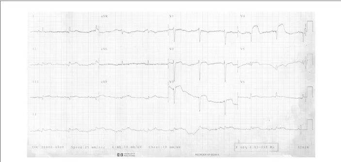
The patient had received a diagnosis of ischemic heart disease 10 years before. She had been submitted to myocardial revascularization in 1991 and was unable to give any specific details about the treatment or the grafts used at the surgical procedure. Five years after the surgery, she presented an episode of prolonged precordial pain. She sought medical assistance and, at this time, alterations in ventricular repolarization were diagnosed at the electrocardiogram (Figure 1), whereas the serum markers of myocardial injury remained unaltered.
The patient remained asymptomatic for five subsequent years, when she started to present recurrent and daily episodes of precordial pain, which lasted a week, until the pain became intense, prolonged and accompanied by dyspnea and intense diaphoresis. The patient then sought emergency care due to precordial pain that had lasted for one hour (March 7, 2001). The patient knew she had arterial hypertension and diabetes mellitus.
At physical examination, the patient exhibited “pain facies”, heart rate (HR) of 63 bpm and blood pressure (BP) of 130/80 mmHg. Pulmonary assessment disclosed rales in the lower third of both hemithoraces, muffled heart sounds without murmurs or pericardial friction rub. Abdominal assessment showed no alterations and the patient presented mild lower-limb edema.
Figure 1 -ECG: ventricular repolarization alterations.
Figure 2 -ECG: Ongoing infarction in the anterior and inferior walls.
the inferior wall associated to typical precordial pain justifies the chemical or mechanical reperfusion therapy, as described in the present case.
The description of the initial approach with the use of tenecteplase (TNK) and non-fractionated heparin can be considered the standard approach for a case as the one described here, as the early reperfusion therapy is the treatment of choice for the condition2. The improvement of
the pain can be considered a reperfusion criterion that favors the opening of the artery, although its significance is uncertain2.
However, the lack of some data in the description hinders a more thorough analysis of the patient’s clinical condition. There is no description of the pulses or chest x-rays that could rule out the presence of an aneurysm in the thoracic aorta leading to the right coronary artery dissection, which is the culprit artery in 80% of the cases of aorta dissection associated with coronary dissection.
Figure 3 -ECG: sinus tachycardia, QS complexes (anterior wall) and persistence of ST-segment elevation.
and death. Cardiogenic shock occurs in 7% of the cases with acute myocardial infarction (AMI)3. In these cases, the main
predictors are older age, systolic blood pressure at admission and Killip classification at admission4.
Cardiogenic shock is present in less than 1% of the patients at admission. However, more than 50% of those who will develop cardiogenic shock do so within the first 24 hours, with a mean time of 5 hours5. Cardiogenic shock mortality is
56% to 74%5 and the most common causes of shock according
to the Shock Trial6are: LV dysfunction in 79% of the cases;
acute mitral regurgitation in 7%; ventricular septal defect in 4%; isolated right ventricular shock in 4% and cardiac tamponade in 7%.
Considering that the patient was adequately monitored and that the second ECG showed tachycardic sinus rhythm without arrhythmias, which can be secondary to the use of dopamine, the most common causes of arrhythmia at the initial post-AMI period become unlikely. Therefore, the differential diagnosis is restricted to other causes of precordial pain, to the associated right ventricular involvement, to the mechanical complications of AMI evolution and the complications inherent to the proposed reperfusion therapy. Each one of these possibilities will be discussed separately.
First, among the clinical pictures of precordial pain associated to ST-segment elevation, which are not basically coronary, only the thoracic aorta dissection with ostium involvement and coronary dissection deserves attention. The acute dissection of the aorta normally affects elderly and hypertensive patients, as the case described here. On the other hand, the involvement of the coronary ostia is rare; only 1% to 2% of the patients present such complication7. Of these,
the great majority presents right coronary involvement, with ST-segment elevation in the inferior wall, as described in this case. In cases where the dissection goes unnoticed and the case is treated with thrombolysis, mortality reaches 71%, due
to the increased risk of cardiac tamponade8. As mentioned
before, the lack of detailed information on pulses, murmurs and chest x-rays does not allow the diagnosis to be confirmed or ruled out.
The involvement of the right ventricle (RV), associated with the left ventricular AMI condition must always be recalled in cases with AMI of the inferior wall, as the case described here. As mentioned before, the absence of right leads in the admission ECG limits the diagnosis, especially when one considers that the presence of 1-mm ST-segment elevation in V4R has a sensitivity of 88% and a specificity of 78% for the presence of or clinical picture evolution to RV involvement9.
Even without the right leads, the association of the ST-segment elevation in DII and DIII are contrary to the hypothesis of RV involvement. The presence of ST-segment elevation in DIII higher than in DII has a sensitivity of 97% and a specificity of 57% for right ventricular AMI; however, its absence does not help to elucidate this possibility10. Moreover, the presence of
crackling rales goes against the condition of right ventricular AMI, as the classic clinical picture courses with infarction and shock in the presence of clear pulmonary auscultation. Other criteria, such as the fact that the patient did not respond to volume replacement or dopamine, which are part of the optimized therapeutics for right ventricular AMI, would make it even less likely for a right ventricular AMI to be responsible for the dramatic evolution11. Not only that, but there have
Iatrogenesis must not be ruled out as a diagnostic possibility, secondary to clinical conditions where early aggressive therapy such as chemical thrombolysis is part of the routine therapeutic arsenal. In patients that are thrombolyzed with TNK, as the case described here, it is necessary to emphasize that this is a safe thrombolytic drug, with an incidence of bleeding lower than streptokinase or r-TPA (26 vs 29% in the ASSENT II study, which compared TNK with r-TPA)13. However, the presence of clinically
significant bleedings is lower than 2% in studies that tested TNK. On the other hand, older age, as in the present case, is one of the most important factors that predispose patients to major bleedings. Additionally, women present a higher risk of bleeding than men. Still, even in cases of major bleeding after thrombolysis, the dramatic clinical evolution and the absence of response to volume replacement are very unlikely, causing such diagnosis to be laid aside.
Finally, in the presence of a picture of cardiogenic shock with a dramatic evolution, one must fully evaluate any signs that can be associated with mechanical complications after the AMI. Among them are: ischemic mitral regurgitation, LV free wall rupture leading to tamponade, ventricular septal defect (VSD) due to septal rupture and previous aneurysm or pseudoaneurysm rupture.
Mitral regurgitation classically courses with acute left heart failure with important pulmonary congestion14, which
is very dissimilar from the patient’s clinical evolution, being highly unlikely.
The ventricular septal defect is a rare complication, with an incidence of 2% in the pre-therapeutic era of reperfusion. With the effective use of reperfusion techniques, the incidence of VSD has become lower than 0.2%, as described in the GUSTO-I study15. Traditionally, the onset of VSD takes place
between the 3rd and 5th days, but it can be present in less
than 24 hours after the initial event15. The clinical evolution
courses with shock due to biventricular dysfunction with a predominance of the RV. More than 80% of the patients present murmurs and more than 50% present thrills.
The absence of the description of these signs makes such diagnosis unlikely. Nevertheless, the presence of inferior wall segment elevation associated with anterior wall ST-segment elevation favors this diagnosis, due to the possibility that the picture is associated with AMI in the territory of the anterior interventricular branch (AIB) of the left coronary artery that surpasses the apex and due to the absence of collaterals, leading to important ischemia of the septum and its rupture. On the other hand, the presence of anterior QS complexes, as previously described, makes the possibility of anterior wall aneurysm more probable, thus making the hypothesis of AMI due to AIB injury leading to VSD unlikely. Although no echocardiogram or pulmonary artery catheter introduction was carried out for diagnostic clarification, VSD is an unlikely diagnosis.
Left ventricular aneurysms are more commonly found in the anterior wall16. Currently, the incidence of aneurysms
in thrombolyzed patients is less than 10%16. In spite of
the lack of precise data, the presence of anterior QS complexes associated to the ST-segment elevation in the
same leads raises the possibility of anterior wall aneurysm. The only complication associated with the true aneurysm that can course with such a dramatic evolution, as the one described here, is its rupture. However, a true aneurysm is a fibrotic scar with a stable structure and presents low risk of rupture. Although such diagnosis is uncommon and the clinical evolution is compatible with the described clinical picture, such hypothesis cannot be ruled out, considering the available data.
Pseudoaneurysms are false cavities that establish a communication between the left ventricle and the pericardial cavity. Although they are more common in the inferior wall, their occurrence in the anterior wall is also possible. Although many of them present murmurs, their absence does not rule out this hypothesis. Pseudoaneurysms have a risk of rupture of up to 40% and a mortality rate > 50% when rupture occurs17.
As the clinical evolution is quite similar to our patient’s evolution, this hypothesis cannot be ruled out either, based on the available data.
The last mechanical complication to be remembered is the LV free wall rupture, which occurs in up to 1% of the cases of AMI18. Of the fatal cases of AMI, the LV free wall rupture is the
cause of death in 12% of the patients that are thrombolyzed and in 7% of the patients that are not18. Among the risk factors
for the rupture, the clinical case patient presented: age older than 70 years; female sex; ST-segment elevation and Q wave at the admission ECG.
Although it normally appears between the third and the fifth days after the AMI, recent data have shown that, with the advent of reperfusion therapy, the occurrence of free wall rupture has become rarer, albeit earlier, and that it can occur within the first hours after the AMI19. Two distinctive
presentations of rupture have been described19 - an early one,
in cases of anterior wall AMI with a sudden clinical picture, as the one described here. This form occurs within the first 2 or 3 days after the AMI. The other form usually occurs after 4 days, as an evolution of the infarction area and usually has a slower evolution. Cases presenting the early form usually develop hemopericardium, leading to tamponade with a preferential clinical presentation of right ventricular AMI, followed by respiratory arrest with pulseless electrical activity (PEA). The clinical presentation of PEA without a previous clinical picture of heart failure in patients without a previous history of AMI has an accuracy of 95% for free wall rupture19. When the
previous presentation included heart failure, the accuracy of the arrest with PEA is only 17%. Although it is not possible to completely rule out the possibility of a previous AMI, these data suggest free wall rupture as a probable diagnosis of the clinical picture, even though it is not possible to state that this was the patient’s first AMI.
Dr. Márcio Sommer Bittencourt and Dr. Henrique Lane Staniak
Diagnostic hypothesis
Acute myocardial infarction, complicated by free wall rupture and tamponade.
Necropsy
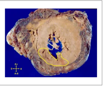
The pericardial sac was filled with a large amount of partially coagulated blood, demonstrating the occurrence of cardiac tamponade, which was the immediate cause of death. Transmural myocardial infarction and rupture were observed, with approximately 3 days of evolution, in the anterosuperior and septal walls of the LV (Figure 4), including the apical wall. The rupture occurred exactly at the apex of the heart and there was also hematic infiltration of epicardial fat (Figure 5). The LV myocardium exhibited moderate hypertrophy.
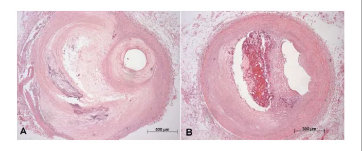
The study of the coronary arteries showed recent occlusive thrombosis of the bypass graft to the anterior interventricular branch of the left coronary artery. Moreover, this saphenous vein also showed the presence of intimal fatty plaques without severe luminal occlusion (Figure 6). Histologically, the coronary
arteries showed the presence of atheroma plaques, with various degrees of occlusion, being greater than > 70% in the initial and mid-thirds of the anterior interventricular branch of the left coronary artery and the mid-third of the right coronary artery (Figure 7).
Advanced aortic atherosclerosis was also observed (Figure 8), with dilation of the ascending segment of the aorta. Other necropsy findings were pulmonary emphysema and benign nephrosclerosis associated with systemic arterial hypertension.
Dr. Vera Demarchi Aiello
Anatomopathological diagnoses
Ischemic heart disease, with ruptured acute myocardial infarction and cardiac tamponade.
Dr. Vera Demarchi Aiello
Comments
The post-infarction ventricular rupture can affect the free wall, the ventricular septum or the papillary muscles.
Figure 6 -Photomicrographs of the bypass graft in two segments. A- proximal segment with intimal thickening (artifactually enhanced intima); B- mid segment, with recent, partially occlusive thrombosis. Hematoxylin-eosin stain. Magniication: 5X and 2.5X, respectively.
Figure 4 -Cross-section of the ventricular mass (short axis), showing irregular area delimited by the yellow line that corresponds to recent myocardial infarction.
Figure 7 -Photomicrographs of two segments of coronary arteries with important atherosclerotic obstruction. Anterior interventricular branch of the left coronary artery, 4th cm; B - Right coronary artery, 8th cm. Hematoxylin-eosin stain. Magniication: 5X.
No association has been described between the size of the infarction and the risk of rupture; and it is not unusual for the infarction to be small. Data collected at the Laboratory of Pathology of the Heart Institute (InCor)20 in cases of necropsy
have shown that most ruptures occur at the first infarction (72%), after an average of 5.4 days of evolution20. The most
commonly affected site is the lateral wall, as shown in the aforementioned series and also others in the lietarture20,21.
The rupture of the ventricular apex is rarely described. A Forensic Medicine series reported a prevalence of 6% concerning this rupture site22.
There is some controversy regarding the role of systemic arterial hypertension in the outcome as ventricular rupture. Whereas some authors indicate arterial hypertension as a risk indicator for cardiac rupture23, others have shown, in series
of infarcted patients with hypertension, a lower prevalence of rupture in comparison with normotensive patients24. Mann
and Roberts observed similar proportions of hypertensive patients between those with and without post-infarction cardiac rupture in an anatomical series25.
An immunohistochemical study in humans26 showed that
the patients with myocardial infarction that died due to cardiac rupture had lower levels of catenin alpha-1 than those that died due to other causes. Catenin is a cell adhesion protein present in the intercalated disks of the contractile cardiac cells, the cardiomyocytes. The same finding was also observed outside the infarcted area, which raised the hypothesis that such individuals would carry a genetic defect or at least, a genetic polymorphism25.
This hypothesis has been tested, but has yet to be proven.
References
1. Grines CL, Booth DC, Nissen SE, Gurley JC, Bennett KA, O’Connor WN, et al. Mechanism of acute myocardial infarction in patients with prior coronary artery bypass grafting and therapeutic implications. Am J Cardiol. 1990; 65: 1292-6.
2. Antman EM, Hand M, Armstrong PW, Bates ER, Green LA, Halasyamani LK, et al. 2007 Focused Update of the ACC/AHA 2004 Guidelines for the Management of Patients With ST-Elevation Myocardial Infarction: a report of the American College of Cardiology/American Heart Association Task Force on Practice Guidelines: developed in collaboration With the Canadian Cardiovascular Society endorsed by the American Academy of Family Physicians: 2007 Writing Group to Review New Evidence and Update the ACC/AHA 2004 Guidelines for the Management of Patients With ST-Elevation Myocardial Infarction, Writing on Behalf of the 2004 Writing Committee. Circulation. 2008; 117: 296-329.
3. Holmes DR Jr. Cardiogenic shock: a lethal complication of acute myocardial infarction. Rev Cardiovasc Med. 2003; 4: 131-5.
4. Mendes LA, Picard MH, Sleeper LA, Thompson CR, Jacobs AK, Hochman JS, et al. Cardiogenic shock: predictors of outcome based on right and left ventricular size and function at presentation. Coron Artery Dis. 2005; 16: 209-15. 5. Matos VA. Cardiogenic shock complicating acute myocardial
infarction--etiologies, management and outcome: a report from the SHOCK Trial Registry. Rev Port Cardiol. 2001; 20: 349-50.
6. Hochman JS, Sleeper LA, White HD, Dzavik V, Wong SC, Menon V, et al. One-year survival following early revascularization for cardiogenic shock. JAMA. 2001; 285: 190-2.
7. Spittell PC, Spitteli JA Jr, Joyce JW, Tajik AJ, Edwards WD, Schaff HV, et al. Clinical features and differential diagnosis of aortic dissection: experience with 236 cases (1980 through 1990). Mayo Clin Proc. 1993; 68: 642-51. 8. Melchior TD, Hallam D, Johansen BE, Aortic dissection in the thrombolytic
era: early recognition and optimal management is a prerequisite for increased survival. Int J Cardiol. 1993; 42: 1-6.
9. Jacobs AK, Leopold JA, Bates E, Mendes LA, Sleeper LA, White H, et al., Cardiogenic shock caused by right ventricular infarction: a report from the SHOCK registry. J Am Coll Cardiol. 2003; 41: 1273-9.
10. Zimetbaum PJ, Krishnan S, Gold A, Carrozza JP 2nd, Josephson ME. Usefulness of ST-segment elevation in lead III exceeding that of lead II for identifying the location of the totally occluded coronary artery in inferior wall myocardial infarction. Am J Cardiol. 1998; 81: 918-9.
11. Pfisterer M. Right ventricular involvement in myocardial infarction and cardiogenic shock. Lancet. 2003; 362: 392-4.
12. Kaul P, Armstrong PW, Chang WC, Naylor CD, Granger CB, Lee KL, et al. Long-term mortality of patients with acute myocardial infarction in the United States and Canada: comparison of patients enrolled in Global Utilization of Streptokinase and t-PA for Occluded Coronary Arteries (GUSTO)-I. Circulation. 2004; 110: 1754-60.
13. Van De Werf F, Adgey J, Ardissimo D, Armstrong PW, Aylward P, Barbash G, et
al. Single-bolus tenecteplase compared with front-loaded alteplase in acute myocardial infarction: the ASSENT-2 double-blind randomised trial. Lancet. 1999; 354: 716-22.
14. Thompson CR, Buller CE, Sleeper LA, Antonelli TA, Webb JG, Jaber WA, et al. Cardiogenic shock due to acute severe mitral regurgitation complicating acute myocardial infarction: a report from the SHOCK Trial Registry. SHould we use emergently revascularize Occluded Coronaries in cardiogenic shocK? J Am Coll Cardiol. 2000; 36 (Suppl A): 1104-9.
15. Birnbaum Y, Fishbein MC, Blanche C, Siegel RJ. Ventricular septal rupture after acute myocardial infarction. N Engl J Med. 2002; 347: 1426-32. 16. Tikiz H, Balbay Y, Atak R, Terzi T, Genç Y, Kutuk E. The effect of thrombolytic
therapy on left ventricular aneurysm formation in acute myocardial infarction: relationship to successful reperfusion and vessel patency. Clin Cardiol. 2001; 24: 656-62.
17. Becker RC, Gore JM, Lambrew C, Weaver WD, Rubison RM, French WJ, et al. A composite view of cardiac rupture in the United States National Registry of Myocardial Infarction. J Am Coll Cardiol. 1996; 27: 1321-6.
18. Pollak H, Nobis H, Mlczoch J, Frequency of left ventricular free wall rupture complicating acute myocardial infarction since the advent of thrombolysis. Am J Cardiol. 1994; 74: 184-6.
19. Honan MB, Harrell FE Jr, Reimer KA, Califf RM, Mark DB, Pryor DB, et al., Cardiac rupture, mortality and the timing of thrombolytic therapy: a meta-analysis. J Am Coll Cardiol. 1990; 16: 359-67.
20. Macedo AVS, Moffa PJ, Grupi CJ, Martinez Filho EE, Libby P, Gutierrez PS. Correlação anatomoclínica. Caso 4/2005 – Infarto fatal do miocárdio em mulher de 88 anos de idade. Arq Bras Cardiol. 2005; 85: 139-46. 21. Mann JM, Roberts WC. Rupture of the left ventricular free wall during acute
myocardial infarction: analysis of 138 necropsy patients and comparison with 50 necropsy patients with acute myocardial infarction without rupture. Am J Cardiol. 1988; 62: 847-59.
22. Hutchins KD, Skurnick J, Lavenhar M, Natarajan GA. Cardiac rupture in acute myocardial infarction: a reassessment. Am J Forensic Med Pathol. 2002; 23: 78-82.
23. Wehrens XH, Doevendans PA. Cardiac rupture complicating myocardial infarction. Int J Cardiol. 2004; 95: 285-92.
24. Abrignani MG, Dominguez LJ, Biondo G, Di Girolamo A, Novo G, Barbagallo M, et al. In-hospital complications of acute myocardial infarction in hypertensive subjects. Am J Hypertens. 2005; 18: 165-70.
25. Mann JM, Roberts WC. Rupture of the left ventricular free wall during acute myocardial infarction: analysis of 138 necropsy patients and comparison with 50 necropsy patients with acute myocardial infarction without rupture. Am J Cardiol. 1988; 62 (13): 847-59.