© 2013 Sociedade Brasileira de Hemodinâmica e Cardiologia Intervencionista. Published by Elsevier Editora Ltda. All rights reserved.
ST-Segment Resolution after Primary Percutaneous
Coronary Intervention: Characteristics, Predictors of
Failure, and Impact on Mortality
Pedro Beraldo de Andrade
1, Fábio Salerno Rinaldi
2, Marcos Henriques Bergonso
3,
Marden André Tebet
4, Ederlon Ferreira Nogueira
5, Vinícius Cardozo Esteves
6,
Igor Ribeiro de Castro Bienert
7, Mônica Vieira Athanazio de Andrade
8, Robson Alves Barbosa
9,
Luiz Alberto Piva e Mattos
10, André Labrunie
11ABSTRACT
Background: ST-segment resolution is an important predictor of infarct-related artery patency and effective microcircula-tory perfusion. The aim of this study was to compare patients with and without ST-segment elevation resolution and iden-tify variables that are associated with failure of ST-segment resolution. Methods: Between March 2012 and July 2013, 61 patients with ST-segment elevation acute myocardial infarction underwent primary percutaneous coronary intervention (PCI) and were included in a prospective registry. To compare the groups with and without ST-segment resolution, the chi-square test or Fisher’s exact test were used for qualitative variables and Student’s t test or Mann-Whitney’s test for quantitative variables. Simple and multiple logistic regression models were used to identify variables associated with failure of ST-segment resolution. Results: The group of patients without ST-segment resolution had higher heart rate, higher prevalence of diabe-tes mellitus, chronic renal failure and a trend towards longer ischemia time. There was a higher mortality rate in patients who did not achieve ST-segment elevation resolution. In the univariate analysis, heart rate, diabetes mellitus, chronic renal failure and ischemia time were associated with increased odds of not obtaining ST-segment resolution, losing signiicance in the multivariate model. Conclusions: Failure of ST-segment resolution is observed in up to one-third of patients
undergo-1 Postgraduate student (Doctoral). Interventionist Cardiologist Physician
at Santa Casa de Marília. Marília, SP, Brazil.
2 Interventionist Cardiologist Physician at Santa Casa de Marília.
Marília, SP, Brazil.
3 Cardiologist Trainee at Santa Casa de Marília. Marília, SP, Brazil. 4 Interventionist Cardiologist Physician at Rede D’Or São Luiz. São
Paulo, SP, Brazil.
5 Interventionist Cardiologist Physician at Hospital do Coração de
Londrina. Londrina, PR, Brazil.
6 Interventionist Cardiologist Physician at Rede D’Or São Luiz. São
Paulo, SP, Brazil.
7 Technician Director of the Interventionist Cardiology Service of
Hospital das Clínicas da Faculdade Estadual de Medicina de Marília. Marília, SP, Brazil.
8 Nurse, Specialist in Cardiovascular Nursing at Santa Casa de Marília.
Marília, SP, Brazil.
9 Nurse, Specialist in Cardiovascular Nursing at Santa Casa de Marília.
Marília, SP, Brazil.
10 Doctor. Interventionist Cardiologist Physician at Rede D’Or São Luiz.
São Paulo, SP, Brazil.
11 Doctor. Interventionist Cardiologist Physician at Coração de Londrina.
Londrina, PR, Brazil.
Correspondence to: Pedro Beraldo de Andrade. Av. Vicente Ferreira, 828 – Marília, SP, Brazil – CEP 17515-900
E-mail: [email protected]
Received on: 7/1/ 2013 • Accepted on: 9/4/2013
Original Article
RESUMO
Resolução do Segmento ST após Intervenção Coronária Percutânea Primária: Características, Preditores de Insucesso e Impacto na Mortalidade
Introdução: A resolução do segmento ST é um importante preditor de patência da artéria relacionada ao infarto e de perfusão efetiva da microcirculação. O objetivo deste trabalho foi comparar pacientes com e sem resolução do segmento ST e identiicar variáveis que se associam à não resolução do ST. Métodos: Entre março de 2012 e julho de 2013, 61 pacientes com infarto agudo do miocárdio com elevação do segmento ST foram submetidos à intervenção coronária percutânea (ICP) primária e incluídos em um registro pro-spectivo. Para comparar os grupos com e sem resolução do segmento ST, foi utilizado o teste qui-quadrado ou exato de Fisher, para as variáveis qualitativas, e o teste t de Student ou de Mann-Whitney, para variáveis quantitativas. Para identiicar variáveis associadas à não resolução do ST, foram ajustados modelos de regressão logística simples e múltipla. Resulta-dos: O grupo de pacientes sem resolução do segmento ST apresentou maior frequência cardíaca, maior prevalência de
diabetes mellitus, de insuiciência renal crônica e tendência
ing primary PCI and requires new pharmacologic strategies or interventions to minimize it.
DESCRIPTORS: Myocardial infarction. Percutaneous coronary intervention. Myocardial reperfusion.
This study aimed to compare demographic and procedure-related characteristics of patients with and without complete ST-segment resolution after perform-ing primary PCI, and to identify the variables that were associated to non-ST-resolution. The secondary objective was to determine its impact on infarct size, measured by maximum peak of CK-MB, length of stay, and in-hospital mortality after 30 days, six months and one year of follow-up.
Ischemia time was deined as the interval between the onset of symptoms and the crossing of the lesion with a pre-dilation balloon, manual thrombus aspiration catheter, or stent. Door-to-balloon time was deined as the interval between hospital admission and the crossing of the lesion with a pre-dilation balloon catheter, manual thrombus aspiration catheter, or stent. A CK-MB analysis was performed every six hours until the occurrence of a reduction in the levels of this biomarker. The choice of antiplatelet therapy, use of prophylactic intracoronary adenosine, manual aspiration thrombectomy, and the access route were left to the discretion of the surgeon.
Statistical Analysis
Qualitative variables were summarized as absolute frequencies and percentages. The quantitative data were expressed as means ± standard deviation or medians and interquartile ranges (25th percentile –75th per-centile) according to distribution for each variable. To compare the groups with and without resolution of the ST-segment, the chi-squared test or Fisher’s exact test were used for qualitative variables, and Student’s t-test
or the Mann-Whitney test were used for quantitative variables.
To identify variables associated with ST-segment resolution, simple (univariate approach) and multiple (multivariate analysis) logistic regression models were adjusted. The results were expressed as odds ratios (OR) and 95% conidence intervals (95% CI). The level of signiicance was 0.05. The statistical program used to perform the calculations was the SPSS for Windows, version 19.0.
ST. Pela análise univariada, as variáveis frequência cardíaca,
diabetes mellitus, insuiciência renal crônica e tempo de
isquemia associaram-se à maior chance de não resolução do ST, perdendo signiicância no modelo multivariado. Conclusões:
A não resolução do segmento ST pode ocorrer em até um terço dos pacientes submetidos à ICP primária, requerendo o advento de novas estratégias farmacológicas ou de intervenção capazes de minimizá-la.
DESCRITORES: Infarto do miocárdio. Intervenção coronária percutânea. Reperfusão miocárdica.
C
ompared to ibrinolysis, percutaneous coronary intervention (PCI) promotes a more early and sustained restoration of epicardial low in the target vessel, and is widely accepted as the preferred strategy in the treatment of acute myocardial infarc-tion (AMI).1,2 The resolution of the ST-segment afterthe institution of reperfusion therapy is an important predictor of patency of the artery related to the event and of effective microcirculatory perfusion.3
The analysis of ST-segment resolution on the elec-trocardiogram (ECG) consists of a simple tool, of easy use and low cost, able to document success of the epi-cardial reperfusion and tissue reperfusion after primary PCI.4 The persistence of ST-segment elevation on the
ECG, despite the restoration of a normal epicardial low, signiies a poor prognosis; such persistence is known to be associated with larger infarct size and higher combined rate of severe cardiovascular adverse events.5
In a contemporary context for the application of primary PCI, characterized by an aggressive anti-thrombotic therapy, manual aspiration thrombectomy, intracoronary adenosine, and ample use of the radial access, this study aimed to determine the percentage of failure in ST-segment resolution, its predictors, and the impact on in-hospital and late mortality.
METHODS
Between March 2012 and July 2013, patients with AMI with ST-segment elevation ≥ 1 mm in two contigu-ous leads of the frontal plane or ≥ 2 mm in precordial leads, with symptoms duration < 12 hours, underwent primary PCI and were included in a local controlled prospective registry. A 12-lead ECG, plus leads V3R, V4R, V7, and V8 in the case of lower AMI, was per-formed at admission and 30 to 60 minutes after the conclusion of the procedure. Patients with left branch block or pacemaker rhythm, surgical myocardial re-vascularization, cardiogenic shock, or life expectation
< six months were excluded. The electrocardiographic analysis followed the prevalent normative guidelines,6
RESULTS
Comparisons among patients with and without ST-segment resolution
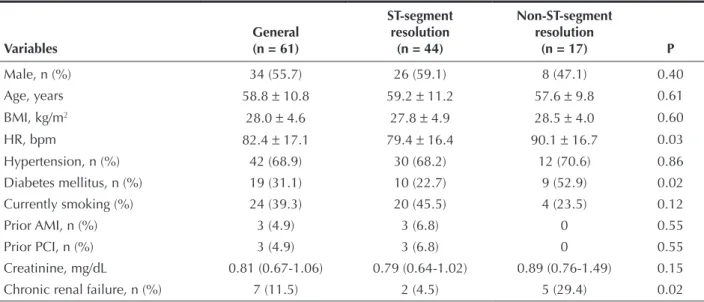
Table 1 shows a comparison among patients with and without ST-segment resolution regarding baseline clinical characteristics. It was observed that the group of patients without ST-segment resolution had, on average, a higher heart rate and higher prevalence of diabetes mellitus and of chronic renal failure.
With respect to the procedure and angiographic characteristics (Table 2), a statistically signiicant dif-ference was observed only in the number of stents used. A trend of longer ischemia time among patients without ST-segment resolution (4.7 ± 2.0 hours vs. 6.3
± 3.1 hours; P = 0.07) was detected.
Regarding the results and the clinical outcome, a higher in-hospital mortality rate among patients who did not achieve ST-segment resolution was observed (Table 3).
Identification of variables associated with non-ST segment resolution
This part of the analysis sought to identify, among the clinical, angiographic and procedural characteristics, the factors associated with the increased probability of non-ST-segment resolution. Initially, the isolated effect of each variable were investigated by simple logistic regression models (univariate analysis), considering as dependent variable the non-ST-segment resolution. The results are shown in Table 4.
As expected, according to the comparison between groups, the variables heart rate, diabetes mellitus, and chronic renal failure were associated with increased probability of non ST-segment resolution. In addition, the ischemia time was also signiicant. The variables prior AMI, prior PCI, route of access, and therapeutic catheters could not be analyzed due to the low fre-quency in some of their categories.
Variables with P < 0.20 in the univariate analysis were selected for the multivariate model, in which the effects were analyzed simultaneously (Table 5). In this model, all variables lost their importance and ceased to be signiicant according to the usual signiicance level of 0.05. This can be explained by the low statistical power to detect differences, caused by the increase in the number of parameters in the model.
In order to obtain a more consistent model, the non-signiicant variables were excluded, step-by-step, until the achievement of a reduced model. In this model, only diabetes (OR = 3.83, 95% CI, 1.17 to 12.51, P = 0.19) and chronic renal failure (OR = 8.75, 95% CI, 1.50 to 50.90, P = 0.15) remained signiicant. Importantly, although they were signiicant, the estimates were not very accurate (very wide CIs), and the ORs may be overestimated. Still, it is clear that diabetes mellitus and chronic renal failure are factors associated with non-ST-segment resolution.
DISCUSSION
Incomplete ST-segment resolution after primary PCI correlates strongly with mortality and reinfarction,
TABLE 1
Baseline clinical characteristics
Variables
General (n = 61)
ST-segment resolution
(n = 44)
Non-ST-segment resolution
(n = 17) P
Male, n (%) 34 (55.7) 26 (59.1) 8 (47.1) 0.40
Age, years 58.8 ± 10.8 59.2 ± 11.2 57.6 ± 9.8 0.61
BMI, kg/m2 28.0
± 4.6 27.8 ± 4.9 28.5 ± 4.0 0.60
HR, bpm 82.4 ± 17.1 79.4 ± 16.4 90.1 ± 16.7 0.03
Hypertension, n (%) 42 (68.9) 30 (68.2) 12 (70.6) 0.86
Diabetes mellitus, n (%) 19 (31.1) 10 (22.7) 9 (52.9) 0.02
Currently smoking (%) 24 (39.3) 20 (45.5) 4 (23.5) 0.12
Prior AMI, n (%) 3 (4.9) 3 (6.8) 0 0.55
Prior PCI, n (%) 3 (4.9) 3 (6.8) 0 0.55
Creatinine, mg/dL 0.81 (0.67-1.06) 0.79 (0.64-1.02) 0.89 (0.76-1.49) 0.15
Chronic renal failure, n (%) 7 (11.5) 2 (4.5) 5 (29.4) 0.02
TABLE 2
Angiographic and procedure characteristics
Variables
General (n = 61)
ST-segment resolution
(n = 44)
Non-ST-segment resolution
(n = 17) P
Killip I, n (%) 57 (93.4) 41 (93.2) 16 (94.1) > 0.99
Location of AMI, n (%) 0.72
Anterior 30 (49.2) 21 (47.7) 9 (52.9)
Inferior 31 (50.8) 23 (52.3) 8 (47.1)
Hemoglobin, g/dL 13.7 ± 1.9 13.9 ± 1.9 13.1 ± 2.0 0.15
CK-MB activity, U/L 51.5 (26.9-115) 52.3 (27-107.5) 48.2 (23-121) 0.94
Ischemia time, hours 5.1 ± 2.4 4.7 ± 2.0 6.3 ± 3.1 0.07
Door-to-balloon time, min 55 (42-75) 56.5 (43.5-80) 50.0 (42-65) 0.40 Manual aspiration
thrombectomy, n (%)
33 (54.1) 23 (52.3) 10 (58.8) 0.45
Direct stent implantation, n (%) 27 (44.3) 21 (47.7) 6 (35.3) 0.38
Visible aspirated, n (%) 19 (31.1) 14 (31.8) 5 (29.4) 0.86
Prophylactic adenosine, n (%) 22 (36.1) 16 (36.4) 6 (35.3) 0.94
TIMI pre-0 or 1, n (%) 49 (80.3) 37 (84.1) 12 (70.6) 0.29
Radial access, n (%) 60 (98.4) 43 (97.7) 17 (100) > 0.99
GPI IIb/IIIa, n (%) 35 (59.3) 24 (57.1) 11 (64.7) 0.59
Left ventriculography, n (%) 0.55
Not evaluated 8 (13.1) 6 (13.6) 2 (11.8)
Normal 18 (29.5) 13 (29.5) 5 (29.4)
Mild dysfunction 16 (26.2) 11 (25.0) 5 (29.4)
Moderate dysfunction 16 (26.2) 13 (29.5) 3 (17.6)
Severe dysfunction 3 (4.9) 1 (2.3) 2 (11.8)
Culprit vessel, n (%) > 0.99
Left anterior descending artery 29 (47.6) 21 (47.7) 8 (47.1)
Right coronary artery 19 (31.1) 14 (31.8) 5 (29.4)
Left circumlex artery 12 (19.7) 8 (18.2) 4 (23.5)
Intermediate branch 1 (1.6) 1 (2.3) 0
P2Y12 blocker, n (%) 0.70
Clopidogrel 300 mg 4 (6.6) 3 (6.8) 1 (5.9)
Clopidogrel 600 mg 38 (62.3) 26 (59.1) 12 (70.6)
Ticagrelor 180 mg 19 (31.1) 15 (34.1) 4 (23.5)
Number of diagnostic catheters 1 (1-2) 1 (1-2) 1 (1-2) 0.91
Diagnostic catheters, n (%) > 0.99
1 44 (72.1) 32 (72.7) 12 (70.6)
2 or more 17 (27.9) 12 (27.3) 5 (29.4)
Number of therapeutic catheters 1 (1-1) 1 (1-1) 1 (1-1) 0.15
Therapeutic catheters, n (%) 0.31
1 56 (91.8) 39 (88.6) 17 (100.0)
2 or 3 5 (8.2) 5 (11.4) 0
Number of stents 1 (1-1) 1 (1-1) 1 (1-1) 0.01
Number of stents, n (%) < 0.01
0 4 (6.6) 0 4 (23.5)
1 50 (82.0) 38 (86.4) 12 (70.6)
2 or 3 7 (11.5) 6 (13.6) 1 (5.9)
Contrast volume, mL 185.8 ± 64.1 187.8 ± 66.4 180.6 ± 59.2 0.70
resolution. Initially, the sample of patients undergoing the procedure was restricted to within 12 hours after onset of symptoms. The mean ischemia time was 5.1 hours and the median door-to-balloon time was only 55 minutes, favored by the logistical conditions set forth in a previous publication.10 Moreover, manual
aspira-tion thrombectomy was used in 54% of the sample, and glycoprotein IIb/IIIa inhibitors and adenosine were administered prophylactically to 59% and 36% of cases, respectively.
In fact, in the electrocardiographic sub-analysis of the trial Platelet Inhibition and Patient Outcomes
(PLATO),11 70% of patients randomized in the interval
of 3-6 hours from the onset of symptoms exhibited ST-segment resolution, compared to 51% of those randomized after 6 hours. Meanwhile, a meta-analysis including 18 randomised trials and 3,936 patients showed that manual thrombus aspiration, compared to conventional primary PCI, promotes higher TIMI 3 inal myocardial blush (63.6% vs. 48.5%; P < 0.0001) and higher complete ST-segment resolution (55.8% vs. 44.3%; P < 0.0001).12
Glycoprotein IIb/IIIa inhibitors, despite lack of consistent evidence to justify their routine indication, ind an application niche in patients classiied as high risk.13,14 Finally, in the “Intracoronary Nitroprusside Versus
Adenosine in Acute Myocardial Infarction”(REOPEN-AMI) randomized trial, 71% of patients who received intra-coronary adenosine after thrombus aspiration exhibited ST-segment resolution > 70%, compared to 54% in the group receiving sodium nitroprusside and to 51% of regardless of the degree of patency of the target vessel.5
In a series from real-world practice, characterized by a mean age of 59 years, 31% diabetics, 11% with chronic renal failure, ischemia time of 5 hours, and median door-to-balloon time of 55 minutes, the rate of obtain-ing a inal Thrombolysis in Myocardial Infarction (TIMI) low 3 was 84%, with ST-segment resolution > 70% in 72% of the sample. Diabetes mellitus, chronic renal failure, elevated heart rate, longer ischemic time, and fewer implanted stents were the characteristics most commonly observed in patients who did not achieve ST-segment resolution; the irst two factors are important predictors of failure.
In pre-speciied analysis of the randomized trial “Harmonizing Outcomes with Revascularization and Stents in Acute Myocardial Infarction” (HORIZONS-AMI), comprising 2,484 of 3,345 patients undergoing primary PCI, the rate of resolution of ST-segment >
70% was 50.5%, with a lower incidence of death, reinfarction, target vessel revascularization, and stent thrombosis at three years in this group.7 In the same
clinical trial, inal TIMI low 3 was achieved in 87.1% of the sample, and age, anterior wall AMI, TIMI pre-0/1, and extent of injury were independent predictors of inal TIMI low < 3 by multivariate analysis.8 In an
Argentinean multicenter registry, despite obtaining a inal TIMI low 3 in 90% of the sample, the rate of complete ST-segment resolution was 48.5%.9
The present series displays characteristics that differ from those previously mentioned, which could explain the high percentage of complete ST-segment
TABLE 3
Outcomes of efficacy, safety, and evolution
Variables
General (n = 61)
ST-segment resolution
(n = 44)
Non-ST-segment resolution
(n = 17) P
TIMI 3post, n (%) 51 (83.6) 39 (88.6) 12 (70.6) 0.12
CK-MB peak, U/L 285.9 ± 206.2 295.2 ± 194.6 261.9 ± 238.4 0.58
Length of hospital stay, nights 3 (3-4) 3 (3-4) 4 (3-5) 0.53
Complications, n (%) 6 (9.8) 3 (6.8) 3 (17.6) 0.34
In-hospital mortality, n (%) 4 (6.6) 0 4 (23.5) < 0.01
Clinical events at 30 days, n (%)
Patients 51 38 13
Events 1 (1.9) 1 (2.6) 0 > 0.99
Clinical events at six months, n (%)
Patients 37 28 9
Events 3 (8.1) 3 (10.7) 0 0.55
Clinical events at one year, n (%) > 0.99
Patients 24 17 7
TABLE 4
Factors associated with non-ST-segment resolution (univariate analysis)
Variables OR 95% CI P
Male 0.62 (0.20-1.90) 0.40
Age, years 0.99 (0.94-1.04) 0.61
Body mass index, kg/m2 1.03 (0.92-1.17) 0.59
Heart rate, ×10 bpm 1.47 (1.03-2.10) 0.03
Hypertension 1.12 (0.33-3.80) 0.86
Diabetes mellitus 3.83 (1.17-12.51) 0.03
Current smoking 0.37 (0.10-1.31) 0.12
Creatinine, mg/dL 1.10 (0.74-1.62) 0.64
Chronic renal failure 8.75 (1.50-50.90) 0.02
Killip I 1.17 (0.11-12.10) 0.90
Infarction, anterior location 1.23 (0.40-3.78) 0.72
Hemoglobin on admission, g/dL 0.80 (0.60-1.08) 0.15
CK-MB activity on admission, U/L 1.00 (0.10-1.01) 0.33
Ischemia time, hours 1.31 (1.03-1.68) 0.03
Door-to-balloon time, min 0.10 (0.98-1.02) 0.86
Manual aspiration thrombectomy 1.30 (0.42-4.05) 0.65
Direct stent implantation 0.60 (0.19-1.90) 0.38
Macroscopic aspirate visible 0.89 (0.26-3.03) 0.86
Prophylactic adenosine 0.96 (0.30-3.07) 0.94
TIMI pre-2 or 3 2.20 (0.59-8.24) 0.24
Glycoprotein IIb/IIIa inhibitor 1.38 (0.43-4.42) 0.59
Moderate or severe LV 0.89 (0.26-3.03) 0.86
Culprit vessel
Right coronary 1.00 – –
Left Circumlex artery/intermediate branch 1.24 (0.26-5.92) 0.78
Left anterior Descending artery 1.07 (0.29-3.94) 0.92
P2Y12 blocker on admission
Ticagrelor 180 mg 1.00 – –
Clopidogrel 300 mg 1.25 (0.10-15.50) 0.86
Clopidogrel 600 mg 1.73 (0.47-6.34) 0.41
2 or more diagnostic catheters 1.11 (0.32-3.83) 0.87
2 or more stents 0.40 (0.04-3.56) 0.41
OR = odds ratio; IC 95% = conidence interval of 95%; bpm = beats per minute.
TABLE 5
Factors associated with non-ST-segment resolution. Results of simple (univariate analysis) and multiple (multivariate analysis) logistic regression models
Factors
Univariate Multivariate
OR 95% CI P OR 95% CI P
Heart rate, ×10 bpm 1.47 (1.03-2.10) 0.03 1.42 (0.93-2.18) 0.11
Diabetes mellitus 3.83 (1.17-12.51) 0.03 2.55 (0.63-10.31) 0.19
Current smoking 0.37 (0.10-1.31) 0.12 0.55 (0.12-2.46) 0.43
Chronic renal failure 8.75 (1.50-50.90) 0.02 4.41 (0.57-33.97) 0.15
Hemoglobin at admission, g/dL 0.80 (0.60-1.08) 0.15 0.84 (0.55-1.28) 0.41
Ischemia time, hours 1.31 (1.03-1.68) 0.03 1.22 (0.90-1.65) 0.21
the saline group (P = 0.009), limiting the reperfusion injury, as measured by the occurrence of angiographic microvascular obstruction (TIMI low ≤ 2 or 3 with TIMI myocardial blush < 2).15
Limitations of the study
Apart from the non-random sampling, the main limitation of this study resided in the small size of their sample, making it impossible to identify potential determinant variables for failure of complete ST-segment resolution after primary PCI.
CONCLUSIONS
Incomplete resolution of ST-segment may occur in up to one-third of patients undergoing primary percutane-ous coronary intervention, regardless of the restoration of TIMI 3 inal epicardial low. The predictor variables diabetes mellitus, chronic renal failure, heart rate, and ischemia time are associated with incomplete resolution of ST-segment elevation, requiring new pharmacological or interventional strategies to minimize this condition.
CONFLICTS OF INTEREST
The authors declare no conlicts of interest.
REFERENCES
1. Steg PG, James SK, Atar D, Badano LP, Lundqvist CB, Borger MA, et al. ESC Guidelines for the management of acute myocardial infarction in patients presenting with ST-segment elevation. The Task Force on the management of St-segment elevation acute myocardial infarction of the European Society of Cardiology (ESC). Eur Heart J. 2012;33(20):2569-619.
2. O’Gara PT, Kushner FG, Ascheim DD, Casey Jr DE, Chung MK, Lemos JA, et al. 2013 ACCF/AHA guideline for the manage-ment of ST-elevation myocardial infarction: a report of the American College of Cardiology Foundation/American Heart Association Task Force on Practice Guidelines. Circulation. 2013;127(4):e362-425.
3. Hallén J, Sejersten M, Johanson P, Atar D, Clemmensen PM. Inluence of ST-segment recovery on infarct size and ejection fraction in patients with ST-segment elevation myocardial in-farction receiving primary percutaneous coronary intervention. Am J Cardiol. 2010;105(9):1223-8.
4. Feldman LJ, Coste P, Furber A, Dupouy P, Slama MS, Monassier JP, et al. Incomplete resolution of ST segment elevation is a marker of transient microcirculatory dysfunction after stenting for acute myocardial infarction. Circulation. 2003;107(21):2684-9. 5. McLaughlin MG, Stone GW, Aymong E, Gardner G, Mehran R,
Lansky AJ, et al. Prognostic utility of comparative methods for assessment of ST-segment resolution after primary angioplasty
for acute myocardial infarction The Controlled Abciximab and Device Investigation to Lower Late Angioplasty Complications (CADILLAC) trial. J Am Coll Cardiol. 2004;44(6):1215-23. 6. Pastore CA, Pinho C, Germiniani H, Samesima N, Mano R,
et al.; Sociedade Brasileira de Cardiologia. Diretrizes da So-ciedade Brasileira de Cardiologia sobre Análise e Emissão de Laudos Eletrocardiográicos (2009). Arq Bras Cardiol. 2009; 93(3 Supl.2):1-19.
7. Farkouh ME, Reiffel J, Dressler O, Nikolsky E, Parise H, Cristea E, et al. Relationship between ST-segment recovery and clinical outcomes after primary percutaneous coronary intervention: the HORIZONS-AMI ECG substudy report. Circ Cardiovasc Interv. 2013;6(3):216-23.
8. Caixeta A, Lansky A, Mehran R, Brener SJ, Claessen B, Généreux P, et al. Predictors of suboptimal TIMI low after primary angioplasty for acute myocardial infarction: results from the HORIZONS-AMI trial. EuroIntervention. 2013;9(2):220-7. 9. Damonte AA, Lasave L, Kozak F, Rossi M, Gamen M, Cura F,
et al. Avaliação da resolução do supradesnivelamento do segmento ST após angioplastia primária: Registro Multicêntrico de Infarto Agudo do Miocárdio com Supradesnivelamento do Segmento ST na Argentina. Rev Bras Cardiol Invasiva. 2009;17(4):470-5.
10. Andrade PB, Tebet MA, Nogueira EF, Rinaldi FS, Esteves VC, Andrade MVA, et al. Impacto da transferência inter-hospitalar nos resultados da intervenção coronária percutânea primária. Rev Bras Cardiol Invasiva. 2012;20(4):361-6.
11. Armstrong PW, Siha H, Fu Y, Westerhout CM, Steg PG, James SK, et al. ST-elevation acute coronary syndromes in the Platelet Inhibition and Patient Outcomes (PLATO) trial: insights from the ECG substudy. Circulation. 2012; 125(3):514-21. 12. Kumbhani DJ, Bavry AA, Desai MY, Bangalore S, Bhatt DL.
Role of aspiration and mechanical thrombectomy in patients with acute myocardial infarction undergoing primary angio-plasty: an updated meta-analysis of randomized trials. J Am Coll Cardiol. 2013 May 8. [Epub ahead of print].
13. Ortolani P, Marzocchi A, Marrozzini C, Palmerini T, Saia F, Taglieri N, et al. Long-term effectiveness of early administration of glycoprotein IIb/IIIa agents to real-world patients undergoing primary percutaneous interventions: results of a registry study in an ST-elevation myocardial infarction network. Eur Heart J. 2009;30(1):33-43.
14. Sethi A, Bajaj A, Bahekar A, Bhuriya R, Singh M, Ahmed A, et al. Glycoprotein IIb/IIIa inhibitors with or without thienopyridine pretreatment improve outcomes after primary percutaneous coronary Intervention in high-risk patients with ST elevation myocardial infarction:a meta-regression of randomized con-trolled trials. Catheter Cardiovasc Interv. 2013; 82(2):171-81. 15. Niccoli G, Rigattieri S, Vita MRD, Valgimigli M, Corvo P,