Heart Rate Variability, Blood Lipids and Physical Capacity of Obese
and Non-Obese Children
Mário Augusto Paschoal, Patrícia Fernandes Trevizan, Natália Folco Scodeler
Grupo de Pesquisa - Função autonômica cardíaca e atividade física na saúde e na doença - Pontifícia Universidade Católica de Campinas, Campinas, SP - Brazil
Summary
Background: Obesity can affect the cardiac autonomic modulation, blood lipid levels and the physical capacity.
Objective: To study the effect of obesity on the heart rate variability (HRV), blood lipid levels and physical capacity of obese children.
Methods: Thirty children aged 9 to 11 years were studied, divided in two groups: a) 15 obese children (O) aged 10.2 ± 0.7 years and body mass index (BMI) between the 95th and the 97th percentiles; b) 15 non-obese children (NO) aged 9.8 ± 0.7 years and BMI between the 5th and 85th percentiles. All children were submitted to an anthropometric and clinical assessment, analysis of the HRV at rest and a physical exercise protocol (PEP). Non-parametric tests were used to compare the variables between the groups. The level of significance was set at p < 0.05.
Results: The waist circumference and levels of triglycerides were higher in O. The cardiac sympathetic activity in the standing position in normalized units (LF n.u.) was higher in O, 71.4%, when compared to NO, 56.3%. The low frequency/ high frequency ratio (LF/HF) was 3.8 in O and 1.7 in NO. The PEP showed differences between the groups, with higher values observed in NO children regarding the total distance, time of exposure to PEP, peak oxygen consumption (VO2 peak) and metabolic equivalent (MET).
Conclusion: Obesity in children resulted in alterations in cardiac autonomic control in the standing position and decreased the physical capacity. (Arq Bras Cardiol 2007;88(6):624-628)
Key Words: Autonomic nervous system; obesity; heart rate; exertion.
Mailing address: Mário Augusto Paschoal •
Rua Ferreira Penteado, 1242/72 - Cambuí - 13010-041 - Campinas, SP - Brazil
E-mail: [email protected]
Manuscript received June 16, 2008; revised manuscript received August, 05 2008; accepted August, 15 2008.
Introduction
Obesity, considered a worldwide epidemic, is characterized by the excessive accumulation of fat tissue in the body and its causes are multifactorial, such as genetic susceptibility, sex, age, occupation, diet and others1.
Basically, the type of obesity of which occurrence is not associated to endocrinological diseases is strongly related to the dietary imbalance resulting from the disequilibrium between the ingestion and the daily need of nutrients to supply the metabolism of a genetically susceptible individual2.
This specific type of obesity, called sociogenic or exogenous, is caused by sociocultural factors, such as the modern lifestyle itself, which predisposes to a lower energy consumption and inadequate dietary habits and sedentary lifestyle3.
Considering that obesity is a pandemic, recognized at the start of this century and with discouraging perspectives on objective and effective programs directed at its control, all investigations related to obesity become relevant4.
In this context, there are studies5 that emphasize the very
likely fact that children with excess weight might have a decrease in their total number of years of life, mainly due to the development of cardiovascular diseases in adult life, which originate during infancy and childhood6.
Among these research lines, the ones that are currently highlighted are those involving the accumulation of fat tissue in the abdominal region7,8, alteration in blood lipid
levels5,9, low physical capacity10 and alterations in the cardiac
autonomic control11,12.
The accumulation of fat tissue in the abdominal region has definitely shown to be a better risk factor predictor for cardiovascular disease and type II diabetes mellitus than total obesity7. Inversely, but supporting this theory, recent studies
have shown that moderate to high levels of cardiorespiratory functional capacity in children are associated to presence of lower abdominal adiposity10.
Thus, one of the most interesting possibilities for the decrease in abdominal fat tissue content and, by extension, for the prevention of type II diabetes mellitus and total obesity is the regular practice of physical exercises.
been largely used in the cardiovascular assessment of obese adults and children16,17.
The cardiac autonomic alterations, as observed regarding the accumulation of fat tissue in the abdominal region, have also been related to the future occurrence of important functional and metabolic disorders, such as systemic arterial hypertension, diabetes mellitus, cerebrovascular accident, acute myocardial infarction, among others11,12.
Based on these arguments, the present study sought to comprehensively assess these main problems observed in childhood obesity, emphasizing the investigation of the body mass index (BMI), the distribution of body fat, blood lipid levels, HRV at rest and physical capacity. The objective was to address the most relevant current paradigms of the early influence of obesity on the future onset of cardiovascular and metabolic diseases. Therefore, it is believed that studies comprehensively addressing all these questions can provide more information on the effect of this important dysfunction on the cardiovascular system, so that new therapeutic actions can be developed and applied to the benefit of the affected population.
Methods
Thirty children aged 9 to 11 years were divided in two groups: group A with 15 obese (O) children (8 boys and 7 girls), with a mean age of 10.2 ± 0.7 years and body mass index (BMI) between the 95th and the 97th percentiles18; and group B, with 15
non-obese (NO) children (7 boys and 8 girls), with a mean age of 9.8 ± 0.7 years and BMI between the 5th and 85th percentiles.
As inclusion criteria, the obese children presented BMI in accordance with the previously established values and did not present heart or metabolic diseases. They did not use any medication that could interfere with the data. Additionally, they had not practiced sports activities for at least two months, except for physical education classes twice a week.
More information on the children’s daily activities was obtained from interviews carried out with parents or tutors. They were informed about the objectives of the study and signed a free and informed consent form. The study was approved by the Ethics Committee in Research involving human beings of PUC-Campinas, Protocol number 138/06.
Procedures
Phase 1
Anthropometric and clinical assessments were carried out, which consisted of weight, height and waist circumference
After advising the parents or tutors as well as the children that the latter should not practice sports activities in the 24 hours prior to the assessment, nor consume stimulants such as coffee, tea, chocolate and carbonated beverages and that they should have a good night’s sleep (of at least 8 hours), the heart rate recording was performed, which corresponded to the heart rate frequency at rest.
This recording was divided in two parts and carried out between 4 and 5 PM to avoid the interference of the circadian rhythm of the HR on the HRV. In the first part, the recording lasted 12 minutes and was carried out in a relaxing and quiet environment, with controlled temperature between 21°C and 23°C, with the volunteer lying on a couch in the supine position. In the second part, with the volunteer in the standing position, the recording lasted seven minutes. In both situations, the volunteer was advised not to move or speak.
The calculation of the HRV was avoided in the first two minutes of the recordings in the supine as well as for the standing position, as problems in signal stability and the onset of artifacts that occur during this time period due to posture adaptation can impair their interpretation20.
All heart beats were recorded by a Polar S810i heart rate
monitor and directed to an interface (IR interface) through
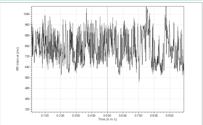
infrared signals. The data obtained were sent to the computer and the HRV analysis was carried out by the software programs Polar Precision Performanceand Nevrokard (Figure 1).
Phase 3
The submaximal progressive physical exercise protocol (PEP) was carried out on a treadmill (super ATL – Inbrasport) with the
initial velocity of 2.0 km/h, for two minutes, without inclination and 0.5 km/h increments every subsequent minute.
During the protocol, all children had to achieve the submaximal HR calculated using the formula: submaxHR = 195 – age. When the test was completed, the treadmill velocity was progressively decreased for 2 minutes, to allow the adequate recovery of the cardiorespiratory and metabolic functions. The VO2 peak was calculated by obtaining the metabolic equivalent values (MET) recorded on the display of the treadmill at the moment of the peak exertion, multiplied by 3.5, as 1 MET = 3.5 ml/O2/kg/min-1
Statistical analysis
Figure 1 -Tachogram of the RR interval recording of a non-obese child in the supine position.
was considered statistically significant.
Regarding the HRV parameters, the non-parametric Mann-Whitney test was applied, with a level of significance of p < 0.05, for the comparison between the groups.
Similarly, the data obtained from the PEP were analyzed. The analysis of the HRV was based on the assessment of the normal RRi extracted from the HR tachogram recordings.
Based on these recordings, the analysis of the time domain (TD) and frequency domain (FD) were carried out. Fourier’s rapid-transformation (FRT) algorithm was selected for the analysis performed in the FD. For the TD, we calculated: a) the mean RRi; b) the standard deviations of RRi in milliseconds (ms) – SDNN; c) the root mean square successive difference (RMSSD) between the RRi; d) the percentage of adjacent RRi with a difference > 50 ms – pNN50.
For the FD, we calculated: a) the total power in ms2; b) the
low frequency (LF) and high frequency (HF) spectral component values in ms2 and in normalized units (n.u.); c) LF/HF ratio.
To calculate the normalized LF and HF units, the Task Force21 methodology was used.
Results
Table 1 shows the anthropometric and clinical characteristics. It is noteworthy the significant differences observed regarding the BMI, AC, levels of HDL and triglycerides.
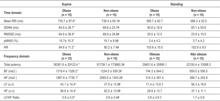
Table 2 shows the mean values and standard deviations of HRV of O and NO children, compared intra-groups and between groups, in the supine and standing position.
Table 1 – Mean values and standard deviations of the
anthropometric and clinical assessment of obese and non-obese children
Variables Obese(N = 15) Non-obese(N = 15) t Test
BMI (kg/m2) 23.9 ± 1 17.7 ± 1.6 0.0001*
SAP (mmHg) 114.6 ± 8.5 112.3 ± 10.1 0.3
DAP (mmHg) 72.8 ± 7.9 71.3 ± 9.1 0.7
HR (bpm) 84.8 ± 11.2 80.2 ± 7.4 0.1
WC (cm) 74.8±4.5 57.8±8.1 0.0001*
Total Cholesterol
(mg/dL) 156.1 ± 25.9 150.0 ± 29.0 0.4
HDL (mg/dl) 49.8 ± 7.7 59.4 ± 9.3 0.04*
LDL (mg/dl) 91.7 ± 29.6 79.1 ± 21.1 0.1
Triglycerides (mg/dl) 107.3 ± 81.1 64.6 ± 25.6 0.01*
*p < 0.05; BMI - body mass index; SAP - systolic arterial pressure; DAP - diastolic arterial pressure; HR - heart rate; WC - waist circumference; HDL - high-density lipoprotein; LDL - low-density lipoprotein.
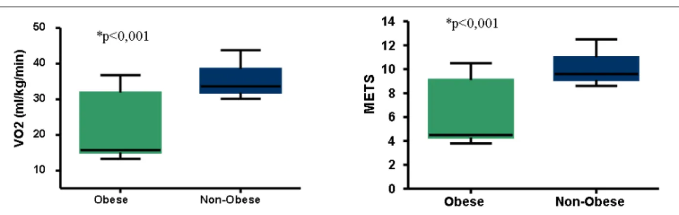
Regarding the variables obtained during the PEP, the data are shown as box plots below (Figure 2 and Figure 3). The NO children presented higher physical capacity determined by the following variables in medians: a) distance achieved at the progressive test, with 1.35 km in NO children when compared to 0.96 km in O children; b) time of exposure to the protocol, respectively, of 15 min and 37 s in NO, versus
Frequency domain (n = 15)Obese Non-obese (n = 15) Obesas(n = 15) Não obesas(n = 15)
Total potency 36381.6 ± 29122.4 * 81739.1 ± 173660.3# 29401.6 ± 35995.1 22150.4 ± 10588.3
BF (ms2 ) 1319.4 ± 1326.2* 1234.5 ± 939.2# 744.3 ± 644.2 659.0 ± 506.5
HF (ms2 ) 1887.6 ± 1730.1* 2065.8 ± 1343.2# 314.3 ± 391.4 388.7 ± 242.8
LF (u.n) 43.1 ± 14.4* 37.9 ± 13.9# 71.4 ± 13.8 † 56.3 ± 18.8
HF (u.n) 56.8 ± 14.4* 62.0 ± 13.9# 28.9 ± 13.7 37.1 ± 11.1
LF/HF Ratio 0.8 ± 0.5* 0.6 ± 0.4# 3.8 ± 3.9 † 1.7 ± 0.9
*p<0,05 - obese supine x obese standing; # p < 0.05 - non-obese supine x non-obese standing; † p<0.05 - obese standing x non-obese standing.
O2/kg/min-1 in NO and 15.7 ml/O 2/kg/min
-1 in O children; d)
metabolic equivalent (MET) value, which was 9.6 for NO and 4.5 for O children.
Discussion
Childhood obesity of sociogenic cause has progressively increased in the last years in practically all countries and it has been observed that among the several factors responsible for that, the association of bad dietary habits and sedentary life style has a predominant role1-3.
In this context, several studies have appeared with different lines of research, although with a single objective, i.e., contribute to a better quality of life of obese individuals. These studies seek to bring new information on the development of mechanisms to improve the prevention and treatment of
Figure 2 -Values of the total distance walked (kilometers) and the time of protocol exposure (seconds) presented by the group of obese (n=15) and non-obese children (n=15) during the performance of the submaximal continuous progressive exercise test. The horizontal line in the center of the box plot represents the median value and the 1st and 3rd quartiles are present, delimitating the size of the box plots. The maximum and minimum values are represented by the vertical
lines, delimitated in their extremities by horizontal lines.
cardiovascular and metabolic diseases caused by obesity. In the present study, important factors regarding the early detection of problems resulting from obesity and potential causes of future diseases, such as the accumulation of fat tissue in the abdominal region and blood lipid levels, HRV and reduced physical capacity were studied.
Measurements of abdominal fat have shown to be excellent risk predictors for cardiovascular disease and type 2 diabetes mellitus, being even more emphasized than total fat and BMI7. It is known, for instance, that the WC values, measured
according to Fernández et al22,have the same assessment
precision for the intra-abdominal and subcutaneous fat as those measurements obtained through magnetic resonance imaging used in children and adolescents.
Figure 3 -Estimated peak oxygen consumption (VO2 peak) values and respective metabolic equivalents (MET) obtained at the moment of peak exertion developed
by the obese (n=15) and non-obese children (n=15) during the performance of the submaximal continuous progressive exercise test. The horizontal line inside the box plot represents the median value and the 1st and 3rd quartiles are present, delimitating the size of the box plots. The maximum and minimum values are
represented by the vertical lines, delimitated in their extremities by horizontal lines.
abdominal fat through the analysis of the WC measurements in O children and we observed that they were higher when compared to those in NO children. Considering that the height was not different between the groups, one can conclude that the fact that the O group presented higher WC values significantly contributed to the increase in the BMI values.
Table 1 also shows blood lipid levels of all volunteers. The concern about such values is based on the fact that the atherosclerotic disease is one of the main causes of morbidity and mortality in adult individuals, although with a tendency to affect increasingly younger individuals in developing countries, without effective prevention programs9. Equally,
the known cause-effect relation between high levels of LDL and atherosclerosis, demonstrated in clinical trials, emphasizes the importance that must be given to such values since childhood23.
According to Willians et al24, childhood obesity is known for
being associated with abnormal blood lipid levels, including total cholesterol levels > 170 mg/dl, high levels of triglycerides and low HDL cholesterol levels.
The levels of triglycerides presented by the children in the O group were significantly higher; however, total cholesterol levels were not different from those observed in the NO group. On the other hand, HDL levels were significantly higher in children from the NO group. In summary, we concluded that: a) both groups presented lipid levels that were compatible with normality; b) LDL levels did not differ between the groups and c) the children from the NO group presented higher HDL and lower triglyceride levels.
Still regarding the values shown in Table 1, we can verify that, although there are studies which show higher values of HR as well as SAP15 and DAP25 at res in O children when
compared to NO children, such fact was not observed in the present study. Perhaps the number of studied children, the methodology used to classify them as obese and the lack of equality between the analyzed age ranges might explain the occurrence of differences in the results among the studies.
Regarding the HRV, it is known that RR interval (RRi) variations present during controlled resting conditions represent a powerful and sensitive method to analyze the
mechanisms of cardiac autonomic control beat by beat26,27.
Therefore, and for being capable of expressing the cardiac sympathetic and parasympathetic action21, the HRV was
analyzed in two body positions that require different autonomic adjustments.
Table 2 shows a significant difference in the comparison of HRV in the two body positions, disclosing that the standing position reduced the parasympathetic tonus and increased the cardiac sympathetic tonus in both groups.
The HRV in the supine position was not different between the groups, which differs from the study carried out by Sekine et al28. In this study, carried out with 7 obese and 9 non-obese
children, aged between 8 and 9 years of age, a significant decrease in the cardiac parasympathetic nervous activity in O children was observed, represented by the decrease in the value of the high frequency component in normalized units (HF n.u.), whereas the values of cardiac sympathetic component (LF n.u.) showed to be significantly higher when compared to NO children.
Other studies with a higher number of volunteers, such as the one by Rabbia et al29, which studied 50 children with a mean
age of 13.9 ± 1.7 years, also showed significantly higher values of the LF component in the obese individuals, when compared to 12 healthy adolescents, thus suggesting that obesity can cause an increase in the cardiac sympathetic tonus.
Riva et al14 suggested that the obese adolescents can present
a sympathovagal dysfunction, characterized by a decrease in the parasympathetic activity and increase in the sympathetic activity, results that are similar to those obtained by Brunetto et al15, who found lower HF (n.u.) and higher LF (n.u.) values
in obese adolescents, when compared to eutrophic ones. The study carried out by Martini et al11, however, using a
long-term recording (24 hours), observed lower values of HF components (n.u.) and of the LF/HF ratio during the 24 hours in the obese individuals. The time domain measurements concerning the vagal activity were also lower.
The study of the HRV in the two functional conditions allowed us to infer that: a) they can be used for clinical investigation on the cardiac autonomic modulation of children in this age range; b) the O children, in spite of having a different autonomic modulation in the standing position, presented HR values that were similar to those observed in the NO group.
The PEP used to evaluate the cardiorespiratory functional capacity (CRFC) of the children was the submaximal progressive type. We decided to use a submaximal protocol to avoid exposing the children to extreme exertion, as studies developed by our group had shown differences in CRFC between O and NO children, even at submaximal exertion intensities25.
All the variables studied during the PEP were significantly higher in the NO group, even when considering that, although both groups had the same number of children, the NO group had one boy less. In this case, in particular, it is important to mention that the literature is clear when stating that boys in the studied age range have a higher VO2 peak or VO2max
than girls32.
Although the children from the O group reached the same HR peak values as the ones from the NO group, that fact happened during a shorter time of exposure to the PEP, showing that their HR increased faster and that the chronotropic reserve was used earlier, consequently resulting
Of the main factors related to childhood obesity and the future development of cardiovascular diseases, this study showed obese children with a significant accumulation of fat tissue in the abdominal region, decreased HDL cholesterol and increased levels of triglycerides, in addition to the presence of a higher cardiac sympathetic tonus in the standing position and significant decrease in the physical capacity documented in a progressive physical exercise protocol.
Acknowledgement
To Dr. Mila Pontes Ramos Cunha, Head of the Ambulatory of Endocrinology of Hospital e Maternidade Celso Pierro of PUC-Campinas, for the analyses of triglycerides and cholesterol.
Potential Conflict of Interest
No potential conflict of interest relevant to this article was reported.
Sources of Funding
There were no external funding sources for this study.
Study Association
This study is not associated with any post-graduation program.
References
1. McArdle WD, Katch FI, Katch VL. Essentials of exercise physiology. Philadelphia - USA. Lea & Febiger; 1994.
2. Marques-Lopes I, Marti A, Moreno-Aliaga MJ, Martinez A. Aspectos genéticos na obesidade. Revista de Nutrição. 2004; 17 (3): 327-38.
3. Giugliano R, Carneiro EC. Fatores associados à obesidade em escolares. J Pediatr. 2004; 80 (1): 17-22.
4. Miller J, Rosembloom A, Silverstein J. Childhood obesity. J Clin Endocrinol Metabol. 2004; 89 (9): 4211-8.
5. Ortega FB, Ruiz JR, Castillo MJ, Sjöström M. Physical fitness in childhood and adolescence: a powerful marker of health. Int J Obesity. 2008; 32: 1-11.
6. Berenson GS, Srinivasan SR, Bao W, Newman III WP, Tracy RE, Wattigney WA. Association between multiple cardiovascular risk factor and atherosclerosis in children and young adults. The Bogalusa Heart Study. N Eng J Med. 1998; 338: 1650-6.
7. Kuk JL, Katzmarzyk PT, Nichaman MZ, Church TS, Blair SN, Ross R. Visceral fat is an independent predictor of all-cause mortality in men. Obes Res. 2006; 14: 336-41.
8. Brambilla P, Bedogni G, Moreno LA, Goran MI, Gutin B, Fox KR, et al. Crossvalidation of anthropometry against magnetic resonance imaging for the assessment of visceral and subcutaneous adipose tissue in children. Int J Obes (Lond). 2006; 30: 23-30.
9. Giuliano ICB, Coutinho MSSA, Freitas SFT, Pires MMS, Zunino JN, Ribeiro RQC. Lípides séricos em crianças e adolescentes de Florianópolis, SC: estudo floripa saudável, 2040. Arq Bras Cardiol. 2005; 85: 85-91.
10. Ortega FB, Tresaco B, Ruiz JR, Moreno LA, Martin-Matillas M, Mesa JL, et al. Cardiorespiratory fitness and sedentary activities are associated with adiposity in adolescents. Obesity. 2007; 15: 1589-99.
variability in childhood obesity. Clin Auton Res. 2001; 11 (2): 87-91.
12. Faulkner MS, Hathaway D, Tolley B. Cardiovascular autonomic function in healthy adolescents. Heart Lung. 2003; 32 (1): 10-22.
13. Nagai N, Moritani T. Effect of physical activity on autonomic nervous system function in lean and obese children. Int J Obes Relat Metab Disord. 2004; 28 (1): 27-33.
14. Riva P, Martini G, Rabbia F, Milan A, Paglieri C, Chiandussi L, et al. Obesity and autonomic function in adolescence. Clin Exp Hypertens. 2001; 23 (1-2): 57-67.
15. Brunetto AF, Roseguini BT, Silva BM, Hirai DM, Guedes DP. Respostas autonômicas cardíacas à manobra de tilt em adolescentes obesos. Rev Assoc Med Bras. 2005; 51 (5): 256-60.
16. Montano N. Heart rate variability as a clinical tool. Ital Heart J. 2002; 3: 439-45.
17. Zahorska-Markiewicz B, Kuagowsa E, Kucio C, Klin M. Heart rate variability in obesity. Int J Obes Relat Metab Disord. 1993; 17: 21-3.
18. Centers for Disease Control and Prevention 2000 growth charts for the United States: improvements to the 1977 National Center for Health Statistics version. Pediatrics. 2002; 109 (1): 45-60.
19. Friedewald WT, Levy RI, Fredrikson DS. Estimation of the concentration of low-density lipoprotein cholesterol in plasma, without use of the preparative ultracentrifuge. Clin Chem. 1972; 18: 499-502.
20. Paschoal MA, Volanti VM, Pires CS, Fernandes FC. Heart rate variability in different age groups. Rev Bras Fisioter. 2006; 10 (4): 413-9.
21. European Society of Cardiology and the North American Society of Pacing and Electrophysiology. Heart rate variability – standards of measurement, physiological interpretation, and clinical use task force. Circulation. 1996; 93 (5): 1043-65.
22. Fernández JR, Redden DT, Pietrobelli A, Allison DB. Waist circumference percentiles in nationally representative samples of African-American, European-American, and Mexican-American children and adolscents. J Pediatr. 2004; 145:
439-44.
23. Levi F, Lucchini F, Negri E, La Vecchia C. Trends in mortality from cardiovascular and cerebrovascular diseases in Europe and other areas of the world. Heart. 2002; 88: 119-24.
24. Williams DP, Going SB, Lohman TG, Harsha DW, Srinivasan SR, Webber LS, et al. Body fatness and risk for elevated blood pressure, total cholesterol, and serum lipoprotein ratios in children and adolescents. Am J Public Health. 1992; 82: 358-63.
25. Petrelluzzi KFS, Kawamura M, Paschoal MA. Avaliação funcional cardiovascular de crianças sedentárias obesas e não obesas. Rev Cienc Med (Campinas). 2004; 13 (2): 127-36.
26. Akserold S, Gordon D, Madwed JB, Snidman NC, Shannon DC, Cohen RJ. Hemodynamic regulation: investigation by spectral analysis. Am J Physiol. 1985; 249: H867-H875.
27. Saul JP, Rea RF, Eckberg DL, Berger RD, Cohen RJ. Heart rate and muscle sympathetic nerve variability during reflex changes of autonomic activity. Am J Physiol. 1990; 258: H713-H721.
28. Sekine M, Izumi I, Yamagami T, Kagamimori S. Obesity and cardiac autonomic nerve activity in healthy children: results of the Toyama birth cohort study. Env Health and Prevent Med. 2001; 6: 149-53.
29. Rabbia F, Silke B, Conterno A, Grosso T, De Vito B, Rabbone I, et al. Assessment of cardiac autonomic modulation during adolescent obesity. Obes Res. 2003; 11: 541-8.
30. Montano N, Ruscone TG, Porta A, Lombardi F, Pagani M, Malliani A. Power spectrum analysis of heart rate variability to assess the changes in sympathovagal balance during graded orthostatic tilt. Circulation. 1994; 90 (4): 1826-31.
31. Streeten DHP. Variations in clinical manifestation of orthostatic hypotension. Mayo Clin Proc. 1995; 70: 713-4.