Periodontal Disease as a Potential Risk Factor for Acute Coronary
Syndromes
Renata Accarini and Moacir Fernandes de Godoy
Hospital de Base da Fundação Faculdade Regional de Medicina (FUNFARME), Faculdade de Medicina de São José do Rio Preto (FAMERP), Curso de Pós-Graduação em Ciências da Saúde da FAMERP, São José do Rio Preto, SP, Brazil
objective: To investigate whether there is a relationship between active periodontal disease (PD) and acute coronary syndromes (ACS).
Methods: Three hundred and sixty-one patients (57,3% male), ages ranging from 27 to 89 (mean ± DP = 60.5 ± 12.2), were admitted to the Intensive Care Unit of a Teaching Hospital with symptoms and complementary examinations consistent with acute coronary syndrome. All the patients had a complete periodontal examination in the ICU setting, and 325 (90.9%) underwent coronary angiography for diagnostic confirmation and/or treatment planning. Periodontal examination included evaluation of all the teeth in the oral cavity and the following parameters: probing depth, clinical attachment level, plaque index, and gingival index.
Results: Of the 325 patients, 91 (28%) had coronary arteries free of obstruction or with mild obstructions (≤ 50% diameter
stenosis), and the remaining 72% had severe obstructions. Fisher’s exact text yielded a p value of 0.0245 with an odds ratio of 2.571 (95% CI 1.192 to 5.547), meaning that the group with ACS and significant obstructive coronary artery disease was 2.5-fold more likely to have active PD.
Conclusion: A significant association was found between active periodontal disease and severe obstructive coronary artery in patients with acute coronary syndrome, underscoring the importance of prevention and adequate treatment of periodontal disease, which should be considered as a potential risk factor in the etiology and instability of the atherosclerotic plaque.
key words: Periodontal disease, acute coronary syndrome, obstructive coronary artery disease, coronary atherosclerosis, inflammation, risk factor.
Mailing Address: Moacir fernandes de godoy •
Rua Garabed Karabashian, 570 - 15070-600 – São José do Rio Preto, SP, Brazil
In Western countries, cardiovascular disease (CVD) accounts for the majority of deaths, but the so-called classic risk factors do not explain 30% of them, particularly among young adults. Recently, the possible association between oral health (especially the extent and nature of periodontal disease) and cardiovascular diseases has been the focus of several epidemiological and laboratory studies. Although results suggest a positive association, controversies still remain, especially regarding the causal relationship and pathophysiological mechanisms that would explain this association.
Periodontal disease (PD) is moderate in 44% to 57% of adults, 10% of whom, in developed countries, have advanced periodontitis1, most often asymptomatic. Periodontal disease is
caused by anaerobic and Gram-negative bacterial colonization, such as Porphyromonas gingivalis and Actinobacillus actinomycetemcomitans.
A number of cohort studies have identified PD as an important predictor of future cardiovascular events2-7. More
recently, other studies focused on demonstrating that genetic factors influence biological processes involved in both diseases, representing a potential mechanism that may link PD to CVD8-13.
To establish a causal association, several studies have detected the presence of periodontopathogens in atherosclerotic plaques14-16.
Since it has been proved that bacteremias may be asymptomatic and occur on a routine basis, some investigators sought a model for the pathophysiological relationship in which poor periodontal health would affect systemic conditions. In this case, they found positive association between dental plaque and systemic neutrophil inflammatory response17 and
endotoxin release, both responses induced by mastication18.
Notably, studies along the same lines have demonstrated that both the presence of PD and its treatment affect cytokine production, especially acute phase proteins19-29.
3.00 Software using Fisher’s exact test and odds ratio (OR), with 95% confidence interval (95% CI). Alpha error was set at 0.05, and p values ≤ 0.05 were considered significant.
Results
Of the 167 patients who had at least six teeth for assessment, 154 (92.2%) underwent cardiac catheterization with coronary angiography during admission, so that the diagnostic of coronary event or risk stratification could be elucidated. In this group, the association between active periodontal disease and severe obstructive coronary disease was evaluated. Of these 154 patients, 49 failed to meet the diagnostic criteria for periodontal disease as established by the Method, but 105 patients did.
Among the 194 edentulous patients, 171 (88.1%) underwent cardiac catheterization during admission. Obviously, this group did not experience periodontal disease, simply because they had no teeth.
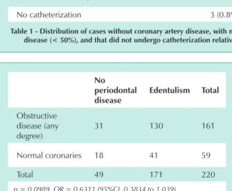
Table 1 shows the distribution of patients in each group. The significant number of edentulous patients (53.7%) may be justified by the fact the hospital where data were collected receives subjects from several regions in the interior of São Paulo state, and many of them belong to lower socioeconomic classes; hence, they are more likely to have poorer oral conditions.
On the other hand, for this very reason, these edentulous patients would be “protected” against active periodontal disease, and according the hypothesis of the study, were expected to have an intermediate behavior between both groups. Thus, among the 49 patients free of periodontal disease and who underwent cardiac catheterization, 28 (57.1%) had severe obstructive coronary disease. Among the 171 edentulous patients who underwent cardiac catheterization, the rate of severe obstructive coronary disease was 73.7% (126 cases), while among the 105 with active periodontal disease, this rate was 76.2% (80 cases). Taking into account the existence of obstructive coronary disease of any degree, these values were 63.3%, 76.0%, and 80.9%, respectively.
Statistical analysis using Fisher’s test yielded a p value of 0.0989 and odds ratio of 0.6311, with a 95% CI ranging from 0.3834 and 1.039 for absence of periodontal disease or edentulism when comparing groups with any degree of coronary disease versus absence of coronary disease (Tab. 2).
Also regarding the presence of active periodontal disease or edentulism, no significant difference was found when groups with coronary disease of any degree versus absence of coronary disease were compared, and Fisher’s test showed a p value of 0.3726 with an odds ratio of 1.340 and a 95% CI from 0.735 to 2.444 (Tab. 3).
Statistical analysis using Fisher’s exact test indicated a p value of 0.0245 (significant) and an odds ratio of 2.571 with a 95% CI ranging from 1.192 to 5.547 for the presence or absence of periodontal disease, when groups of patients with severe obstructive coronary disease (≥ 50%) versus no coronary artery disease were compared (Tab. 4). According to these data, patients with severe obstructive coronary disease are 2.5 times more likely to have periodontal disease than that S sanguis and P gingivalis cause thrombus formation and
platelet aggregation, in addition to ST-segment changes on ECG, which is a dose-dependent response, suggesting that they may trigger cardiac ischemic episodes30-32.
Considering this, the present study was designed to assess the possible association between active periodontal disease and the degree of obstruction in patients with acute coronary syndrome (ACS).
Methods
Three hundred and sixty-one patients with acute coronary syndrome, that is, unstable angina or acute myocardial infarction (with or without ST-segment elevation) admitted to the Intensive Care Unit of a Teaching Hospital in the interior of São Paulo were evaluated. This diagnosis was based both on patients clinical condition and complementary examinations (electrocardiogram and enzyme measurement). Patients whose clinical condition precluded adequate oral examination, such as those with coagulopathies and those with history of or greater risk for infective endocarditis, were excluded. All the others agreed to participate in the study and signed an informed consent. The study received Institutional Ethics Committee approval (File No.5124/2001; Opinion No. 083/2001). Patients underwent specific odontological anamnesis and intraoral clinical examination by a single periodontist in the ICU during admission, in order to further clarify the condition suggestive of acute coronary failure.
T h e f o l l o w i n g p a r a m e t e r s w e r e u s e d f o r P D classification:
-Clinical probing depth or pocket depth (CPD)33.
-Clinical attachment level (CAL)33.
-Bleeding index (BI)34.
-Plaque index (PI)35.
CAL and CPD were assessed using a standard manual probe calibrated in millimeters, in both arches, examining all the teeth on their surfaces: mesial buccal, buccal, distal buccal, mesial lingual, distal lingual, and lingual. Periodontal probing was performed moving the probe through these regions parallel to the long axis of the tooth at its free surfaces. All probing measurements were rounded to the nearest whole millimeter.
Periodontal disease was classified according to the following criteria: at least six teeth for assessment and more than 30% of sites whose CPD and/or CAL exceeded 5 mm or more than 30% of sites with CPD or CAL between 0 and 4 mm, but with ≥ 50% of bleeding on probing (BOP) at six sites per tooth: mesial buccal, buccal, distal buccal, mesial lingual, mid-lingual, and distal lingual.
Of the patients with ACS, 207 (57.3%) were male. Ages ranged from 27 to 89, with mean ± standard deviation and median of age 60.5 ±12.2 years and age 60, respectively. As for the number of teeth, 194 (53.7%) were edentulous (< 6 teeth) and 167 (46.3%) had between 6 and 28 teeth. A total of 2269 teeth were assessed, corresponding to 13,524 surfaces.
those with normal coronaries. The inclusion of the subgroup with mild obstructive coronary disease (less than 50% of luminal stenosis) does not change the results, now with a p value of 0.0265 and an odds ratio of 2.468 with a 95% CI ranging from 1.156 to 5.267 for the presence or absence of periodontal disease when comparing groups of patients with any degree of coronary disease versus normal coronaries. The approximate 2.5-fold likelihood (odds) of concurrent active periodontal disease and any degree of coronary disease was maintained, compared with normal coronaries (Tab. 5).
Discussion
One of the great difficulties in comparing different studies about this subject is the lack of a consistent and consensual classification for periodontal disease. Based on published data, it is difficult to reach a conclusion whether there is or not an association between PD and CVD.
We made a point of evaluating all teeth to prevent any bias in data collection. Additionally, our classification sought to aggregate all periodontal parameters simultaneously, namely, bleeding as clinical evidence of gingival inflammation; plaque index as an indicator of infective burden; probing depth, since periodontal pockets are a reservoir for microorganisms with direct access to the connective tissue and circulatory system; and clinical attachment level, because periodontal recession is the record of past history of PD and its remissions.
In this study, the presence of obstructive coronary artery disease in ACS patients was significantly associated with active periodontal disease. The most promising hypothesis to
explain this association may lie in the analysis of inflammatory markers characteristic or predictive of cardiac ischemic events. Analyzing these studies, in which inflammatory markers decrease when the subject receives periodontal treatment, or even comparing these markers in different groups of subjects with and without PD, one may consider periodontal interventions as adjuvant for preventing cardiac events19-29.
Our findings suggest that the ability of periodontopathogenic bacteria to invade periodontal tissues may offer a basis for such association. Another explanation that would validate the interrelationship between PD and CVD are gene polymorphisms, including frequency of different interleukin-1 (IL-1) and interleukin-6 (IL-6) genotypes, both of which are present in both the progression of PD and CVD. Accordingly, the patient would be hyper-responsive to harmful stimuli in the periodontal tissues and vascular endothelium. It is interesting to note that, despite the polymorphism, after periodontal treatment these patients show a significant decrease in serum C-reactive protein levels36.
In the present study, inflammatory markers were not measured. Czerniuk et al37 studied 50 consecutive patients
with chronic periodontitis (41males) admitted to the Coronary Care Unit with clinical diagnosis of ACS who underwent serial laboratory measurements, in some cases up to six months after the acute event.Although no significant differences were detected in serum tumor necrosis factor (TNFα) or IL-1 in ACS patients with advanced periodontal disease, as compared with those with a less advanced stage of the disease, the first tended to be characterized by higher mean levels of IL-1 in early and later phases of ACS, in addition to higher mean
no periodontal
disease periodontal disease Edentulous
No periodontal disease 18 (5.0%) 20 (5.5%) 41 (11.3%) At least one coronary obstruction, always < 50% 3 (0.8%) 5 (1.4%) 4 (1.1%)
At least one coronary obstruction, ≥ 50% 28 (7.7%) 80 (22.2%) 126 (34.9%) No catheterization 3 (0.8%) 10 (2.8%) 23 (6.4%)
table 1 - distribution of cases without coronary artery disease, with mild obstructive coronary disease (< 50%), with severe obstructive coronary disease (< 50%), and that did not undergo catheterization relative to the presence or not of periodontal disease or edentulous condition
no periodontal disease
Edentulism total
Obstructive disease (any degree)
31 130 161
Normal coronaries 18 41 59
Total 49 171 220
p = 0.0989 OR = 0.6311 (95%CI 0.3834 to 1,039)
table 2 - distribution of cases in groups with obstructive coronary disease of any degree and no coronary disease relative to the
absence of current periodontal disease or edentulism
periodontal
disease Edentulism total
Obstructive disease (any degree)
85 130 215
Normal
coronaries 20 41 61
Total 105 171 276
p = 0.3726 OR = 1.340 (95% CI 0.735 to 2.444)
table 3 - distribution of cases in groups with obstructive coronary disease of any degree and no coronary disease relative to the
levels of TNFα in later phases. Furthermore, in patients with less advanced periodontal inflammatory response decreased more rapidly compared to groups with more advanced periodontal disease.
The statistically significant association between obstructive coronary disease and active periodontal disease strongly suggests that periodontal disease be considered among the risk factors for the development of obstructive coronary disease. Therefore, considering the inflammatory aspects involved, it would represent a potential risk factor in the etiology and atherosclerotic plaque instability and cause
acute coronary syndrome.
Studies that are prospective, controlled and have a larger number of patients are needed to further elucidate this issue.
Acknowledgment
The authors would like to thank the Fundação de Amparo à Pesquisa do Estado de São Paulo (FAPESP) for funding this research (Case No. 01/03808-1).
References
periodontal disease
no periodontal disease
total
Obstructive
disease (≥ 50%) 80 28 108 Normal
coronaries 20 18 38
Total 100 46 146
p = 0.0245 OR = 2.571 (95% CI 1.192 to 5.547)
table 4 - distribution of cases in groups with severe obstructive coronary disease (> 50%) and no coronary disease relative to the
presence or not of periodontal disease
periodontal disease
no periodontal disease
total
Periodontal disease (any
degree) 85
31 116
Normal
coronaries 20 18 38
Total 105 49 154
p = 0.0265 OR = 2.468 (95% CI 1.156 to 5.267)
table 5 - distribution of cases in groups with obstructive coronary disease of any degree and no coronary disease relative to the
presence or not of periodontal disease
1. Brown LJ, Brunelle JA, Kingman A. Periodontal status in the United States, 1988-1991: Prevalence, extent and demographic variation. J Dent Res 1996;75:672-83.
2. DeStefano F, Anda RF, Kahn HS, Williamson DF, Russel CM. Dental disease and risk of coronary heart disease and mortality. Br Med J 1993;306:688-91.
3. Matilla KJ, Valtonen VV, Nieminen M, Hattunen JK. Dental infection and the risk of new coronary events: prospective study of patients with documented coronary artery disease. Clin Infect Dis 1995; 20:588-92.
4. Morrison HI, Ellison LF, Taylor GW. Periodontal disease and risk of fatal coronary heart and cerebrovascular diseases. J Cardiovasc Risk 1999;6:7-11.
5. Genco R, Chadda S, Grossi S. Periodontal disease is a predictor of cardiovascular disease in a native American population. J Dent Res 1997;76:408.
6. Beck J, Garcia R, Heiss G, Vokona PS, Offenbacher S. Periodontal disease and cardiovascular disease. J Periodontol 1996;67(Suppl.):1123-37.
7. Wu T, Trevisan M, Genco RJ, Dorn JP, Falkner KL, Sempos CT. Periodontal disease and risk of cerebrovascular disease. NHANES I. Arch Intern Med 2000;160:2749-55.
8. Kornman KS, Pankow J, Offenbacher S, Beck J, Di Giovine F, Duff GW. Interleukin-1 genotypes and the association between periodontitis and cardiovascular disease. J Periodont Res 1999;34:353-7.
9. Kornman KS, Duff G W. Candidate genes as potential links between periodontal and cardiovascular diseases. J Periodont 2001;6:48-57.
10. Gibson FC, Hong C, Chou H-H, Yumoto H, Chen J, Lien E, et al. Innate immune recognition of invasive bacteria accelerates atherosclerosis in Apolipoprotein E-deficient mice. Circulation 2004;109:2801-06.
11. Li L MD, Messas E, Batista EL, Levine RA, Amar S. Porphyromonas gingivalis infection accelerates the progression of atherosclerosis in a heterozygous apolipoprotein E-deficient murine model. Circulation 2002;105:861-7.
12. Chung HJ, Champagne CME, Southerland JH, et al. Effects of P gingivalis infection on atheroma formation in APoE (±) mice. J Dent Res 2000; 79 (Spec.Issue): 313-(Abstr.1358).
13. Lalla E, Lamster IB, Spessor AL, et al. Oral infection with a periodontal pathogen accelerates atherosclerosis in APoE null mice. Circulation 2000;102(suppl. II):II41-II-42(Abstr.188).
14. Stelzel M, Conrads G, Pankuweit S, Maisch B, Vogt S, Moodsdorf R, et al. Detection of Porphyromonas gingivalis DNA in aortic tissue by PCR. J Periodontol 2002;73:868-70.
15. Okuda K, Ishihara K, Nakagawa T, Hirayama A, Inayama Y. Detection of Treponema denticola in atherosclerotic lesions. J Clin Microbiol 2001; 39:1114-17.
16. Haraszthy VI, Zambon JJ, Trevisan M, Zeid M, Genco RJ. Identification of periodontal pathogens in atheromatous plaques. J Periodontol 2000;71:1554-60.
17. Kowolik MJ, Dowsett SA, Rodrigues J, De La Rosa M, Eckert G. Systemic neutrophil response resulting from dental plaque accumulation. J Periodont 2001;72:146-51.
18. Geerts S O, Nys M, de Mol P, Charpentier J, Albert A, Legrand V, et al. Systemic release of endotoxins induced by gentle mastication: association with periodontitis severity. J Periodont 2002;73:73-8.
20 Ebersole JL, Capelli D, Mathys EC, Steffen MJ, Singer RE, Montgomery M, et al. Periodontitis in humans and non-human primates: oral-systemic linkage inducing acute phase proteins. Ann Periodontol 2002;7:102-11.
21. Matilla K, Versanen M, Valtonen V, Nieminem M, Palosuo T, Rasi V, et al. Effect of treating periodontitis on C-reactive protein levels: a pilot study. BMC Infect Dis 2002;2:30.
22. Slade GD, Ghezzi EM, Heiss G, Beck JD, Riche E, Offenbacher S. Relationship between periodontal disease and C-reactive protein among adults in the atherosclerosis risk in community study. Arch Intern Med 2003;163:1172-79.
23. Meurman JH, Janket S, Qvarnström M, Nuutinen P. Dental infections and serum inflammatory markers in patients with and without severe heart disease. Oral Surg Oral Med Oral Pathol Oral Radiol Endod 2003;96:695-700.
24. D’Aiuto F, Parkar M, Andreou G, Suvan J, Brett PM, Ready R, et al. Periodontitis and systemic inflammation: control of the local infection is associated with a reduction in serum inflammatory markers. J Dent Res 2004;83(2):156-60.
25. D’Aiuto F, Ready D, Tonetti MS. Periodontol disease and C-reactive protein-associated cardiovascular risk. J Periodontol Res 2004;39:236-41.
26. Montebugnoli L, Servidio D, Miaton RA, Prati C, Tricoci P, Melooni C. Poor oral health is associated with coronary heart disease and elevated systemic inflammatory and haemostatic factors, J Clin Periodontol 2004;31:25-9.
27. Deliagyris EN, Madianos PN, Kadoma W, Marron I, Smith S, Beck JD, et al. Periodontal disease in patients with acute myocardial infarction: Prevalence and contribution to elevated C-reactive proteins levels. Am Heart J 2004;147:1005-9.
28. Nakamura T, Nitta H, Ishikawa I. Effect of low dose Actinobacillus actinomycetemcomitans lipopolysaccharide pretreatment on cytokine production by human whole blood. J Periodontol 2004;39:129-35.
29. Joshipura KJ, Wand HC, Merchant AT, Rimm EB. Periodontal disease and biomarkers related to cardiovascular disease. J Dent Res 2004;83(2):151-5.
30. Meyer MW, Gong K, Herzberg MC, Streptococcus sanguis –induced platelet clotting in rabbits and hemodynamic and cardiopulmonary consequences. Infect Immun 1998;66:5906-14.
31. Herzberg MC, Meyer MW. Dental plaque, platelets and cardiovascular diseases. Ann Periodontol 1998;3:151-60.
32. Herzberg MC. Coagulation and thrombosis in cardiovascular disease: plausible contributions of infectious agents. Ann Periodontol 2001;6:16-9.
33. Papanou NP, Lindhe J. Epidemiologia da Doença Periodontal. In : Lindhe J , Karring T, Lang NP, editores . Tratado de Periodontia Clínica e Implantologia Oral . 3º ed. Rio de Janeiro ( RJ) : Guanabara-Koogan, 1999.
34. Ainamo J, Bay I. Problems and Proposals for recording gingivitis and plaque. Int Dental J. 1975; 25(4):229-35.
35. Löe H. The gingival index, the plaque index and the retention index systems. J Periodont 1964;38:610-67.
36. DÁiuto F, Parkar M, Andreou G, Brett PM, Ready D, Tonetti MS. Periodontitis and atherogenesis: causal association or simple coincidence? J Clin Periodontol 2004;31:402-11.