Ischemic left ventricle systolic dysfunction: An evidence-based
approach in diagnostic tools and therapeutics
EDUARDO GOMES LIMA1*, FELIPE PEREIRA CÂMARADE CARVALHO2, JAIME PAULA PESSOA LINHARES FILHO2, FABIO GRUNSPUN PITTA2,
CARLOS VICENTE SERRANO JR1
1MD, PhD, Department of Atherosclerosis, Instituto do Coração do Hospital das Clínicas da Faculdade de Medicina da Universidade de São Paulo (InCor-HC-FMUSP), São Paulo, SP, Brazil 2MD, Department of Atherosclerosis, InCor-HC-FMUSP, São Paulo, SP, Brazil
S
UMMARYStudy conducted at the Department
of Atherosclerosis, Instituto do Coração do Hospital das Clínicas da Faculdade de
Medicina da Universidade de São Paulo (InCor-HC-FMUSP), São Paulo, SP, Brazil
Article received: 3/13/2017
Accepted for publication: 4/3/2017
*Correspondence: Address: Av. Dr. Enéas de Carvalho Aguiar,
44, 2º andar, sala 2 São Paulo, SP – Brazil
Postal code: 05403-000 [email protected]
http://dx.doi.org/10.1590/1806-9282.63.09.793
Funding: There is no relationship
with industry. Funding was provided by the Zerbini Foundation.
Coronary artery disease (CAD) associated with left ventricular systolic dysfunction is a condition related to poor prognosis. There is a lack of robust evidence in many aspects related to this condition, from definition to treatment. Ischemic cardiomyopathy is a spectrum ranging from stunned myocardium associated with myocardial fibrosis to hibernating myocardium and repetitive episodes of ischemia. In clinical practice, relevance lies in identifying the myocardium that has the ability to recover its contractile reserve after revascularization. Methods to evaluate cellular integrity tend to have higher sensitivity, while the ones assessing contractile reserve have greater specificity, since a larger mass of viable myocytes is required in order to generate contractility change. Since there are many methods and different ways to detect viability, sensitivity and specificity vary widely. Dobutamine-cardiac magnetic resonance with late gadolinium enhancement has the best accuracy is this setting, giving important predictors of prognostic and revascularization benefit such as scar burden, contractile reserve and end-systolic volume index. The latter has shown differential benefit with revascularization in some recent trials. Finally, authors discuss interventional procedures in this population, focusing on coronary artery bypass grafting and evolution of evidence from CASS to post-STICH era.
Keywords: coronary artery disease, heart failure, coronary artery bypass graft.
I
NTRODUCTIONApproximately 5.1 million persons in the United States have clinically manifest heart failure (HF),1 while the
lifetime risk of developing HF is 20% for Americans ≥ 40 years of age.2 Heart failure with reduced ejection fraction
(HF-REF) and preserved ejection fraction each make up about half of the overall HF burden.3 The most common
etiology of HF-REF in the developed world is ischemic heart disease, which is associated with more than 60% of diagnoses.4 Coronary artery disease (CAD) associated with
left ventricular systolic dysfunction is a condition related to poor prognosis.5,6 Despite the fact that this condition
has been studied for over 30 years, there is a lack of robust evidence in many aspects related to this condition, from definition to treatment.
D
EFINITIONSAND CONCEPTSSince the CASS trial,7 we know that the left ventricle
ejec-tion fracejec-tion impacts on CAD prognosis and treatment. However, a correct definition for “ischemic myocardi-opathy” has not been established in all these years. The concept that this condition is an association between significant coronary obstruction and systolic dysfunction of the left ventricle is too simplistic and disregards im-portant pathophysiologic and causative mechanisms. Even LVEF cut-off values considered as dysfunctional vary among different trials8 and need consensus.
lies in identifying the myocardium that has the ability to recover its contractile reserve after revascularization.
The concept of hibernating myocardium is often con-fused with viable myocardium. Currently, the term “viable myocardium” has a prospective aspect; it is the one that has potential recovery following re-established coronary flow. On the other hand, “hibernating myocardium” can only be used retrospectively, representing the myocardial contractile reserve that recovered after revascularization.9
Ventricular dysfunction in chronic coronary artery disease is known to be an important prognostic predictor. Discrepancy between loss of left ventricular function caused by an infarct (necrosis/fibrosis) and that resulting from potentially reversible chronic ischemic insult (hibernating myocardium) may have relevant clinical implications.
Several studies have shown contractile recovery after restored blood flow, in both the acute and chronic settings. Kim et al.,10 using cardiac magnetic resonance imaging
(MRI) before and after revascularization documented contractile recovery in dysfunctional myocardial segments in patients with chronic CAD.
Association between myocardial viability and favor-able clinical outcomes has been suggested in several studies. In a meta-analysis of 24 studies, Allman et al.1
demonstrated that the presence of viability is correlated with reduction in mortality when these patients under-went revascularization. While in the absence of viability, there was no difference regarding mortality depending on the treatment performed (revascularization or med-ical treatment).
There are several non-invasive methods of myocar-dial viability identification, which is based on three pa-rameters: metabolism evaluation and cell integrity, pres-ence of tissue non-viable by determining the extent of fibrosis and/or necrosis, and, finally, evaluation of con-tractile reserve after inotropic stimulation.11
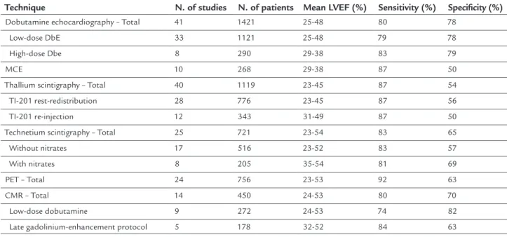
Methods to evaluate cellular integrity tend to have higher sensitivity (single-photon emission computed tomography – SPECT, positron emission tomography – PET and myocardial contrast echocardiography – MCE), while those assessing contractile reserve (dobutamine stress echocardiography – DbE and dobutamine stress cardiac magnetic resonance – DbCMR) have greater spec-ificity, since a larger mass of viable myocytes is required to generate a contractility change. Since there are many methods and different ways of viability detection, sensitiv-ity and specificsensitiv-ity vary widely (Table 1).
The use of complementary test examinations for vi-ability assessment, therefore, may provide crucial informa-tion for the identificainforma-tion of patients who could possibly benefit from the indication of myocardial revascularization.
D
IAGNOSIS SPECTThis method evaluates technetium-99 or thallium-201 radioisotope uptake by viable myocytes, which depends on cellular and mitochondrial integrity. Both protocols have good sensitivity to predict contractile recovery after revascularization (thallium-201, 87%, versus tech-netium-99, 83%). However, in both cases, specificity is
TABLE 1 Comparison of imaging techniques for viable myocardium assessment.21
Technique N. of studies N. of patients Mean LVEF (%) Sensitivity (%) Speciicity (%) Dobutamine echocardiography – Total 41 1421 25-48 80 78
Low-dose DbE 33 1121 25-48 79 78
High-dose Dbe 8 290 29-38 83 79
MCE 10 268 29-38 87 50
Thallium scintigraphy – Total 40 1119 23-45 87 54
TI-201 rest-redistribution 28 776 23-45 87 56
TI-201 re-injection 12 343 31-49 87 50
Technetium scintigraphy – Total 25 721 23-54 83 65
Without nitrates 17 516 23-52 83 57
With nitrates 8 205 35-54 81 69
PET – Total 24 756 23-53 92 63
CMR – Total 14 450 24-53 80 70
Low-dose dobutamine 9 272 24-53 74 82
below other available methods (thallium-201, 54%, tech-netium-99, 65%).12
Although widespread and available in most centers, low spatial resolution and exposure to radiation can limit its usefulness.
PET
PET viability evaluation is becoming increasingly common in clinical practice. The combination of a tracer for eval-uation of blood flow and fluorine-18 fluorodeoxyglucose (18F-FDG) to detect cellular metabolism proved to be a promising approach in viability assessment.
The method can provide four result patterns, and the three main ones related to ischemic cardiomyopathy are: low blood flow with preserved metabolism (mismatch compatible with hibernating myocardium), decrease in both blood flow and metabolism (match compatible with fibrosis/necrosis), flow and metabolism preserved (normal tissue).12
Studies have shown that the use of PET in viability assessment has good sensitivity, about 92%, but with mod-erate specificity of 63%.13 It has better spatial resolution
and less radiation exposure compared with SPECT, but is still an expensive test, little available and has limited util-ity in diabetic patients, especially in type 1, which depends on the sensitivity of the glucose transporters.
Echocardiography
The use of echocardiography in the myocardial viability approach is based on three parameters: wall thickness, contrast enhancement by myocytes, and contractile reserve with inotropic stimulation.
The decrease in ventricular wall thickness (end-diastol-ic wall th(end-diastol-ickness < 6 mm), since it is associated with loss of tissue due to fibrosis/necrosis, showed a high negative pre-dictive value for contractile recovery after revascularization.14
In recent years, the use of contrast echocardiography (MCE) has increased. Contrast enhancement assesses myo-cardial perfusion and, subsequently, cellular integrity.
The evaluation of contractile reserve by dobutamine stress, however, was further studied. The dysfunctional segment at rest, after inotropic stimulation, presents con-tractile recovery. Low dose dobutamine (5-10 mcg/kg) is enough to assess the contractile reserve. After an initial improvement, contractility worsens at higher doses of dobutamine (20 mcg/kg), which is the so called “biphasic response,” highly suggestive of viable myocardium.15
Despite presenting good sensitivity (80%) and specific-ity (78%), this method has limitations, such as poor acous-tic window and being an operator-dependent technique.13
Cardiac MRI
Cardiac magnetic resonance has been gaining importance in ischemic myocardiopathy. Good spatial resolution, lack of exposure to radiation and acoustic window indepen-dence are advantages of resonance compared to other methods such as echocardiography and SPECT.16
Viability assessment by resonance is based on three main parameters: end-diastolic wall thickness (EDWT), low-dose dobutamine inotropic stimulation and late gadolinium enhancement (LGE) imaging.
The evaluation of EDWT constitutes the measure of maximum thickness of myocardial wall at rest. In com-parison with PET (FDG uptake), Baer et al.17
demon-strated that a measure ≥ 5.5 mm was associated with vi-ability. In turn, thicknesses < 5.5 mm had low uptake in PET, representing low probability of viability.
The low-dose dobutamine stress resonance (≤ 10 mg/kg per minute) has proven a useful tool in clinical practice. The inotropic stimulation promotes an improvement of myocardial contractility in viable segments, which was associated with increased likelihood of contractile recovery after revascularization.18
The gold standard technique for viability assessment is LGE. It relies on a greater distribution of gadolinium in the extracellular space (i.e. in the areas of fibrosis/ne-crosis), resulting in delayed washout. The transmural extension of scars showed correlation with the potential contractile recovery, described by Kim et al.10 Infarcted
areas are < 50% more likely to functional improvement after revascularization, while those > 50% are associated with poorer outcomes.19
In a meta-analysis of 24 studies, Romero et al. compared these three techniques. The use of dobutamine stress showed better specificity and positive predictive value, while LGE was associated with better sensitivity and negative predictive value.20As a result, the best approach to select
patients eligible for revascularization might be the use of two techniques combined. Some authors propose the ini-tial realization of LGE, and areas of infarction between 50-75% would then be evaluated with inotropic dobutamine stress. Improved contractile function would help in predict-ing viable areas. Scars < 50% and > 75% have a high and low probability of functional recovery, respectively.21
Although many studies point to the benefit of reso-nance and described tests in the management of ischemic cardiomyopathy, the lack of randomized controlled trials with hard clinical endpoints has not yet established the routine use of these methods in clinical practice.
and observational studies. However, prospective trials were conducted in this scenario. Despite yielding neutral results, these studies have several critical and methodological limitations that lead to more controversy in this regard.22,23
T
REATMENTThe importance of treatment lies on the fact that patients with ischemic causes of left ventricular systolic dysfunc-tion have significantly higher mortality rates than those with non-ischemic etiologies.24
The treatment of ischemic HF-REF can be didactically divided into medical and interventional therapies, the main goals being relief of symptoms and prognostic improvement. Medical therapy is the cornerstone of patient manage-ment and is associated with significant improvemanage-ment in survival and quality of life. In terms of interventional procedures, the most important is coronary artery bypass graft surgery (CABG), sometimes combined with surgical ventricular reconstruction (SVR) or surgery mitral valve repair. Other intervention procedures that can be used include insertion of implantable cardioverted-defibrilla-tors (ICD), cardiac resynchronization therapy (CRT) among those with left bundle branch block, and ortho-tropic heart transplantation and ventricular assist de-vices in highly selected patients with advanced disease. Percutaneous coronary intervention (PCI) has been some-what less studied.25,26
Medical therapy
Medical therapy is a priority in the management of CAD with systolic dysfunction, mainly because it is the only treatment directed to the disease itself, not only the lesion, acting on fundamental pathophysiologic pathways and improving outcomes.
The main classes of drugs include aspirin, statins, al-dosterone inhibitors, beta-blockers, angiotensin-converting-enzyme inhibitors (ACEI) or angiotensin receptor blockers (ARB).25,26 However, it is important to emphasize that the
utility and outcome benefits of these drugs are different. Beta-blockers are very useful for the relief of angina in CAD patients. However, among those with left ven-tricle dysfunction this class of drug has prognostic im-plications. Treatment with beta-blockers was evaluated in the CIBIS-II (The Cardiac Insufficiency Bisoprolol Study II)27 and MERIT-HF (Metoprolol CR/XL
Random-ized Intervention Trial in Congestive Heart Failure)28
trials, which showed that bisoprolol and metoprolol therapy had survival benefits among stable heart failure patients. In these two trials, 65% (n=2,606) and 50% (n=1,316) of patients had ischemic HF, respectively.
Similar to therapy with beta-blockers, ACEI are recom-mended for patients with CAD and HF-REF. The SOLVD trial29 showed reduced mortality and hospitalization in
patients with heart failure using enalapril. Among the pa-tients, 70% were ischemic. As for ARB, candesartan was gen-erally well tolerated and significantly reduced cardiovascular deaths and hospital admissions due to heart failure. Ejection fraction or treatment at baseline did not alter these effects.30
Aldosterone is important in the pathophysiology of heart failure. It is well known that blockade of aldosterone receptors by spironolactone, in addition to standard therapy, substantially reduces the risk of both morbidity and death among patients with severe heart failure.31 In
addition, eplerenone reduces morbidity and mortality among patients with acute myocardial infarction com-plicated by left ventricular dysfunction and heart failure.32
Surgical revascularization
The most important thing in CAD and HF-REF is to select patients that would benefit from revascularization pro-cedures in terms of prognosis.
The first observational studies comparing survival in patients treated surgically versus medically suggested that CABG improves survival in patients with HF-REF and CAD. Reductions in mortality with surgery compared with medical therapy ranged from 10 to > 50%. However, most of these studies were conducted before the advent of beta-blockers and inhibitors of the renin-angiotensin-aldosterone system, or failed to provide sufficient details to determine if medical management would be optimal by current standards.33-35
One of the first randomized clinical trials, the Coro-nary Artery Surgery Study (CASS),7 allocated 780 patients
to an initial strategy of coronary surgery or medical ther-apy. In a subgroup analysis, patients with an ejection fraction of less than 0.50 exhibited better survival with initial surgery treatment (medical, 61% vs. surgical, 79%; p=0.01). Conversely, patients with an ejection fraction greater than or equal to 0.50 exhibited a higher propor-tion of individuals free of death and myocardial infarcpropor-tion with initial medical therapy (medical, 75% vs. surgical, 68%; p=0.04), even though long-term survival remained unaffected (medical, 84% vs. surgical, 83%; p=0.75). It should be noted that the CASS was randomized in the 1970s, when more than half of the patients did not use beta-blockers. The number of arterial grafts in the study was only 16%. LVEF < 35% and/or New York Heart As-sociation functional classes III to IV were excluded.
with severe LV dysfunction. The MASS-II (Medicine, Angioplasty or Surgery Study)36 and the COURAGE
(Clinical Outcomes Utilizing Revascularization and Aggressive Drug Evaluation)37 excluded patients with
severe LV dysfunction. The BARI 2D trial (Bypass An-gioplasty Revascularization Investigation in Type 2 Diabetes)38 included patients with LV dysfunction but
only enrolled 17.5% with LVEF < 50%.
The STICH trial (Surgical Treatment of Ischemic Heart failure)39 is the only prospective, randomized,
con-trolled trial to specifically investigate the role of CABG in patients with LVEF < 35% who were also receiving OMT. Between 2002 and 2007, a total of 1,212 patients with an ejection fraction of 35% or less and CAD amenable to CABG were randomly assigned to medical therapy alone (602 patients) or medical therapy plus CABG (610 pa-tients). Primary outcome was the rate of death from any cause. The first publication, comprising a 5-year follow-up, did not show significant difference between medical therapy alone and medical therapy plus CABG with respect to the primary end point of death from any cause. Patients assigned to CABG, as compared with those assigned to medical therapy alone, had lower rates of death from cardiovascular causes and death from any cause or hos-pitalization for cardiovascular causes.
Additional analyses of the STICH trial have been per-formed to identify subsets of patients with CAD and severe LV dysfunction most likely to benefit from revascularization. In a post-hoc analysis, the Extent of Coronary and Myocardial Disease and Benefit From Surgical Revascu-larization in LV Dysfunction,40 all 1,212 patients in the
STICH surgical revascularization trial were included. This study focused on three prognostic factors: presence
of 3-vessel CAD, EF below the median (27%) and end-sys-tolic volume index (ESVI) above the median (79 mL/m2).
Patients were categorized as having 0 to 1 or 2 to 3 of these factors. Although 30-day risk with CABG was higher, a net beneficial effect of CABG compared with OMT was observed at > 2 years in patients with 2 to 3 factors (HR: 0.53; 95CI: 0.37 to 0.75; p<0,001), but not in those with 0 to 1 factor (HR: 0.88; 95CI: 0.59 to 1.31; p=0.535). Pa-tients with more advanced ischemic cardiomyopathy achieve greater benefit with CABG. This supports the indication for surgical revascularization in patients with more extensive CAD and poorer myocardial dysfunction and remodeling.
However, more recently, the 10-year follow-up of the STICH trial has been published and the rates of death
from any cause, death from cardiovascular causes and death from any cause or hospitalization for cardiovascu-lar causes were over 10 years lower among patients who underwent CABG in addition to receiving medical ther-apy compared to those who received medical therther-apy alone.41 These results are not included in any guideline,
but will certainly change our current practice in the man-agement of ischemic cardiomyopathy.
Percutaneous coronary intervention (PCI)
There is little evidence available regarding percutaneous treatment in patients with CAD and HF-REF. Two large trials that included patients with LV dysfunction were the BARI (Bypass Angioplasty Revascularization Investi-gation),42 in which 22% of patients had LVEF < 50%, and
the AWESOME43 (Angina With Extremely Serious
Op-erative Mortality Evaluation), in which 21% had LVEF < 35%. Subgroup analyses in patients with LV dysfunction from these trials suggest no difference in outcomes be-tween PCI and CABG. However, these analyses involved less than 500 patients and included PCI with both balloon angioplasty and bare-metal stents.44,45
Bangalore et al.46 have recently published a
registry-based study from New York registries including 4,616 sub-jects with LVEF ≤ 35% and multivessel CABG that under-went to CABG or everolimus eluting stent. They observed a comparable long-term survival (median 2.9 years), but a higher risk of myocardial infarction (HR 2.16; 95CI 1.42-3.28; p=0.0003), a lower risk of stroke (HR 0.57; 95CI 0.33-0.97; p=0.04) and a higher risk of repeat revascular-ization (HR 2.54; 95CI 1.88-3.44; p<0.0001) associated to PCI. These data must be interpreted with caution, main-ly because of the design of this study (observational, registry-based), the population studied and the device used (only everolimus-eluting stents).
C
ONCLUSIONFIGURE 1 Suggested algorithm based on the most recent evidence in the post-STICH era.
*In the presence of akinetic segments.
Multivessel CAD + LVEF < 50%
0 or 1 criteria above
OMT
LVEF < 27% 3-vessel disease ESVLV ≥ 79 mL/m2
Viability assessment*
LVEF ≤ 35% LVEF 36-50%
2 or more criteria above
CABG recommended
If surgical risk is high, consider PCI
Viability documented
Heart failure management No viability documented
A
CKNOWLEDGMENTSFinancial support for the present study was provided in part by a research grant from the Zerbini Foundation, São Paulo, Brazil.
The authors are solely responsible for the drafting and editing of the paper and its final contents.
C
ONFLICT OF INTERESTThe authors declare no conflict of interest.
R
ESUMOMiocardiopatia isquêmica: uma abordagem diagnóstica e terapêutica baseada em evidências
miócitos viáveis para gerar uma mudança de contratili-dade é necessária. Tendo em vista que existem muitos métodos e diferentes formas de detecção de viabilidade, a sensibilidade e a especificidade variam amplamente. O uso da ressonância magnética cardíaca com detecção de realce tardio associada a estresse com dobutamina tem a melhor acurácia na avaliação de viabilidade, além de fornecer importantes preditores de benefício prognósti-co prognósti-com a revascularização, tais prognósti-como carga de cicatriz, reserva contrátil e índice de volume sistólico final. Final-mente, os autores discutem sobre procedimentos inter-vencionistas nessa população, com foco na revasculari-zação cirúrgica do miocárdio e na evolução da evidência desde o estudo CASS até os trials da era pós-STICH.
Palavras-chave: doença arterial coronariana, insuficiên-cia cardíaca, revascularização do miocárdio.
R
EFERENCES1. Allman KC, Shaw LJ, Hachamovitch R, Udelson JE. Myocardial viability testing and impact of revascularization on prognosis in patients with coronary artery disease and left ventricular dysfunction: a meta-analysis. J Am Coll Cardiol. 2002; 39(7):1151-8.
2. Rizzello V, Poldermans D, Biagini E, Schinkel AF, Boersma E, Boccanelli A, et al. Prognosis of patients with ischaemic cardiomyopathy after coronary revascularisation: relation to viability and improvement in left ventricular ejection fraction. Heart. 2009; 95(15):1273-7.
3. Senior R, Kaul S, Lahiri A. Myocardial viability on echocardiography predicts long-term survival after revascularization in patients with ischemic congestive heart failure. J Am Coll Cardiol. 1999; 33(7):1848-54.
4. Gheorghiade M, Sopko G, De Luca L, Velazquez EJ, Parker JD, Binkley PF, et al. Navigating the crossroads of coronary artery disease and heart failure. Circulation. 2006; 114(11):1202-13.
5. Nestico PF, Hakki AH, Iskandrian AS. Left ventricular dilatation. Prognostic value in severe left ventricular dysfunction secondary to coronary artery disease. Chest. 1985; 88(2)215-20.
6. Cohn PF, Herman MV, Gorlin R. Ventricular dysfunction in coronary artery disease. Am J Cardiol. 1974; 33(2):307-10.
7. Alderman EL, Bourassa MG, Cohen LS, Davis KB, Kaiser GG, Killip T, et al. Ten-year follow-up of survival and myocardial infarction in the randomized Coronary Artery Surgery Study. Circulation. 1990; 82(5):1629-46. 8. Choudhury L, Gheorghiade M, Bonow RO. Coronary artery disease in
patients with heart failure and preserved systolic function. Am J Cardiol. 2002; 89(6):719-22.
9. Underwood SR, Bax JJ, vom Dahl J, Henein MY, Knuuti J, van Rossum AC, et al. Imaging techniques for the assessment of myocardial hibernation. Report of a Study Group of the European Society of Cardiology. Eur Heart J. 2004; 25(10):815-36.
10. Kim RJ, Wu E, Rafael A, Chen EL, Parker MA, Simonetti O, et al. The use of contrast-enhanced magnetic resonance imaging to identify reversible myocardial dysfunction. N Engl J Med. 2000; 343(20):1445-53.
11. Schuster A, Morton G, Chiribiri A, Perera D, Vanoverschelde JL, Nagel E. Imaging in the management of ischemic cardiomyopathy: special focus on magnetic resonance. J Am Coll Cardiol. 2012; 59(4):359-70.
12. Bogaert J, Gheysens O, Dymarkowski S, Goetschalckx K. Comprehensive evaluation of hibernating myocardium: use of noninvasive imaging. J Thorac Imaging. 2014; 29(3):134-46.
13. Schinkel AF, Bax JJ, Poldermans D, Elhendy A, Ferrari R, Rahimtoola SH. Hibernating myocardium: diagnosis and patient outcomes. Curr Probl Cardiol. 2007; 32(7):375-410.
14. Cwajg JM, Cwajg E, Nagueh SF, He ZX, Qureshi U, Olmos LI, et al. End-diastolic wall thickness as a predictor of recovery of function in myocardial hibernation:
relation to rest-redistribution T1-201 tomography and dobutamine stress echocardiography. J Am Coll Cardiol. 2000; 35(5):1152-61.
15. Senior R, Lahiri A. Enhanced detection of myocardial ischemia by stress dobutamine echocardiography utilizing the “biphasic” response of wall thickening during low and high dose dobutamine infusion. J Am Coll Cardiol. 1995; 26(1):26-32.
16. Morgan RB, Kwong R. Role of cardiac MRI in the assessment of cardiomyopathy. Curr Treat Options Cardiovasc Med. 2015; 17(11):53. 17. Baer FM, Voth E, Schneider CA, Theissen P, Schicha H, Sechtem U. Comparison
of low-dose dobutamine-gradient-echo magnetic resonance imaging and positron emission tomography with [18F]fluorodeoxyglucose in patients with chronic coronary artery disease. A functional and morphological approach to the detection of residual myocardial viability. Circulation. 1995; 91(4):1006-15. 18. Wellnhofer E, Olariu A, Klein C, Gr, K M, Wahl A, Fleck E, et al. Magnetic resonance low-dose dobutamine test is superior to SCAR quantification for the prediction of functional recovery. Circulation. 2004; 109(18):2172-4. 19. Pegg TJ, Selvanayagam JB, Jennifer J, Francis JM, Karamitsos TD,
Dall’Armellina E, et al. Prediction of global left ventricular functional recovery in patients with heart failure undergoing surgical revascularisation, based on late gadolinium enhancement cardiovascular magnetic resonance. J Cardiovasc Magn Reson. 2010; 12(1):56.
20. Romero J, Xue X, Gonzalez W, Garcia MJ. CMR imaging assessing viability in patients with chronic ventricular dysfunction due to coronary artery disease: a meta-analysis of prospective trials. JACC Cardiovasc Imaging. 2012; 5(5):494-508.
21. Shah BN, Khattar RS, Senior R. The hibernating myocardium: current concepts, diagnostic dilemmas, and clinical challenges in the post-STICH era. Eur Heart J. 2013; 34(18):1323-36.
22. Bonow RO, Maurer G, Lee KL, Holly TA, Binkley PF, Desvigne-Nickens P, et al. Myocardial viability and survival in ischemic left ventricular dysfunction. N Engl J Med. 2011; 364(17):1617-25.
23. Beanlands RS, Nichol G, Huszti E, Humen D, Racine N, Freeman M, et al.; PARR-2 Investigators. F-18-fluorodeoxyglucose positron emission tomography imaging-assisted management of patients with severe left ventricular dysfunction and suspected coronary disease: a randomized, controlled trial (PARR-2). J Am Coll Cardiol. 2007; 50(20):2002-12. 24. Felker GM, Shaw LK, O’Connor CM. A standardized definition of ischemic
car-diomyopathy for use in clinical research. J Am Coll Cardiol. 2002; 39(2):210-8. 25. Yancy CW, Jessup M, Bozkurt B, Butler J, Casey DE Jr, Drazner MH, et al. 2013 ACCF/AHA guideline for the management of heart failure: a report of the American College of Cardiology Foundation/American Heart Association Task Force on Practice Guidelines.; American College of Cardiology Foundation; American Heart Association Task Force on Practice Guidelines. J Am Coll Cardiol. 2013; 62(16):e147-239.
26. McMurray JJ, Adamopoulos S, Anker SD, Auricchio A, BA, M, Dickstein K, et al.; ESC Committee for Practice Guidelines. ESC Guidelines for the diagnosis and treatment of acute and chronic heart failure 2012: The Task Force for the Diagnosis and Treatment of Acute and Chronic Heart Failure 2012 of the European Society of Cardiology. Developed in collaboration with the Heart Failure Association (HFA) of the ESC. Eur Heart J. 2012; 33(14):1787-847. 27. The Cardiac Insufficiency Bisoprolol Study II (CIBIS-II): a randomised trial.
Lancet. 1999; 353(9146):9-13.
28. Effect of metoprolol CR/XL in chronic heart failure: Metoprolol CR/XL Randomised Intervention Trial in Congestive Heart Failure (MERIT-HF). Lancet. 1999; 353(9169):2001-7.
29. The SOLVD Investigators, Yusuf S, Pitt B, Davis CE, Hood WB, Cohn JN. Effect of enalapril on survival in patients with reduced left ventricular ejection fractions and congestive heart failure. N Engl J Med. 1991; 325(5):293-302. 30. Pfeffer MA, Swedberg K, Granger CB, Held P, McMurray JJ, Michelson EL,
et al.; CHARM Investigators and Committees. Effects of candesartan on mortality and morbidity in patients with chronic heart failure: the CHARM-Overall programme. Lancet. 2003; 362(9386):759-66.
31. Pitt B, Zannad F, Remme WJ, Cody R, Castaigne A, Perez A, et al. The effect of spironolactone on morbidity and mortality in patients with severe heart failure. Randomized Aldactone Evaluation Study Investigators. N Engl J Med. 1999; 341(10):709-17.
33. Manley JC, King JF, Zeft HJ, Johnson WD. The “bad” left ventricle. Results of coronary surgery and effect on late survival. J Thorac Cardiovasc Surg. 1976; 72(6):841-8.
34. Vlietstra RE, Assad-Morell JL, Frye RL, Elveback LR, Connolly DC, Ritman EL, et al. Survival predictors in coronary artery disease. Medical and surgical comparisons. Mayo Clin Proc. 1977; 52(2):85-90.
35. Faulkner SL, Stoney WS, Alford WC, Thomas CS, Burrus GR, Frist RA, et al. Ischemic cardiomyopathy: medical versus surgical treatment. J Thorac Cardiovasc Surg. 1977; 74(1):77-82.
36. Hueb W, Lopes NH, Gersh BJ, Soares P, Machado LA, Jatene FB, et al. Five-year follow-up of the Medicine, Angioplasty, or Surgery Study (MASS II): a randomized controlled clinical trial of 3 therapeutic strategies for multivessel coronary artery disease. Circulation. 2007; 115(9):1082-9.
37. Boden WE, O’Rourke RA, Teo KK, Hartigan PM, Maron DJ, Kostuk WJ, et al.; COURAGE Trial Research Group. Optimal medical therapy with or without PCI for stable coronary disease. N Engl J Med. 2007; 356(15):1503-16. 38. BARI 2D Study Group, Frye RL, August P, Brooks MM, Hardison RM, Kelsey
SF, et al. A randomized trial of therapies for type 2 diabetes and coronary artery disease. N Engl J Med. 2009; 360(24):2503-15.
39. Velazquez EJ, Lee KL, Deja MA, Jain A, Sopko G, Marchenko A, et al.; STICH Investigators. Coronary-artery bypass surgery in patients with left ventricular dysfunction. N Engl J Med. 2011; 364(17):1607-16.
40. Panza JA, Velazquez EJ, She L, Smith PK, Nicolau JC, Favaloro RR, et al. Extent of coronary and myocardial disease and benefit from surgical revascularization in ischemic LV dysfunction [Corrected]. J Am Coll Cardiol. 2014; 64(6):553-61.
41. Velazquez EJ, Lee KL, Jones RH, Al-Khalidi HR, Hill JA, Panza JA, et al. Coronary-artery bypass surgery in patients with ischemic cardiomyopathy. N Engl J Med. 2016; 374(16):1511-20.
42. The Bypass Angioplasty Revascularization Investigation (BARI) Investigators. Comparison of coronary bypass surgery with angioplasty in patients with multivessel disease. N Engl J Med. 1996; 335(4):217-25.
43. Morrison DA, Sethi G, Sacks J, Henderson W, Grover F, Sedlis S, et al.; Angina With Extremely Serious Operative Mortality Evaluation (AWESOME). Percutaneous coronary intervention versus coronary artery bypass graft surgery for patients with medically refractory myocardial ischemia and risk factors for adverse outcomes with bypass: a multicenter, randomized trial. Investigators of the Department of Veterans Affairs Cooperative Study #385, the Angina With Extremely Serious Operative Mortality Evaluation (AWESOME). J Am Coll Cardiol. 2001; 38(1):143-9.
44. Berger PB, Velianou JL, Aslanidou Vlachos H, Feit F, Jacobs AK, Faxon DP, et al.; BARI Investigators. Survival following coronary angioplasty versus coronary artery bypass surgery in anatomic subsets in which coronary artery bypass surgery improves survival compared with medical therapy. Results from the Bypass Angioplasty Revascularization Investigation (BARI). J Am Coll Cardiol. 2001; 38(5):1440-9.
45. Sedlis SP, Ramanathan KB, Morrison DA, Sethi G, Sacks J, Henderson W; epartment of Veterans Affairs Cooperative Study #385, Angina With Extremely Serious Operative Mortality Evaluation (AWESOME) Inves-tigators. Outcome of percutaneous coronary intervention versus coro-nary bypass grafting for patients with low left ventricular ejection frac-tions, unstable angina pectoris, and risk factors for adverse outcomes with bypass (the AWESOME Randomized Trial and Registry). Am J Car-diol. 2004; 94(1):118-20.