1
Arquivos Brasileiros de Cardiologia - Volume 84, Nº 3, Março 2005Volume 84, Nº 3, Março 2005Volume 84, Nº 3, Março 2005Volume 84, Nº 3, Março 2005Volume 84, Nº 3, Março 2005
Original Article
Initial Analysis of the Use of the L-D-Hydro (Eato
L-D-Hydro) Organic Tubular Graft for Performing the
Modified Blalock-Taussig Procedure in Congenital
Heart Diseases with Decreased Pulmonary Blood Flow
Wilson Luiz da Silveira, Mirna de Sousa, Fernanda A. Oliveira Peixoto, Rogério Souza Lobo,
Mailza A. Costa Rios, Carlos César Elias de Souza, Fabiana A. Penachi Bosco Ferreira,
Lincoln Henrique Costa, João Alberto Pansanni, Adélio Ferreira Leite
Goiânia, GO - Brazil
Objective
To analyze the initial results of the use of an organic tubular graft for systemic-pulmonary anastomoses.
Methods
From March 2002 to April 2003, 10 patients underwent sys-temic-pulmonary shunt of the modified Blalock-Taussig type, using a new type of biological graft originating from the bovine mesente-ric artery treated with polyglycol, the so-called L-D-Hydro. The patients’ ages ranged from 3 days to 7 years, and 60% of them were of the male sex. The diagnoses of heart disease were deter-mined on echocardiography. All patients had clinical signs of severe hypoxia (cyanosis). The heart diseases were as follows: tetralogy of Fallot (40%), tricuspid atresia (50%), and atrioventricular septal defect (10%).
Results
One patient died due to sepsis and 9 had an immediate im-provement in O2 saturation on pulse oximetry and in the partial oxygen pressure on arterial blood gas analysis. The intensive care unit length of stay ranged from 2 to 6 days. No patient had obs-truction of the shunt on the immediate postoperative period or any other complication. All patients had a patent shunt on the echocardiographic studies performed in the immediate postope-rative period and later, in the third postopepostope-rative month. No blee-ding occurred during surgery or in the postoperative period.
Conclusion
The tubular L-D-Hydro graft proved to be promising for perfor-ming systemic-pulmonary shunt as an alternative for the inorganic products available in the market, however, we need a greater number of implantations and late follow-up for definitive assessment.
Key words
systemic-pulmonary anastomosis, modified Blalock-Taussig
Grupo CentroCardio - Hospital Santa Genoveva, Goiânia
Mailing address: Wilson Luiz da Silveira - Rua 9, nº 504/1301 - Setor Oeste - 74110-100 - Goiânia, GO, Brazil - E-mail: [email protected] Received for publication: 09/12/2003
Accepted for publication: 07/01/2004 English version by Stela Maris Costalonga
The systemic-pulmonary shunt using blood flow from the sub-clavian artery to the ipsilateral pulmonary artery was clinically introdu-ced by Blalock and Taussig 1. Potts et al 2 have reported the
perfor-mance of a shunt between the descending aorta and the pulmonary artery, Waterston 3 has created a shunt between the ascending
aorta and the pulmonary artery, and Redo and Ecker 4 have introduced
the use of a prosthesis for performing a systemic-pulmonary shunt. We present the initial analysis of the results with 10 patients undergoing systemic-pulmonary shunt with a new type of organic graft (L-D-Hydro) originating from the bovine mesenteric artery treated with polyglycol.
Methods
From March 2002 to April 2003, 10 patients underwent sys-temic-pulmonary shunt of the modified Blalock-Taussig type, using a new type of biological graft originating from the bovine mesenteric artery treated with polyglycol, the so-called L-D-Hydro. The pa-tients’ ages ranged from 3 days to 7 years, and 60% of them were of the male sex.
The inclusion criterion was children of any age with congenital heart disease and decreased pulmonary blood flow, who required systemic-pulmonary shunt. The diagnosis of the heart diseases regarding pulmonary blood flow was determined through echo-cardiographic study as follows: 6 patients had pulmonary atresia and 4 patients had severe stenosis of the right ventricular outflow tract. All patients had clinical signs of severe hypoxia (cyanosis) confirmed on pulse oximetry and arterial blood gas analysis. The heart diseases were as follows: tetralogy of Fallot (40%); tricuspid atresia (50%); and atrioventricular septal defect (10%). All patients were followed up after the surgical procedure by the clinical team with clinical and echocardiographic assessments (tab. I).
incorpo-2
Arquivos Brasileiros de Cardiologia - Volume 84, Nº 3, Março 2005Volume 84, Nº 3, Março 2005Volume 84, Nº 3, Março 2005Volume 84, Nº 3, Março 2005Volume 84, Nº 3, Março 2005
Initial Analysis of the Use of the L-D-Hydro (Eato L-D-Hydro) Organic Tubular Graft for Performing the Modified Blalock-Taussig Procedure in Congenital Heart Diseases with Decreased Pulmonary Blood Flow
ration of a nonsteroidal anti-inflammatory agent and an antithrombo-tic agent to the tissue. The third stage consists of sterilization of the tissue in the aqueous phase of hydrogen peroxide. The fourth stage is the D-Hydro process, which consists of lyophilization and replacement of the water molecules in the matrix and extracellular space by glycerol, a flexible polymer that replaces water.
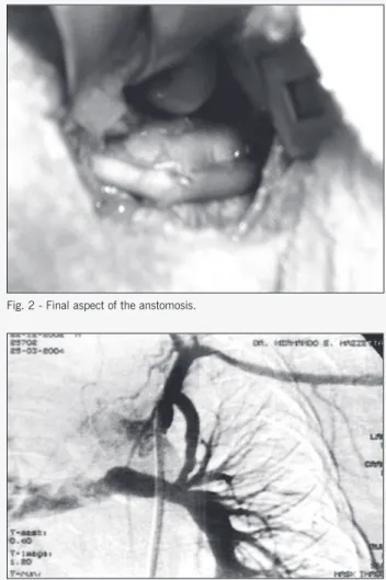
The surgical technique was as follows: 8 patients were placed in the right lateral decubitus position; they underwent left lateral thoracotomy according to the anatomic location of the aortic arch; opening was performed in the third or fourth intercostal space; anastomosis of the graft to the subclavian artery and the pulmonary artery was performed with Prolene 7-0 thread, taking care not to clamp the graft, according to the recommendation of the manufacturer (fig. 1). The other 2 patients underwent right thoracotomy, and the shunt was performed with the right subcla-vian artery and the right pulmonary artery, due to the location of the aortic arch. In all patients, the anastomoses were preceded by heparinization at the dosage of 1 mg/kg, and no patient required heparin reversion (fig. 2).
In the postoperative period, the immediate follow-up protocol was similar to that of the procedure used for patients undergoing the Blalock-Taussig shunt with expanded polytetrafluoroethylene (PTFE) graft, which is as follows: all patients were heparinized with 400 to 600 U/kg/day of sodium heparin in continuous infusion beginning 2 hours after admission at the ICU; their APTT was maintained 1.5 to 2.5 times the normal value, and heparin was
T T T T
Table I - Distribution of patients according to age, sex, diagnosis, surgerable I - Distribution of patients according to age, sex, diagnosis, surgerable I - Distribution of patients according to age, sex, diagnosis, surgerable I - Distribution of patients according to age, sex, diagnosis, surgerable I - Distribution of patients according to age, sex, diagnosis, surgeryyyyy, and graf, and graf, and graf, and graf, and graft size.t size.t size.t size.t size.
Patient Age Sex Diagnosis Surgery Graft size (mm)
1 2 months M AVSD + RVOT stenosis MBT to the L 4
2 8 days F T4F + Pulmonary atresia MBT to the R 4
3 3 days F Pulmonary and tricuspid atresia MBT to the L 4
4 2 months F Pulmonary and tricuspid atresia MBT to the L 4
5 3 months M Tricuspid atresia + TGA + PVS MBT to the L 4
6 4 days F Pulmonary and tricuspid atresia MBT to the R 4
7 7 years M Pulmonary and tricuspid atresia +
Coronary cavitary fistula RC-RV MBT to the L 5
8 12 days M T4F + Pulmonary atresia MBT to the L 4
9 10 months M T4F MBT to the L 4
10 10 months M T4F MBT to the L 4
M- male; F- female; AVSD- atrioventricular septal defect; RVOT- right ventricular outflow tract; TGA- transposition of the great arteries; PVS- pulmonary valve stenosis; T4F- tetralogy of Fallot; RC- right coronary; RV- right ventricle; MBT- modified Blalock-Taussig; R- right; L- left; mm- millimeter.
Fig. 1 - Re-hydration of the D-hydro graft in saline solution and heparin at the time of implantation.
Fig. 2 - Final aspect of the anstomosis.
Fig. 3 - Arteriography showing a patent Blalock Taussig shunt to the left, with a well-developed pulmonary tree.
kept for 24 hours, being then replaced by acetylsalicylic acid, at the dosage of 5 mg/kg/day, for 3 months.
In late follow-up, our protocol included clinical, echocardio-graphic, and arteriographic assessments in all patients on the occasion of indicating definitive correction (fig. 3).
Results
3
Arquivos Brasileiros de Cardiologia - Volume 84, Nº 3, Março 2005Volume 84, Nº 3, Março 2005Volume 84, Nº 3, Março 2005Volume 84, Nº 3, Março 2005Volume 84, Nº 3, Março 2005 Initial Analysis of the Use of the L-D-Hydro (Eato L-D-Hydro) Organic Tubular Graft for Performing the Modified Blalock-Taussig Procedure in Congenital Heart Diseases with Decreased Pulmonary Blood Flow
improvement in oxygen saturation on pulse oximetry and arterial blood gas analysis. The mean initial arterial oxygen saturation was 69.4% ± 1.17%, with a significant increase to 90.1% ± 1.85% immediately after the procedure. The length of stay in the intensive care unit ranged from 2 to 6 days.
No patient had obstruction of the shunt in the immediate postoperative period or any other complication, although the clas-sical modified Blalock-Taussig murmur could not be heard with its usual intensity, which can be explained by the elasticity of the graft. All patients showed, on the echocardiographic study, a patent shunt with turbulent flow (fig. 4). In the immediate post-operative period, the diameter of the graft ranged from 4.3 to 5.4 mm (4.68±0.29), and, in the third month, from 4.2 to 5.2 mm (4.48±0.29) (tab. II).
No bleeding was observed during surgery or in the postoperative period.
Discussion
Usually, the indications for the use of the systemic-pulmonary shunt varied among the institutions as follows: cyanogenic complex defects; hypoplasia of the pulmonary arteries; hypoplasia of the pulmonary ring, which required a transannular flap for complete repair; abnormality of the pulmonary arteries; neonates with tetra-logy of Fallot and pulmonary atresia; and young low-weight neo-nates. In addition, the systemic-pulmonary shunt is used when the mortality of the total correction is greater than that of the 2-stage repair 5-12.
Currently, the most used shunt in most services is the modified Blalock-Taussig, which has a mortality rate lower than 1% 12. De
Leval and Stark 5, McKay et al 6, and other authors reported
excellent results with the modified Blalock Taussig. The shunt has adequate patency, a low index of surgical complications, low mortality, and allows the growth of the pulmonary tree, without the risks of the secondary repair 13-21. In our study, no patient died
in the immediate postoperative period; all patients had an imme-diate improvement in oxygen saturation on pulse oximetry and arterial blood gas analysis (from 69.4% ± 1.17% to 90.1% ± 1.85%). The diameter and characteristic of the flow, shown on echocardiography, confirmed the efficacy of the shunt.
Berger 19 and other authors 22-26 have reported high postoperative
morbidity and mortality in the procedures of Potts et al 2 and
Waterston 3. Kirklin et al 10 have identified age (below 3 months)
as a risk factor for postoperative mortality, mainly when other risk factors coexisted. Arciniegas et al 29 have also identified age as a
risk factor for postoperative mortality as follows: 6% for one month, 4% for 3 months, 3% for 6 months, and 2.5% for 12 months 6,28.
Alkhulaifi et al 30 have identified low weight and preoperative
ventilation as risk factors for mortality. Khalid et al 31 have related
low weight (below 3 kg) to the early failure of the shunt, and identified the use of intra- and postoperative heparin as a protective
Fig. 4 - Echocardiographic image during late postoperative period on continuous color Doppler, flow by the Blalock Taussig.
T T T T
Table II - Pable II - Pable II - Pable II - Postoperative assessment through Doppler echocardiography in patients undergoing systemic-pulmonarable II - Postoperative assessment through Doppler echocardiography in patients undergoing systemic-pulmonarostoperative assessment through Doppler echocardiography in patients undergoing systemic-pulmonary anastomosis with the Lostoperative assessment through Doppler echocardiography in patients undergoing systemic-pulmonarostoperative assessment through Doppler echocardiography in patients undergoing systemic-pulmonary anastomosis with the Ly anastomosis with the Ly anastomosis with the Ly anastomosis with the L-D Hydro graf-D Hydro graf-D Hydro graf-D Hydro graf-D Hydro grafttttt
Patient Diagnosis Graft diameter Type of flow
Immediate (mm) 3 months (mm) Immediate 3 months
1 AVSD + RVOT stenosis 4.6 4.5 Turbulent Turbulent
2 T4F + Pulmonary atresia 4.5 4.3 Turbulent Turbulent
3 Pulmonary and tricuspid atresia 4.8 - Turbulent
-4 Pulmonary and tricuspid atresia 4.3 4.2 Turbulent Turbulent
5 Tricuspid atresia + TGA + PVS 4.6 4.4 Turbulent Turbulent
6 Pulmonary and tricuspid atresia 4.8 4.5 Turbulent Turbulent
7 Pulmonary and tricuspid atresia + Coronary cavitary fistula RC-RV 5.4 5.2 Turbulent Turbulent
8 T4F + Pulmonary atresia 4.7 4.5 Turbulent Turbulent
9 T4F 4.5 4.3 Turbulent Turbulent
10 T4F 4.6 4.2 Turbulent Turbulent
4
Arquivos Brasileiros de Cardiologia - Volume 84, Nº 3, Março 2005Volume 84, Nº 3, Março 2005Volume 84, Nº 3, Março 2005Volume 84, Nº 3, Março 2005Volume 84, Nº 3, Março 2005
Initial Analysis of the Use of the L-D-Hydro (Eato L-D-Hydro) Organic Tubular Graft for Performing the Modified Blalock-Taussig Procedure in Congenital Heart Diseases with Decreased Pulmonary Blood Flow
1. Blalock A. and Taussig H. The surgical treatment of malformations of the heart in which there is pulmonary stenosis or pulmonary atresia. JAMA. 1945; 128: 189. 2. Potts W, Smith S, Gibson S. Anastomosis of aorta to pulmonary artery. Certain
types in congenital heart disease. JAMA. 1946; 132: 627.
3. Waterston D. Treatment of Fallot’s tetralogy in children under one year of age. Rozhl Chir. 1962; 41: 181.
4. Redo S. Ecker R. Intrapericardiac aorto-pulmonary artery shunts. Circulation. 1963; 28: 520.
5. De Leval M, J Stark. Systemic to Pulmonary and Cavopulmonary Shunts. Surgery for Congenital Heart Defects. WB Saunders. Philadelphia. 1983; 11: 175-86. 6. McKay R, Leval M, Rees P, et al. Postoperative angiographic assessment of
modi-fied Blalock-Taussig shunts using expanded polytetrafluoroethylene (Gore-Tex). Ann Thorac Surg. 1980; 30: 137-45.
7. Arciniegas E, Farooki ZQ, Hakimi M, et al. Results of two-stage surgical treatment of tetralogy of Fallot. J Thorac Cardiovasc Surg. 1980; 79: 876-83.
8. Kirklin JW, Blackstone CH, Pacifico AD, et al. Routine primary repair vs two-stage repair of tetralogy of Fallot. Circulation. 1979; 60: 373-86.
9. Kirklin JW, Ksro RB. The Tetralogy of Fallot from a Surgical Viewpoint. Philadel-phia, Saunders WB. 1970; 18-19: 119-53.
10. Kirklin JW, Barrat-Boyes BG, Kbe MD. Morphology, diagnostic criteria, natural history. Techniques, results and indication. In Cardiac Surgery. New York, John Wiley & Sons, inc. 1986; 699-819.
11. Siwik ES, Patel CR, Zanka KG. Tetralogy of Fallot. Moss and Adam’s. Heart Disea-se in Infants, Children and Adolescents. 6ª ed. 2001; 42: 880-902.
12. Karl TR. Tetralogy of Fallot. Thoracic and Cardiovasc Surg. 6ª ed. 1995; 81: 1345-68. 13. Arciniegas E. Tetralogy of Fallot. Pediatric Cardiac Surg. 1985; 13: 203-18. 14. Park MK. Tetralogy of Fallot. Pediatric Cardiology for Pratitioners. 4ª ed. 2002; 14:
189-95.
15. Kurosa WA, Himai Y, Taksnsdhi Y, et al. Standartized patch infundibuloplasty for tetralogy of Fallot. In second World congress of pediatric cardiology. New York, Springer-Verlag. 1986; 5.
16. Garsen A. Tetralogy of Fallot and pulmonary atresia. Pediatr Cardiol.1997; 1: 1401-02. 17. Haworth SG. Pulmonary hypertension in child hood. Eur Respir J. 1993; 6:1037-43 18. Rabinovitch M. Pulmonary hypertension: updating a mysterious disease.
Cardio-vasc Res. 1997; 34: 268-72.
References
19. Berger RMF. Possibilities and impossibilities in evaluation of pulmonary vascular disease in congenital heart defects. Eur Heart J. 2000; 21: 17-27.
20. Chiariello L, Meyer J, Wukasch DC, et al. Intracardiac repair of tetralogy of Fallot. Five-years of 403 patients. J Thorac Cardiovasc Surg. 1975; 70: 529-35. 21. Kay PH, Capuani A, Franks R, et al. Experience with modified Blalock-Taussig
operation using polytetrafluoroethylene (Impra) grafts. Bri Heart J. 1983; 65: 403-10.
22. De Leval M, McKay R, Jones M, et al. Modified Blalock-Taussig shunt. Use of sub-clavian artery orifice as flow regulation in prosthetic systemic-pulmonary shunts. J Thorac Cardiovasc surg. 1981; 81: 112-9.
23. Diaz AF, Sanz E, Sanchez PA, et al. Fallot’s tetralogy – palliation and repair with a previous shunt. Pediatr Cardiol. 1977; 273-82.
24. Somerville J, Barbosa R, Ross D, et al. Problems with radical corrective surgery after ascending aorta to right pulmonary artery shunt (Waterston’s anastomosis) for cyanotic congenital heart disease. Bri Heart J. 1975; 37: 1105-12. 25. Riker WL. Intracardiac lesions from common congenital heart lesions. Surg Clin of
North Am. 1963; 43: 133-45.
26. White BD, McNamara DG, Baversfeld SR, et al. Five years post operative results of first 500 patients with Blalock-Taussig anastomosis for pulmonary stenosis or atresia. Circulation. 1956; 14: 515-9.
27. Bircks W, Ostermeyer V. Surgical correction of Tetralogy of Fallot after palliative operations. J Thorac Cardiovasc Surg. 1984; 32: 224-7.
28. Kirklin JW, Blackstone CH, Jonas RA, et al. Morphologic and surgical determi-nants of outcome events after repair of tetralogy of Fallot and pulmonary steno-sis. A two-institution study. J Thorac Cardiovasc Surg.1992; 103: 706-23. 29. Arciniegas E, Blackstone EH, Pacifico AD, et al. Classical shunts operations as part
of two-stage repair for tetralogy of Fallot. Ann Thorac Surg. 1979; 27: 514-8. 30. Alkhulaifi AM, Lacour-Gayet F, Planché C, et al. Systemic pulmonary shunts in
neo-nates: early clinical outcome and choice of surgical approach. Ann Thorac Surg. 200; 69: 1499-504.
31. Khalid A, Al Jubair, Sawyer W, et al. Results of 546 Blalock-Taussig shunts perfor-med in 478 patients. Cardiol Young. 1998; 8: 486-90.
32. Berger RMF. Heparin as a risk factor for perigraft seroma complicating the modified Blalock-Taussig shunt. J Thorac Cardiovasc surg. 1998; 116:286-92.
factor for reducing shunt failure and early obstruction in 1.6% of the modified Blalock Taussig. They have also reported that patients with shunts with a greater caliber and those who received no heparin had a greater incidence of occlusion. In our study, no obstruction was observed in the period assessed, and permeability of the shunt was shown on Doppler echocardiographic study.
In our case series, one patient died due to sepsis on the third postoperative day, and, therefore, a cause not directly related to the graft.
Berger et al 32, using the PTFE graft, reported 12% of serum
collection around the graft in patients receiving heparin in the
postoperative period, a complication that was not observed in our case series, although some cases may have remained undetected. On clinical assessment, we observed that the intensity of the murmur was lower than usual, which may be explained by the elasticity of the graft.