Heart Institute (InCor) of São Paulo University - Brazil. Maddress: Mauricio I. Scanavacca
Unidade de Arritmias Cardíacas do Instituto do Coração - HC- FMUSP Av. Dr. Enéas de Carvalho Aguiar, 44 - São Paulo (SP) - Cep 05488-000 E-mail: [email protected]
Arq Bras Cardiol, volume 82 (nº 2), 160-4, 2004
Mauricio Scanavacca, Raul Sartini, Fernando Tondato, André d’Ávila, Denise Hachul, Francisco Darrieux, Sissy Lara, Eduardo Sosa
São Paulo, SP - Brazil
Pulmonary Veins Isolation to Treat Patients with Refractory
Paroxysmal Atrial Fibrillation. Clinical Results After a Single
Procedure
Radiofrequency (RF) ablation of ectopic beats from pulmonary veins that trigger atrial fibrillation has been re-cently introduced in the clinical practice1,2. Although
effec-tive, this technique can only be used for few patients who present quite active foci, which allow electrophysiological mapping and precise localization3-5. The strategy for
pa-tients with focal atrial fibrillation without active foci during electrophysiological mapping was to apply provocative maneuvers to induce their activity6. Using this strategy
pa-tients presented frequent recurrences, as many ectopic foci were present in other veins not identified in the first study7.
Therefore, the empirical isolation of the four pulmonary veins has been suggested as the most effective method to treat those patients8-10. However, there are some
controver-sies about the clinical results and necessity of new procedu-res to obtain a good clinical control of these patients11, 14.
The aim of this study was to assess the clinical results ob-tained from patients with refractory paroxysmal atrial fibril-lation submitted to a single radiofrequency procedure to empirically isolate three or four pulmonary veins.
Methods
From January 2001 to August 2002, forty-nine consecu-tive patients were prospecconsecu-tively studied. They presented re-fractory paroxysmal atrial fibrillation and were referred to ra-diofrequency catheter ablation, aiming to empirically isolate pulmonary veins of the left atrium. Thirty-six men and 13 women, mean age of 54±10 years old (32 to 76 y.o.) presenting atrial fibrillation for more than two years and having unsuc-cessfully used at least three antiarrhythmic drugs before con-sidering atrial fibrillation ablation were included in this study. Patients presenting frequent atrial premature contractions (APC) or atrial tachycardia triggering atrial fibrillation during the electrophysiological mapping were excluded. In those patients the procedures were referred for specific foci mapping and focal ablation. Atrial fibrillation was regarded Objective - The purpose of this study was to access the
clinical outcome of patients submitted to a single proce-dure of radiofrequency pulmonary veins (PV) isolation to treat refractory paroxysmal atrial fibrillation (AF).
Methods - This study included 49 consecutive patients (36 male; mean age 54±10 years old) who had frequent symptomatic paroxysmal AF refractory to at least three antiarrhythmic drugs. We used a circular decapolar ca-theter for mapping PVs - left atrial connections and a 4-mm distal tip catheter for ablation (30 W and 50 C), aiming to achieve electrical isolation of 3 -4 PVs.
Results - Twenty-five patients (51%) did not present any AF recurrence in a mean follow-up of 12±5 months. Twenty-four (49%) had at least one recurrence during out-come; twenty (83%) of them within the first month after the procedure and four after two to nine months. After introdu-cing antiarrhythmic drugs 15 (63%) patients were under control, 10 were asymptomatic and five complained of sporadic short duration AF episodes. Nine (37%) pa-tients remained very symptomatic despite the use of antiar-rhythmic drugs and were referred to a new procedure of PV isolation. No patient presented major complications. At the end of the follow-up, 35 (71%) patients remained in stable sinus rhythm with no AF recurrences after a single procedure, 50% of them without antiarrhythmic drugs.
Conclusion - Most patients who present symptomatic paroxysmal AF refractory to antiarrhythmic drugs obtain a good clinical control after a single PV isolation procedure.
paroxysmal when episodes were spontaneously reverted be-fore 48 hs. Patients submitted to electrical (ECV) or chemical (CCV) cardioversion were enrolled in this study if ECV or CCV were performed within a period of 48 hs from the beginning of the crises. Among 49 patients, five presented chronic coro-nary disease (two with previous myocardial infarction); one presented mild dilated cardiomyopathy; one assymetric hy-pertrophic myocardiopathy and the others did not present structural heart disease. Seven patients had systemic arterial hypertension under clinical control. The mean left atrium size was 40 ± 6 mm (29 to 50 mm). Patients underwent oral anticoa-gulation from four to six weeks (RNI between 2.5 and 3.5) or performed transesophageal echocardiography within 24 hs to assure there was not any atrial thrombus before the proce-dure. Pulmonary magnetic resonance angiography (MRI) with Gadolinium was suggested but not mandatory to per-form the study.
Patients were taken to the electrophysiologic room after 8 hs of fasting state, and sedated with intravenous propofol. Arterial blood pressure, digital oximetry and CO2 exhaled were
non-invasively monitored. Three multipolar electrode cathe-ters were inserted into the femoral vein and placed under fluoroscopy guidance. One 6 F decapolar catheter was inser-ted into the coronary sinus; one 6 F circular decapolar (“Lasso” – J&J) catheter was introduced by a long sheath in the left atrium through the foramem oval or by transeptal puncture. One 7 F or 8 F (EPT or J&J) regular catheter for RF ablation with a 4-mm distal tip electrode was also introduced in the left atrium by a second transeptal puncture if necessary. Heparin was infused (10.000 IU) and the activated coagula-tion time (ACT) was evaluated after 15 minutes, repeated each hour, aiming to maintain ACT between 250 and 350 se-conds. Pulmonary veins angiography was performed with Meglumine loxalate (32,5 g/100ml) using one of the long tran-septal sheathes, except for the right inferior pulmonary vein that was not systematically evaluated. The pulsed fluorosco-pic images in a 7 square / second were obtained (Fisher sys-tem) in the left anterior oblique position (400) for the left
pul-monary veins and in the right anterior oblique position (300)
for the right pulmonary veins. In this system the fluoroscopic time measurement is restricted to X-ray pulsed-time15.
Elec-trophysiological data were digitally recorded by a PC Electro-physiologic Measurement System (EMS 4.2 – University of Limburg - The Netherlands).
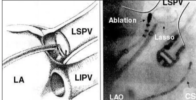
The pulmonary veins isolation technique consisted in mapping the left veno-atrial junction using the “Lasso” ca-theter to localize pulmonary veins in connection with the left atrium and disconnect them by applying RF pulse of 30 W and 500 C from 30 to 60 seconds. The “Lasso” decapolar
ca-theter was introduced in the pulmonary vein after angiogra-phy and then removed until being nearly five mm of the os-tium (Figure 1). Pulmonary vein isolation was performed if potentials were present. The vein isolation criterion was the disappearance of electrical potentials in the vein during si-nus rhythm or continuous atrial stimulation (entrance block) (Figure 2). The isolation of such veins was performed under continuous electrical stimulation of the distal
coronary sinus in order to distinguish left vein potentials from left atrial appendage electrograms. The right pulmona-ry veins were mapped during sinus rhythm with no atrial sti-mulation. RF ablation was initially directed towards the most suggestive point regarding the atrial vein connection localization and kept for 30 s. Conduction block through the veno-atrial connection was shown by conduction delay in this specific sector and by the change in activation sequen-ce of the ostium analyzed by the circular catheter activation (figure 2). Applications were repeated in other connection points until complete isolation of pulmonary vein was obtai-ned. After 10 to 12 pulses sequence, we performed evalua-tion of the pulmonary vein permeability by angiography with contrast. Applications were stopped when there was reduction of vein ostium size or still due to the amount of applications, initially and empirically established around 20 to 25 applications. Pulmonary vein isolation was complete when all venous potentials obtained from the circular cathe-ter disappeared. However, when amplitude of such poten-tials was reduced but with no complete disappearance, we considered pulmonary vein connections just as partially modified. After the end of the procedure, infusion of 10 to 40 micrograms of isoproterenol was performed followed by 12 to 18 mg of adenosine, in order to identify ectopic triggers outside the veins. Patients were kept in resting position du-ring 4 hours and in general discharged the day after. Subcuta-neous low molecular weight heparin (1 mg/Kg/ twice a day) was started six hours after the end of the procedure and main-tained until oral coagulation was obmain-tained (INR 2.5 to 3.5). Antiarrhythmic maintenance depended on symptoms re-currence within the first 24 hours or on the discomfort inten-sity provoked by atrial fibrillation crises before ablation.
Clinical follow-up was performed according to clinical appointments in our institution, or still by telephone contact with patients unable to come personally. Patients with pul-monary vein stenosis suspicion were evaluated by pulmo-nary vein angiography, transesophageal echo or pulmona-ry ventilation/perfusion cintilography one to six months after the procedure. Patients were clinically evaluated in one month and each three months. In case of recurrence, we suggested EKG documentation of the crise. Ambulatorial
monitoring (Holter and symptomatic events recordings) was used to clarify suggestive symptoms of atrial fibrilla-tion recurrence not documented in EKG. Typical symptoms of atrial fibrillation recurrence were accepted in the absence of EKG documentation.
As Statistical Analysis we used the Student t-test to compare continuous variables and the Fisher test to com-pare proportions. We considered 5% a significant statistical level.
Results
We performed ostial radiofrequency ablation in 184 veins (49 patients - 3.7 veins per patient). A mean of 21±12 RF pulses per vein was applied in each patient: 25±14 at the left superior pulmonary vein (LSPV) ostium; 22±10 at the right superior pulmonary vein (RSPV); 13±9 at the left inferi-or pulmonary vein (LIPV) and 23±12 at the right inferiinferi-or pul-monary vein (RIPV). Procedure mean time and X ray time exposition were 231±47 min and 12±4 min, respectively. Complete isolation was obtained in 146 out 184 (79%) veins, 40 LSPV (82%), 41RSPV (84%), 35 (81%) and 30 of 45 RIPV (67%). All four pulmonary veins were completely isolated in 18 patients; three veins in 20 patients; two veins in six; one vein in three and two patients did not have any vein
comple-tely isolated. In those cases, just the ostium electrogram amplitude reduction was obtained.
Patients did not present any important complication during hospitalization and follow-up. No patient presented important hematoma or vascular complication. There were no complications related to transeptal puncture, pulmonary vein stenosis or embolic events.
After ablation, 25 patients (51%) did not present recur-rence in a mean follow-up of 12±5 months. Of those, 22 (88%) had three or four pulmonary veins isolated and three (12%) had two or less veins isolated. Nineteen patients mained asymptomatic and six complained of palpitations re-lated to premature contractions. Fifteen (60%) patients were not using antiarrhythmic drugs; four (16%) were using be-ta-blockers, three due to systemic arterial hypertension and one due to premature ectopic beats perception; and six (24%) were under amiodarone.
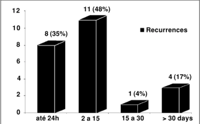
Twenty-four (49%) patients presented at least one re-currence during follow-up; in 20 (83%) rere-currence occurred within the first month after RF ablation, and in four (17%) after two to nine months of follow-up (figure 3). After intro-ducing antiarrhythmic drugs (beta-blockers: six, amiodaro-ne: six, sotalol: three, propafenoamiodaro-ne: two, flecainide: one and diltiazen: one), fifteen (63%) patients presented important symptoms relief, ten became asymptomatic (including four
Fig. 2 - Left inferior pulmonary vein (LIPV) isolation. Basal: Pulmonary vein electrograms from 1 (distal) to 10 (proximal) obtained by the circular (“Lasso”) catheter positioned in the LIPV ostium during atrial pacing (st) in distal coronary sinus (CS). RF: Note the “Lasso” catheter electrogram activation changing during radiofrequency (RF) application secondary to entrance conduction block in a sector of the LIPV ostia. Disconnection: LIPV isolation. Observe that pulmonary vein potentials obtained by “Lasso” catheter disappeared. CS pacing activates the left atrium, but it does not achieve the interior of LIPV.
V1
10
9
8
7
6
5
4
3
2
1
CS LIPV
Basal RF Disconnection
Pulmonary
vein potentials
patients who stopped drugs) and five referred sporadic and short AF episodes. Nine (37%) remained with symptoms unchanged despite the use of antiarrhythmic drugs and seven were submitted to a second procedure. At the end of the follow-up, 35 (71%) of the patients remained in stable si-nus rhythm without atrial fibrillation recurrences after a sin-gle procedure. No patient presented symptoms related to systemic emboli or pulmonary veins stenosis.
Discussion
The electrophysiologic observation that atrial fibrilla-tion is triggered by ectopic foci frequently originated in the pulmonary veins opened a new era in radiofrequency catheter ablation. However, just few patients presented frequent spontaneous ectopic beats enough to allow mapping of spe-cific foci2-5. The attempts to apply the potential clinical
bene-fits of AF triggers by catheter ablation presented major limita-tions. Difficulty to localize all ectopic foci which triggered atrial fibrillation and low reproducibly of the provocative ma-neuvers to induce the triggers resulting in high AF recurrence rate after ablation, consequently, many ablation procedures were necessary to obtain good clinical control6- 8.
Additional-ly, multiples radiofrequency pulses applied in the ostium of pulmonary veins may cause pulmonary vein stenosis, some-times resulting in severe pulmonary hypertension 16 – 17.
Aiming to solve these problems two strategies were develo-ped in order to isolate the four pulmonary veins in a single procedure with low risk of pulmonary veins stenosis: map-ping and ablating specific sectors of pulmonary veins ostia8,9
and circumferential ablation of pulmonary veins ostia using a 3-D electro-anatomic system10, 11.
The pulmonary vein isolation technique developed by Haissaguerre et al aims to electrically isolate pulmonary veins minimizing the stenosis risk8,9. It is based on
recogni-zing veno-atrial junction connections by the circular cathe-ter positioned at the pulmonary veins ostia18, 19. Through
the use of this technique, radiofrequency applications can be optimized and the stenosis risk lower. Haisaguerre et al, reported 3% of symptomatic pulmonary veins stenosis9.
Oral et al, applying the same technique did not report
pulmo-Figure 3 - Atrial fibrillation recurrence rate after pulmonary vein isolation. Observe that 19 (82%) of 23 recurrences occurred in the first 15 days after the procedure.
nary veins complications12. Marrouche et al, reported
pul-monary vein stenosis during high energy radiofrequency application inside the veins through irrigated tip catheters. Pulmonary vein stenosis were not observed after using intracardiac echocardiogram to adequate radiofrequency applications13. In this study we did not observe any
manifestation related to pulmonary veins stenosis. Howe-ver, clinical manifestation occurs just when the pulmonary vein stenosis is very important, mainly when there is more than one vein involved16, 17. Symptoms, in general, occur in
the first days after the procedure, but can occur later20. In
this study, we did not evaluate pulmonary veins systemati-cally during follow-up, however all patients but one were followed for more than six months and none of them reported any symptom related to pulmonary veins stenosis. These initial data confirm other observations suggesting that abla-tion of pulmonary vein ostia applying low energy radiofre-quency pulses presents low risk of promoting pulmonary vein stenosis. Nevertheless, further studies are necessary to confirm the safety of this procedure.
There are some controversies related to the clinical re-sults of pulmonary veins isolation to treat patients with recur-rent atrial fibrillation. Stable sinus rhythm without atrial fibrilla-tion recurrence has been reported from 70% to 94%11 –14, 21.
This success rate variation can be related to patients selection, pulmonary veins isolation technique, number of procedures applied, antiarrhythmic drugs used and patient’s follow-up22.
Ectopic foci located outside the pulmonary vein ostia or related to other atrial structures have been described as a cause of atrial fibrillation recurrence after pulmonary veins isolation23, 24.
In our study, some patients without complete PV isolation had also good clinical evolution and radiofrequency applications around the pulmonary veins ostia could be responsible for that. Those lesions applied around the pulmonary veins ostia could ablate focal triggers of atrial fibrillation and could explain why two of our patients without any PV completely isolated became free of atrial fibrillation recurrence, one of them without an-tiarrhythmic drugs. Pappone et al, have systematically applied RF lesions around the four pulmonary veins. Although pulmo-nary veins isolation was obtained in just 75% of all patients, 85% of them did not present atrial fibrillation, most of them wi-thout antiarrhythmic drugs10,11. However, reconnection of
pul-monary vein previously isolated seems to be the most common cause of recurrence and obtaining a definitive, without increasing the pulmonary veins stenosis risk is one of the technical challenges to be solved. Marrouche et al has suggested that performing deep lesions outside the vein through an 8-mm tip catheter is related to better results13.
Ho-wever, Macle et al, using a 4-mm irrigated tip had to perform two or three procedures in 49% of patients obtaining clinical control in 80% of them, 44% using antiarryhthmic drugs14.
In this study 47% of our patients presented at least one recurrence after pulmonary veins isolation. In most of them recurrences occurred within two weeks after radiofre-quency ablation. But, after the first month they became asymptomatic or oligosymptomatic after antiarrhythmic in-troduction. This observation was also reported by other
12
10
8
6
4
2
0
até 24h 2 a 15 15 a 30 > 30 days 8 (35%)
11 (48%)
Recurrences
1 (4%)
authors13, 25. The radiofrequency lesion repair process could
be responsible for this evolution13. Otherwise, an
inflamma-tory process induced by radiofrequency lesions on the pul-monary veins ostia could induce atrial fibrillation transitory episodes. Such observations were reported by other au-thors that suggested to perform a new intervention for pul-monary vein isolation just after six or eight weeks of the first procedure11, 14. Postponing a new intervention for 4 to 6
weeks and restricting it to symptomatic patients non
con-trolled by antiarrhythmic drugs we indicated a new procedu-re just for 16% of our patients.
In summary, most patients presenting refractory paro-xysmal atrial fibrillation obtain a good clinical control after a single procedure of empirical isolation of four pulmonary veins. Atrial fibrillation recurrences, when occur, are more frequent in first two weeks after the procedure, but, in most patients it is possible to obtain a good clinical control during outcome.
1. Haissaguerre M, Jais P, Shah DC, Takahashi A, Hocini M, Quiniou G, Garrigue S, Le Mouroux A, Le Metayer P, Clementy J. Spontaneous initiation of atrial fibril-lation by ectopic beats originating inthe pulmonary veins. N Engl J Med 1998;339(10):659-66.
2. Chen SA, Hsieh MH, Tai CT, Tsai CF, Prakash VS, Yu WC, Hsu TL, Ding YA, Chang MS. Initiation of atrial fibrillation by ectopic beats originating from the pulmonary veins: electrophysiological characteristics, pharmacological responses, and effects of radiofrequency ablation. Circulation 1999;100(18):1879-86. 3. Scanavacca M, Sosa E, d’Avila A, Tondato F, Darrieux F, Hachul D, Bahia A,
Ca-valcanti P, Oliveira F. Radiofrequency catheter ablation in patients with atrial fi-brillation. Arq Bras Cardiol 1999;72(6):693-708.
4. Rocha Neto AC, Farias RL, de Paola AA. Treatment of atrial fibrillation with ra-diofrequency ablation and simultaneous multipolar mapping of the pulmonary veins. Arq Bras Cardiol 2001 Nov;77(5):407-28.
5. Mehta N, Távora MZ, Takeschita N, Figueiredo E, Lourenco RM, Germiniani H, Precoma D. Useful clinical features for the selection of ideal patients with atrial fi-brillation for mapping and catheter ablation. Arq Bras Cardiol 2002;78(1):1-16. 6. Shah DC, Haissaguerre M, Jais P, Hocini M, Yamane T, Deisenhofer I, Garrigue S, Clementy J. Curative catheter ablation of paroxysmal atrial fibrillation in 200 pa-tients: strategy for presentations ranging from sustained atrial fibrillation to no arrhythmias. Pacing Clin Electrophysiol 2001;24(10):1541-58.
7. Gerstenfeld EP, Guerra P, Sparks PB, Hattori K, Lesh MD. Clinical outcome after radiofrequency catheter ablation of focal atrial fibrillation triggers. J Cardiovasc Electrophysiol 2001;12(8):900-8.
8. Haissaguerre M, Jais P, Shah DC, Garrigue S, Takahashi A, Lavergne T, Hocini M, Peng JT, Roudaut R, Clementy J. Electrophysiological end point for catheter ablation of atrial fibrillation initiated from multiple pulmonary venous foci. Cir-culation 2000;101(12):1409-17.
9. Haissaguerre M, Shah DC, Jais P, Hocini M, Yamane T, Deisenhofer I, Chauvin M, Garrigue S, Clementy J. Electrophysiological breakthroughs from the left atrium to the pulmonary veins. Circulation 2000;102(20):2463-5
10. Pappone C, Rosanio S, Oreto G, Tocchi M, Gugliotta F, Vicedomini G, Salvati A, Dicandia C, Mazzone P, Santinelli V, Gulletta S, Chierchia S. Circumferential ra-diofrequency ablation of pulmonary vein ostia: A new anatomic approach for cu-ring atrial fibrillation. Circulation 2000;102(21):2619-28.
11. Pappone C, Oreto G, Rosanio S, Vicedomini G, Tocchi M, Gugliotta F, Salvati A, Dicandia C, Calabro MP, Mazzone P, Ficarra E, Di Gioia C, Gulletta S, Nardi S, Santinelli V, Benussi S, Alfieri O. Atrial electroanatomic remodeling after circum-ferential radiofrequency pulmonary vein ablation: efficacy of an anatomic ap-proach in a large cohort of patients with atrial fibrillation. Circulation 2001;104 (21):2539-44.
12. Oral H, Knight BP, Tada H, Ozaydin M, Chugh A, Hassan S, Scharf C, Lai SW, Gre-enstein R, Pelosi F Jr, Strickberger SA, Morady F.Pulmonary vein isolation for paroxysmal and persistent atrial fibrillation. Circulation 2002 105(9):1077-81. 13. Marrouche NF, Dresing T, Cole C, Bash D, Saad E, Balaban K, Pavia SV,
References
Schweikert R, Saliba W, Abdul-Karim A, Pisano E, Fanelli R, Tchou P, Natale A. Circular mapping and ablation of the pulmonary vein for treatment of atrial fibril-lation: impact of different catheter technologies. J Am Coll Cardiol 2002;40 (3):464-74.
14. Macle L, Jais P, Weerasooriya R, Hocini M, Shah DC, Choi KJ, Scavee C, Raybaud F. Clementy J, Haissaguerre M. Irrigated-tip catheter ablation of pulmonary veins for treatment of atrial fibrillation. J Cardiovasc Electrophysiol 2002 Nov;13 (11):1067-73.
15. Scanavacca M, d’Avila A, Velarde JL, Reolao JB, Sosa E. Reduction of radiation exposure time during catheter ablation with the use of pulsed fluoroscopy. Int J Cardiol 1998 Jan 5;63(1):71-4.
16. Robbins IM, Colvin EV, Doyle TP, Kemp WE, Loyd JE, McMahon WS, Kay GN. Pulmonary vein stenosis after catheter ablation of atrial fibrillation. Circulation 1998;98(17):1769-75.
17. Scanavacca MI, Kajita LJ, Vieira M, Sosa EA. Pulmonary vein stenosis complica-ting catheter ablation of focal atrial fibrillation. J Cardiovasc Electrophysiol 2000;11(6):677-81.
18. Cabrera JA, Sanchez-Quintana D, Farre J, Navarro F, Rubio JM, Cabestrero F, An-derson RH, Ho SY. Implications for catheter ablation. Ultrasonic characteriza-tion of the pulmonary venous wall: echographic and histological correlacharacteriza-tion. Circulation 2000;101(11):1274-81.
19. Ho SY, Cabrera JA, Tran VH, Farre J, Anderson RH, Sanchez-Quintana D. Archi-tecture of the pulmonary veins: relevance to radiofrequency ablation. Heart 2001;86(3):265-70.
20. Sohn RH, Schiller NB Left upper pulmonary vein stenosis 2 months after radio-frequency catheter ablation of atrial fibrillation Circulation 2000 Apr 4;101(13):E154-5.
21. Shah DC, Haissaguerre M, Jais P, Hocini M, Yamane T, Deisenhofer I, Garrigue S, Clementy. Electrophysiologically guided ablation of the pulmonary veins for the curative treatment of atrial fibrillation. Ann Med 2000;32(6):408-16. 22. J. Turco, P. Atrial fibrillation ablation: Role of patient enrollment and follow-up
in clinical results. J Am Coll Cardiol 2003; 41(7):1232-1233.
23. Tsai CF, Tai CT, Hsieh MH, Lin WS, Yu WC, Ueng KC, Ding YA, Chang MS, Chen SA. Initiation of atrial fibrillation by ectopic beats originating from the superior vena cava: electrophysiological characteristics and results of radiofre-quency ablation. Circulation 2000;102(1):67-74.
24. Hwang C, Wu TJ, Doshi RN, Peter CT, Chen PS. Vein of Marshall cannulation for the analysis of electrical activity in patients with focal atrial fibrillation. Circu-lation 2000;101(13):1503-5.
25. Oral H, Knight BP, Ozaydin M, Tada H, Chugh A, Hassan S, Scharf C, Lai SW, Greenstein R, Pelosi F Jr, Strickberger SA, Morady F. Clinical significance of early recurrences of atrial fibrillation after pulmonary vein isolation. J Am Coll Cardiol 2002;40(1):100-4.